Dr. Treacy's Casebook: Treating Squamous Cell Cancer
•
0 gostou•418 visualizações

Squamous cell carcinoma is the second-most common cancer of the skin (after basal cell carcinoma but more common than melanoma). It usually occurs in areas exposed to the sun. Sunlight exposure and immunosuppression are risk factors for SCC of the skin, with chronic sun exposure being the strongest environmental risk factor
![42 Aesthetic Medicine • May 2015
SPONSORED BYCASE FILES www.aestheticmed.co.uk
S K I N / D E R M AT O L O G Y
CHARACTERISTIC FEATURES OF SCC TUMORS
INCLUDE THE FOLLOWING:
The clinical appearance of SCC is highly variable but
usually presents as an ulcerated lesion with hard, raised
edges or reddish skin plaque that is slow growing
The lesion caused by SCC is often asymptomatic but
may have intermittent bleeding, especially on the lip
SCC may present as a hard plaque or a papule with tiny
blood vessels
The tumor commonly presents on sun-exposed areas
(e.g. back of the hand, scalp, lip, and superior surface
of pinna)
HISTOPATHOLOGICAL TYPES
SCC is a histologically distinct form of cancer that arises
fromtheepithelium,fromcellsshowingtissuearchitectural
characteristicsofsquamouscelldifferentiation,suchasthe
presence of keratin, tonofilament bundles, or desmosomes,
structures involved in cell-to-cell adhesion. SCC typically
initially occurs in the sixth decade of life (the 50s), but is
most common in the eighth decade (the 70s). It is twice
as prevalent in men as in women. People with darker skin
are less at risk to develop SCC. Populations with fair skin,
light hair, and blue/green/grey eyes are at highest risk of
developing the disease. The majority of invasive cutaneous
SCCs are due to exposure to ultraviolet radiation, which
damages the DNA of fair-skinned individuals. SCCs most
often arise within actinic keratoses, and less often within
Bowen’s disease. Other risk factors for invasive SSC
include2
:
Inherited predisposition to skin cancer
Smoking – especially SCC of the lip
Thermal burn scars
Longstanding leg ulcers
Immunosuppression from drugs such as ciclosporin or
azathioprine, especially in organ transplant recipients
Infection with human papillomavirus (HPV causes
carcinoma cuniculatum but rarely causes other forms
of cutaneous SCC.
TREATMENT OF INVASIVE SCC
The treatment for SCC depends upon its size and location,
thenumbertobetreated,andthepreferenceorexpertiseof
thedoctor.Patientswithlargeroraggressivelesions,orone
in a difficult site, may first require imaging with ultrasound,
CTorMRItodeterminetheextentofthetumourandtolook
for metastases in the regional lymph nodes or elsewhere.3
Surgery
Invasive SCCs are usually excised by a full thickness
surgical procedure to cut out the lesion completely. Mohs
micrographic surgery may be necessary for large, ill-
defined, deep or recurrent tumours. After excising a large
tumour, the dermatologic surgeon or plastic surgeon may
create a flap or graft to repair the defect.
Radiotherapy
Radiotherapy is sometimes used for high-risk primary skin
cancers on the face and for metastatic disease.
CONCLUSION
Squamous cell carcinoma is the second-most common
cancer of the skin (after basal cell carcinoma but more
common than melanoma). It usually occurs in areas exposed
to the sun. Sunlight exposure and immunosuppression are
risk factors for SCC of the skin, with chronic sun exposure
being the strongest environmental risk factor.4
There
is a risk of metastasis starting more than 10 years after
diagnosable appearance of squamous cell carcinoma,
but the risk is low, though much higher than with basal
cell carcinoma. The long-term outcome of squamous-cell
carcinomas is dependent upon several factors: the sub-
type of the carcinoma, available treatments, location(s)
and severity, and various patient health-related variables
(accompanying diseases, age, etc.).5-6
Generally, the long-
term outcome is positive, as less than 4% of Squamous-cell
carcinoma cases are at risk of metastasis.7
AM
REFERENCES
1. Squamouscellcancer:apracticalapproach.GoldmanGD.SeminCutanMedSurg.
1998Jun;17(2):80-95.
2. NEnglJMed.2001Mar29;344(13):975-83.Cutaneoussquamous-cellcarcinoma.
AlamM1,RatnerD.
3. http://www.dermnetnz.org/lesions/squamous-cell-carcinoma.html
4. JAmAcadDermatol.1992Jun;26(6):976-90.Prognosticfactorsforlocalrecurrence,
metastasis,andsurvivalratesinsquamouscellcarcinomaoftheskin,ear,andlip.
Implicationsfortreatmentmodalityselection.RoweDE1,CarrollRJ,DayCLJr.
5. CholletA,HohlD,PerrierP(April2012).“[Riskofcutaneoussquamouscell
carcinomas:theroleofclinicalandpathologicalreports]”8(335).pp.743–6.PMID
22545495.
6. Analysisofriskfactorsdeterminingprognosisofcutaneoussquamous-cell
carcinoma:aprospectivestudy.KayDBrantsch,MD,ChristophMeisner,PhD,Birgitt
Schönfisch,PhD,BirgitTrilling,DiplInformMed,JörgWehner-Caroli,MD,Martin
Röcken,ProfMD,HelmutBreuninger,ProfMD.TheLancetOncology.Volume9
7. Maula,Sanna-Mari.Luukkaa,Marjaana.Grénman,Reidar.Jackson,David.Jalkanen,
Sirpa.Ristamäki,Raija.(2003), IntratumoralLymphaticsAreEssentialforthe
MetastaticSpreadandPrognosisinSquamousCellCarcinomasoftheHeadand
NeckRegion](data:image/gif;base64,R0lGODlhAQABAIAAAAAAAP///yH5BAEAAAAALAAAAAABAAEAAAIBRAA7)
Recomendados
Recomendados
Mais conteúdo relacionado
Mais procurados
Mais procurados (20)
Destaque
Destaque (20)
Semelhante a Dr. Treacy's Casebook: Treating Squamous Cell Cancer
Semelhante a Dr. Treacy's Casebook: Treating Squamous Cell Cancer (20)
Mais de Dr. Patrick J. Treacy
Mais de Dr. Patrick J. Treacy (20)
Último
Último (20)
Dr. Treacy's Casebook: Treating Squamous Cell Cancer
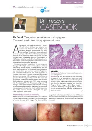
- 1. S K I N / D E R M AT O L O G Y 41Aesthetic Medicine • May 2015 SPONSORED BY CASE FILESwww.aestheticmed.co.uk Dr Patrick Treacy shares some of his most challenging cases. This month he talks about treating squamous cell cancer Dr Treacy’s CASEBOOK A 64-year-old Irish male patient with a history of male pattern baldness and leukemia was referred to Ailesbury with multiple scaly thickened reddened lesions on the area of his scalp and face. These lesions presented mostly onhisnose,templesandforeheadwiththelargestcollection along the vertex of his scalp. He had lived in South Africa for nearly 20 years. There were at least four lesions present on his face and scalp that wouldn’t heal and bled easily when traumatized. More recently his wife had become concerned because her friend had died of skin cancer. On examination there was evidence chronic skin photo- damage, with multiple actinic keratoses (solar keratoses) surrounding multiple eroded, ulcerated lesions that bled easilywhentraumatized.Thepresenceofulceratedborders, and telangiectases gave a clinical suspicion of squamous cell carcinoma and a decision was made to proceed to removal rather than do a biopsy. The author feels that any doctor should consider SCC in any patient with a history of previous chemotherapy and skin anomaly that do not heal occurring on sun-exposed skin. Unlike basal cell carcinoma (BCC), squamous cell carcinoma (SCC) has a substantial risk of metastasis. The risk of metastasis is higher in SCC arising in scars, on the lower lips or mucosa, and occurring in immunosuppressed patients. To evaluate for lymph node metastasis, particular attention should be taken to examine the parotid posterior auricular, suboccipital, and upper cervical groups of lymph nodes.1 TREATMENT (EXCISIONAL SURGERY) Afternumbingtheareawithlocalanesthesia,an11scalpelto remove the entire growth along with a surrounding border of normal skin as a safety margin. The skin around the HISTOLOGY Clinical details: History of Squamous cell carcinoma. Leukaemic patient. Microscopy: A: Skin, left zygoma, excision: Specimen corresponds to a squamous cell carcinoma, well differentiated. Maximum dimension = 2mm. Depth of invasion=1.1mmClarklevel4.Lymphovascularinvasion not identified. Perineural invasion not identified. Margins: Closest margin = 2mm. Deep margin = uninvolved. Pathological stage (TNM 7’h edition): pTl. The second described specimen corresponds to actinic keratosis. surgical site is then closed with a number of stitches, and the excised tissue is sent to the laboratory for microscopic examination to verify that all the malignant cells have been removed.
- 2. 42 Aesthetic Medicine • May 2015 SPONSORED BYCASE FILES www.aestheticmed.co.uk S K I N / D E R M AT O L O G Y CHARACTERISTIC FEATURES OF SCC TUMORS INCLUDE THE FOLLOWING: The clinical appearance of SCC is highly variable but usually presents as an ulcerated lesion with hard, raised edges or reddish skin plaque that is slow growing The lesion caused by SCC is often asymptomatic but may have intermittent bleeding, especially on the lip SCC may present as a hard plaque or a papule with tiny blood vessels The tumor commonly presents on sun-exposed areas (e.g. back of the hand, scalp, lip, and superior surface of pinna) HISTOPATHOLOGICAL TYPES SCC is a histologically distinct form of cancer that arises fromtheepithelium,fromcellsshowingtissuearchitectural characteristicsofsquamouscelldifferentiation,suchasthe presence of keratin, tonofilament bundles, or desmosomes, structures involved in cell-to-cell adhesion. SCC typically initially occurs in the sixth decade of life (the 50s), but is most common in the eighth decade (the 70s). It is twice as prevalent in men as in women. People with darker skin are less at risk to develop SCC. Populations with fair skin, light hair, and blue/green/grey eyes are at highest risk of developing the disease. The majority of invasive cutaneous SCCs are due to exposure to ultraviolet radiation, which damages the DNA of fair-skinned individuals. SCCs most often arise within actinic keratoses, and less often within Bowen’s disease. Other risk factors for invasive SSC include2 : Inherited predisposition to skin cancer Smoking – especially SCC of the lip Thermal burn scars Longstanding leg ulcers Immunosuppression from drugs such as ciclosporin or azathioprine, especially in organ transplant recipients Infection with human papillomavirus (HPV causes carcinoma cuniculatum but rarely causes other forms of cutaneous SCC. TREATMENT OF INVASIVE SCC The treatment for SCC depends upon its size and location, thenumbertobetreated,andthepreferenceorexpertiseof thedoctor.Patientswithlargeroraggressivelesions,orone in a difficult site, may first require imaging with ultrasound, CTorMRItodeterminetheextentofthetumourandtolook for metastases in the regional lymph nodes or elsewhere.3 Surgery Invasive SCCs are usually excised by a full thickness surgical procedure to cut out the lesion completely. Mohs micrographic surgery may be necessary for large, ill- defined, deep or recurrent tumours. After excising a large tumour, the dermatologic surgeon or plastic surgeon may create a flap or graft to repair the defect. Radiotherapy Radiotherapy is sometimes used for high-risk primary skin cancers on the face and for metastatic disease. CONCLUSION Squamous cell carcinoma is the second-most common cancer of the skin (after basal cell carcinoma but more common than melanoma). It usually occurs in areas exposed to the sun. Sunlight exposure and immunosuppression are risk factors for SCC of the skin, with chronic sun exposure being the strongest environmental risk factor.4 There is a risk of metastasis starting more than 10 years after diagnosable appearance of squamous cell carcinoma, but the risk is low, though much higher than with basal cell carcinoma. The long-term outcome of squamous-cell carcinomas is dependent upon several factors: the sub- type of the carcinoma, available treatments, location(s) and severity, and various patient health-related variables (accompanying diseases, age, etc.).5-6 Generally, the long- term outcome is positive, as less than 4% of Squamous-cell carcinoma cases are at risk of metastasis.7 AM REFERENCES 1. Squamouscellcancer:apracticalapproach.GoldmanGD.SeminCutanMedSurg. 1998Jun;17(2):80-95. 2. NEnglJMed.2001Mar29;344(13):975-83.Cutaneoussquamous-cellcarcinoma. AlamM1,RatnerD. 3. http://www.dermnetnz.org/lesions/squamous-cell-carcinoma.html 4. JAmAcadDermatol.1992Jun;26(6):976-90.Prognosticfactorsforlocalrecurrence, metastasis,andsurvivalratesinsquamouscellcarcinomaoftheskin,ear,andlip. Implicationsfortreatmentmodalityselection.RoweDE1,CarrollRJ,DayCLJr. 5. CholletA,HohlD,PerrierP(April2012).“[Riskofcutaneoussquamouscell carcinomas:theroleofclinicalandpathologicalreports]”8(335).pp.743–6.PMID 22545495. 6. Analysisofriskfactorsdeterminingprognosisofcutaneoussquamous-cell carcinoma:aprospectivestudy.KayDBrantsch,MD,ChristophMeisner,PhD,Birgitt Schönfisch,PhD,BirgitTrilling,DiplInformMed,JörgWehner-Caroli,MD,Martin Röcken,ProfMD,HelmutBreuninger,ProfMD.TheLancetOncology.Volume9 7. Maula,Sanna-Mari.Luukkaa,Marjaana.Grénman,Reidar.Jackson,David.Jalkanen, Sirpa.Ristamäki,Raija.(2003), IntratumoralLymphaticsAreEssentialforthe MetastaticSpreadandPrognosisinSquamousCellCarcinomasoftheHeadand NeckRegion