In heart valve disease, one or more of the valves in your heart doesn't work properly.
Your heart has four valves that keep blood flowing in the correct direction. In some cases, one or more of the valves don't open or close properly. This can cause the blood flow through your heart to your body to be disrupted.
Your heart valve disease treatment depends on the heart valve affected and the type and severity of the valve disease. Sometimes heart valve disease requires surgery to repair or replace the heart valve.Your heart has four valves that keep blood flowing in the correct direction. These valves include the mitral valve, tricuspid valve, pulmonary valve and aortic valve. Each valve has flaps (leaflets or cusps) that open and close once during each heartbeat. Sometimes, the valves don't open or close properly, disrupting the blood flow through your heart to your body.
Heart valve disease may be present at birth (congenital). It can also occur in adults due to many causes and conditions, such as infections and other heart conditions.
Heart valve problems may include:
Regurgitation. In this condition, the valve flaps don't close properly, causing blood to leak backward in your heart. This commonly occurs due to valve flaps bulging back, a condition called prolapse.
Stenosis. In valve stenosis, the valve flaps become thick or stiff, and they may fuse together. This results in a narrowed valve opening and reduced blood flow through the valve.
Atresia. In this condition, the valve isn't formed, and a solid sheet of tissue blocks the blood flow between the heart chambers.Several factors can increase your risk of heart valve disease, including:
Older age
History of certain infections that can affect the heart
History of certain forms of heart disease or heart attack
High blood pressure, high cholesterol, diabetes and other heart disease risk factors
Heart conditions present at birth (congenital heart disease)Heart valve disease can cause many complications, including:
Heart failure
Stroke
Blood clots
Heart rhythm abnormalities
Death
2. INTRODUCTION
According to the American Heart Association, about 5 million Americans are diagnosed
with heart valve disease each year. In heart valve disease, one or more of the valves in r
heart doesn't work properly. r heart has four valves that keep blood flowing in the
correct direction. In some cases, one or more of the valves don't open or close properly.
This can cause the blood flow through r heart to r body to be disrupted.
3. DEFINITION
Valvular heart disease occurs when r heart's valves do not work correctly.Valvular heart
disease can be caused by valvular stenosis or valvular insufficiency.
4. PHYSIOLOGY OF HEART
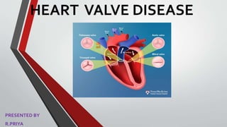
heart has these four valves:
•the tricuspid valve, which is located between the right atrium and the right ventricle
•the pulmonary valve, which is located between the right ventricle and the pulmonary artery
•the mitral valve, which is located between the left atrium and the left ventricle
•the aortic valve, which is located between the left ventricle and the aorta
Blood flows from the right and left atria through the tricuspid and mitral valves, which open to
allow blood to flow into the right and left ventricles. These valves then close to prevent blood
from flowing back into the atria.
Once the ventricles have filled with blood, they begin to contract, forcing the pulmonary and
aortic valves to open. Blood then flows to the pulmonary artery and aorta. The pulmonary
artery carries deoxygenated blood from the heart to the lungs. The aorta, which is the body’s
largest artery, carries oxygen-rich blood to the rest of r body.
The heart valves work by ensuring that blood flows in a forward direction and doesn’t back up
or cause leakage. If have a heart valve disorder, the valve isn’t able to do this job properly.
This can be caused by a leakage of blood, which is called regurgitation, a narrowing of the
valve opening, which is called stenosis, or a combination of regurgitation and stenosis.
5. DISORDER
There are a number of causes for the different heart valve disorders. The causes may include:
•a birth defect
•infective endocarditis, an inflammation of the heart tissue
•rheumatic fever, an inflammatory disease brought on by an infection with group
A Streptococcus bacteria
•age-related changes, such as calcium deposits
•a heart attack
•coronary artery disease, a narrowing and hardening of arteries that supply the heart
•cardiomyopathy, which involves degenerative changes in the heart muscle
•syphilis, a relatively rare sexually transmitted infection
•hypertension, or high blood pressure
•aortic aneurysm, an abnormal swelling or bulging of the aorta
•atherosclerosis, a hardening of the arteries
•myxomatous degeneration, a weakening of connective tissue in the mitral valve
•lupus, a chronic autoimmune disorder
6. CAUSES OFVALVE DISEASE
Heart valve problems may include:
•Regurgitation. In this condition, the valve flaps don't close
properly, causing blood to leak backward in r heart. This
commonly occurs due to valve flaps bulging back, a condition
called prolapse.
•Stenosis. In valve stenosis, the valve flaps become thick or
stiff, and they may fuse together. This results in a narrowed
valve opening and reduced blood flow through the valve.
•Atresia. In this condition, the valve isn't formed, and a solid
sheet of tissue blocks the blood flow between the heart
chambers.
7. VALVULAR STENOSIS
In the valvular heart disease condition valvular
stenosis , the tissues forming the valve leaflets
leaflets become stiffer, narrowing the valve
opening and reducing the amount of blood that
can flow through it. If the narrowing is mild, the
overall functioning of the heart may not be
reduced. However, the valve can become so
narrow (stenotic) that heart function is reduced,
and the rest of the body may not receive
adequate blood flow.
8. REGURGITANTVALVE
Regurgitant Valve
A regurgitant (incompetent, insufficient, or
valve does not close completely, letting blood
backward through the valve.
Some patients may have both valvular
valvular insufficiency in one or more valves.
disease causes the heart muscle to work
circulate the right amount of blood through
Valvular regurgitation may be also called “leaky
valve.” It occurs when any of the heart valves
9. VALVE PROLAPSE
Mitral valve prolapse
A mitral valve prolapse is also called:
•floppy valve syndrome
•click-murmur syndrome
•balloon mitral valve
•Barlow’s syndrome
It occurs when the mitral valve doesn’t close properly, sometimes causing blood to flow back
into the left atrium.
Most people with mitral valve prolapse don’t have symptoms and don’t require treatment as a
result.
10. RISK FACTOR
•Older age
•History of certain infections that can affect the heart
•History of certain forms of heart disease or heart attack
•High blood pressure, high cholesterol, diabetes and other heart disease risk factors
•Heart conditions present at birth (congenital heart disease)
12. SIGNS AND SYMPTOMS
•Abnormal sound (heart murmur) when a doctor is listening to the heart beating with a
stethoscope
•Chest pain
•Abdominal swelling (more common with advanced tricuspid regurgitation)
•Fatigue
•Shortness of breath, particularly when have been very active or when lie down
•Swelling of r ankles and feet
•Dizziness
•Fainting
•Irregular heartbeat
13. DIAGNOSTIC EVALUATION
•Echocardiography. In this test, sound waves directed at r heart from a wandlike device (transducer) held on r chest produce
video images of r heart in motion. This test assesses the structure of r heart, the heart valves and the blood flow through r heart.
An echocardiogram helps r doctor get a close look at the heart valves and how well they're working. Doctors may also use a 3D
echocardiogram.
Doctors may conduct another type of echocardiogram called a transesophageal echocardiogram. In this test, a small transducer
attached to the end of a tube is inserted down the tube leading from r mouth to r stomach (esophagus). This test allows doctors
to have a closer look at the heart valves than is possible with a regular echocardiogram.
•Electrocardiogram (ECG). In this test, wires (electrodes) attached to pads on r skin measure electrical impulses from r heart.
An ECG can detect enlarged chambers of r heart, heart disease and abnormal heart rhythms.
•Chest X-ray. A chest X-ray can help r doctor determine whether the heart is enlarged, which can indicate certain types of heart
valve disease. A chest X-ray can also help doctors determine the condition of r lungs.
•Cardiac MRI. A cardiac MRI uses magnetic fields and radio waves to create detailed images of r heart. This test may be used
to determine the severity of r condition and assess the size and function of r lower heart chambers (ventricles).
•Exercise tests or stress tests. Different exercise tests help measure r activity tolerance and monitor r heart's response to
physical exertion. If are unable to exercise, medications to mimic the effect of exercise on r heart may be used.
•Cardiac catheterization. This test isn't often used to diagnose heart valve disease, but it may be used if other tests aren't able
to diagnose the condition or to determine its severity.
In this procedure, a doctor threads a thin tube (catheter) through a blood vessel in r arm or groin and guides it to an artery in r
heart and injects dye through the catheter to make the artery visible on an X-ray. This provides r doctor with a detailed picture of
r heart arteries and how r heart functions. It can also measure the pressure inside the heart chambers
14. MANAGEMENT
• Treatments for valve disease include:
• Protecting r valve from further damage by reducing the risk for endocarditis
• Medications to ease symptoms and reduce the risk of further valve damage
• Valve repair or replacement surgery, if needed
• Catheter based procedures, if needed
• Following up with r doctor for regular visits. Valve disease can get worse without
any symptoms, so it is important to see r doctor as scheduled to checck r condition
15. SURGERY
Heart Valve Surgery
There are two types of valve surgery - valve repair surgery and valve replacement
Valve repair surgery
During valve repair surgery, the surgeon fixes the damaged or faulty valve, often without
artificial parts.
The mitral valve is the most commonly repaired valve, but repair surgery can also be
problems with the aortic and tricuspid valve.
The potential advantages of heart valve repair versus valve replacement are:
•Lower risk of infection
•Less need for life-long anticoagulant (blood thinning) medication
•Preserved heart muscle strength
16. SURGERY
Common valve repairs
Commissurotomy (aortic valve shown)
Fused valve leaflets are separated to widen the valve opening
Before: Aortic valve leaflets are fused, causing
stenosis
After: Valve leaflets are separated,
widening the valve opening
17. SURGERY
Before: A portion of the mitral valve leaflet is floppy
(flail) and bows back into the left atrium. A
section is cut out.
After: The leaflet is sewn back together, allowing the
valve to close more tightly.
Quadrangle Resection of Leaflet (mitral valve shown)
If a portion of the mitral valve leaflet is flail (floppy), and bows back
into the left atrium, a segment may be cut out and the leaflet sewn
back together, allowing the valve to close more tightly.
18. SURGERY
Before: Valve annulus is too wide; the leaflets lack support and do not close
tightly. This causes the valve to leak.
After: The leaflet is reshaped or tightened by sewing a ring around the annulus
(annuloplasty)
Annulus Support (mitral valve shown)
If the valve annulus is too wide, it may be reshaped or tightened by
sewing a ring around the annulus (annuloplasty). The ring may be made
of tissue, cloth or metal with a clot covering. It acts like a belt
supporting the valve and bringing the leaflets together.
19. SURGERY
Before: Valve leaflet has a hole or tear
After: Tissue patches are used to repair the hole or
tear.
Patched leaflets (mitral valve shown)
The surgeon may patch leaflets with tears
or holes with tissue patches.
20. SURGERY
Bicuspid aortic valve repair (aortic valve shown)
When have aortic valve disease, heart surgery most often requires replacement of the valve. In
aortic valve can be repaired.
Before: A bicuspid aortic valve has two leaflets instead of the normal three. The valve may
not open fully (stenosis) or may not close tightly (regurgitation)
After: The aortic valve leaflets are surgically reshaped allowing the valve to open and close
more easily.
21. TYPES OFVALVE REPLACEMENT SURGERY
Types of Valve Replacement Surgeries
Biological Valves
Biological valves (also called tissue or bioprosthetic valves) are made of cow
pig tissue (porcine) or human tissue (allografts or homografts). Biological
some artificial parts to give the valve support and to make placement easier.
22. TYPES OFVALVE REPLACEMENT SURGERY
Homograft Valve
A homograft (also called allograft) is a human heart valve that comes from a
death. It is frozen and preserved under sterile conditions. A homograft is
replace a diseased aortic valve in children or ng adults, especially when the
diseased or there is infection (endocarditis).
23. TYPES OFVALVE REPLACEMENT SURGERY
Ross Procedure
The Ross procedure involves switching r pulmonary valve to the aortic valve position and
pulmonary homograft. This is a very complex procedure; however it has many benefits,
patients with aortic valve disease. Techniques, such as the Ross Procedure are examples of
surgeons are able to treat valve disease while protecting the heart's natural functioning
There are advantages and drawbacks to biologic valves.
Advantages: Most patients do not need to be on lifelong blood-thinner
medication, unless they have other conditions (such as atrial fibrillation) which
warrant it.
Drawbacks: Traditionally, biological valves were not considered as durable as
mechanical valves, especially in nger people. Previously available biologic valves
usually needed to be re-replaced after about 10 years However, recent studies
show these valves often last 15 - 20+ years without a decline in function.
24. MECHANICALVALVE
Mechanical Valves
Mechanical valves are made of metal or carbon and are
designed to work just like a patient's native valve.
valves, are well-tolerated by the body, very durable and
last a lifetime. The leaflet valve is the most common type
mechanical valve. It is made up of two carbon leaflets
ring covered with polyester knit fabric.
There are advantages and drawbacks to mechanical valves.
Advantages: Mechanical valves are very durable. They are designed to last a lifetime.
Disadvantages: Due to the artificial material involved, patients who receive these valves need
to take a blood-thinning (anticoagulant) medication lifelong. Blood-thinners are medications
(such as warfarin or Coumadin) delay the clotting action of the blood. They help prevent clots
from forming on the replaced valve, which can cause a heart attack or stroke. If take
Coumadin, will need to have regular blood tests to see how well are responding to the
medication and if need a change in dose.
Some patients who have a mechanical valve replacement hear the valve make a clicking noise
25. VALVE SURGERYTECHNIQUES
Valve Surgery Techniques
Traditional Surgical Approach
Traditional heart surgery involved an incision (6-8 inches) through the breastbone. This
open the chest and see the heart and arteries. The surgeon will use the smallest possible
the surgery.
Minimally Invasive Surgical Approach
Minimally invasive heart valve surgery is performed using smaller incisions than those in
traditional heart valve surgery. Other techniques include endoscopic or keyhole approaches
(also called port access, thoracoscopic or video-assisted surgery) and robotic-assisted
surgery. There are many minimally invasive approaches based on the type of valve surgery
need.
The benefits of minimally invasive surgery include a smaller incision (3-4 inches or smaller)
and smaller scar. Other possible benefits include:
•A lower risk of infection
•Less bleeding and trauma
•Shorter hospital stay
26. VALVE SURGERYTECHNIQUES
Percutaneous Valve Procedures
Transcatheter Aortic Valve Replacement (TAVR)
a) Balloon catheter with valve replacement positioned in diseased valve;
b) Balloon inflation to secure the valve;
c) Valve in place
Transcatheter aortic valve replacement is a treatment option for some
patients with severe aortic stenosis who are too ill to have traditional, open-
heart surgery to replace the aortic valve.
The doctor uses a catheter to replace the diseased valve with a biologic
valve. The catheter is inserted into an artery in the groin (transfemoral
approach) or an incision in the chest (transapical, subclavian and direct
aortic approaches).
27. VALVE SURGERYTECHNIQUES
MitraClip in Place; used with Permission Abbot Vascular
The MitraClip is a treatment option for some patients with severe symptomatic mitral
(leaky vale) who are too ill to have tradition, open heart surgery to repair the valve.
The doctor uses a catheter to place the Mitra Clip on the valve leaflets. This helps them
tightly. The catheter is inserted into an artery in the groin or an incision in the chest.
• will get general anesthesia and be "asleep" during the
procedure. Because of this, will have a breathing tube that will
probably be taken out before leave the operating room.
•The MitraClip is put in place with a long, thin tube called a
catheter.
•The catheter is inserted into a vein though an incision at the
top of r thigh. r doctor uses X-rays and echocardiography to
guide the fdevice to r mitral valve. TheMitraClip is placed at the
edges of the valve so they can come together and keep blood
from flowing backward. may need more than one MitraClip to