SlideShare uma empresa Scribd logo
1 de 79
Dr. Pritam Kr Chatterjee
DM PDT
Department of Cardiology
R.G.Kar Medical college
Kolkata
• Definition
• Genetics
• Pathophysiology
• Clinical features
• Evaluation
• Treatment
HCM is a genetic disease state characterized by
unexplained LV hypertrophy associated with nondilated
ventricular chambers in the absence of another cardiac
or systemic disease that itself would be capable of
producing the magnitude of hypertrophy evident in given
patient.
It’s prevalence estimated to be 1:500
IHSS, HOCM, and MSS are older terms
• Beta MHC mutations-clinical presentation apparent by
late adolescents and develop substantial hypertrophy
and more severe diseases.
• MyBPC mutations can have delayed clinical presentation
until age 50 or older.Less severe symptoms.
• cTnT mutations-modest hypertrophy, increased risk of
sudden death
• cTnI mutations- Greater predisposition of apical
hypertrophy
• Alpha tropomyosin-relatively good survival. Variable
degree of hypertrophy
• LV outflow tract obstruction
• Diastolic dysfunction
• Myocardial ischemia
• Mitral regurgitation
• Arrhythmias
• End stage/ burned out
• Produced by SAM of mitral valve
• Explanations for the SAM of the mitral valve
1. Mitral valve is drawn toward the septum
because of the lower pressure that occurs as
blood is ejected at high velocity through a
narrowed outflow tract (Venturi effect)
2. Mitral valve is pulled against the septum by
contraction of the papillary muscles, which
occurs because of the valve's abnormal location
and septal hypertrophy altering the orientation
of the papillary muscles
3. Hydrodynamic “drag” or the “pushing” force of
flow
LV OUTFLOW OBSTRUCTION
• Increase in contractility
VPC
Dobutamine,Isoproterenol
Exercise
• Decrease in afterload/volume
Valsalva maneuver
Standing
Nitroglycerine/amyl nitrite inhalation
Blood loss
Dehydration
Hemodynamic state Conditions Outflow gradient
Basal obstruction Rest >30mmHg
Non obstructive Rest <30mmHg
Physiologically provoked <30mmHg
Dynamic obstruction Rest <30mmHg
Physiologically provoked >30mmHg
• Hyper contractile states
• Anomalous papillary muscle insertion
• Anteroapical infarction with hyperkinetic basal
segments
• Elderly women with LVH/sigmoid septum and
hyperdynamic ventricular function
• After mitral valve repair
•Impaired relaxation
•Decreased compliance
• Hypertrophy
• Disorganized cellular architecture
• Replacement scarring
• Interstitial fibrosis
•Accounts for symptoms of exertional
dyspnea
• Increased filling pressures  increased
pulmonary venous pressure
•Often occurs without atherosclerotic
coronary artery disease
•Postulated mechanisms
• Abnormally small and partially obliterated
intramural coronary arteries as a result of
hypertrophy
• Inadequate number of capillaries for the degree
of LV mass and increased myocardial oxygen
consumption-supply demand mismatch
• Increased filling pressures resulting in
subendocardial ischemia
MYOCARDIAL ISCHEMIA
• Twice the normal size due to elongation of both leaflets
or segmental enlargement of only anterior leaflet or mid
portion of posterior leaflet
• Congenital and anomalous anterolateral papillary muscle
insertion into the anterior leaflet without interposition of
chordae tendineae and produce muscular mid cavity
outflow obstruction >>SAM>>LVOTO
• Variations in leaflet length (posterior/anterior leaflet
length mismatch) – restrict the ability of the posterior
leaflet to follow the anterior leaflet and to coapt effectively
resulting in MR
• Severity of MR directly proportional to LV outflow
obstruction
• Results in symptoms of dyspnea, orthopnea
• Adabag AS1 J Am Coll Cardiol. 2005 Mar 1;45(5):697-704- total 178
patients.
• Majority are asymptomatic
• Dyspnea on exertion (90%), orthopnea, PND
• Palpitations (PAC, PVC, sinus pauses, AF, A flutter,
SVT and VT)
• Congestive heart failure (2o to increased filling
pressures and myocardial ischemia)
• Sudden cardiac death (<1%)
• Angina (70-80%)
• Syncope (20%), Presyncope (50%)
• Outflow obstruction worsens with increased contractility
during exertional activities resulting in decrease in cardiac
output
• Secondary to arrhythmias
• Jugular venous pulse: prominent a- wave
• Double carotid arterial pulse: declines in mid systole
as gradient develop.
• Double apical impulse:
• Forceful left atrial contraction against non-compliant ventricle
• Triple apical impulse:
• Late systolic bulge near isometric contraction
• S1: normal
• S2: normal or paradoxical split
• S3 gallop: decompensated Lt. ventricle
• S4: atrial systole against hypertrophic ventricle
17
• Holosystolic Murmur of MR:
• Retrograde ejection of blood flow into low pressure left
atrium
• Best heard at apex and axilla
• Pt. with SAM* and significant LV outflow gradients
• Systolic ejection crescendo-decrescendo murmur:
crescendo–decrescendo systolic murmur along the
upper left sternal border that increases with the
Valsalva manoeuvre, which is indicative of dynamic
LVOT obstruction.
• Diastolic Decrescendo Murmur of AR: 10% of Pt.
* Systolic anterior motion
19
• Chronic hypertension
• RV hypertrophy
• Cardiac amyloidosis
• Athlete's heart
• Valvular AS
Apical hypertrophy - apical cavity obliteration caused by
hypereosinophilic syndrome or noncompaction.
• Family history of HCM
• Asymmetry
• Right ventricular hypertrophy
• Late gadolinium enhancement at the RV insertion points or
localized to segments of maximum LV thickening on CMR
• Maximum LV wall thickness ≥15 mm (Caucasian); ≥20 mm
(black)
• Severe diastolic dysfunction
• Marked repolarization abnormalities, conduction disease or Q-
waves on 12 lead ECG
• ESC Guidelines2014
Circulation 1995;91.
HCM Fixed
Obstruction
carotid pulse spike and dome parvus
murmur radiate to
carotids
 valsalva, standing
 squatting, handgrip
 passive leg elevation
systolic thrill 4th left ics 2nd right ics
systolic click absent
present
• ECG
• IMAGING
- ECHOCARDIOGRAPHY
- CARDIAC MRI
- OTHER MODALITIES
• CATH DATA
• TESTS TO RISK STRATIFY PATIENTS
• GENETIC TESTING
• FAMILY SCREENING
Abnormal - >90% of pts & >75% of asymptomatic
relatives
• Increased voltages consistent with LV hypertrophy
• ST-T changes - marked T wave inversion in the lateral
precordial leads
• Left atrial enlargement
• Deep and narrow Q waves lateral precordial leads
• Diminished R waves in the lateral precordial leads.
• 1/3rd patients has delayed his purkinjee conduction in EP study
• Yamaguchi disease
Normal ECG - 5% of pts
• Less severe phenotype and favorable course
• Not predictive of future sudden death
• Usually normal or increased
• Can have small LV end-diastolic volumes and
therefore reduced stroke volumes despite having
normal EFs
• Overt LV systolic dysfunction, termed the ‘‘dilated
or progressive phase of HCM,’’ ‘‘end-stage
HCM,’’ or ‘‘burnt-out HCM,’’ is usually defined as
an LV EF < 50% and occurs in a minority (2%–
5%) of patients
• Prognosis is worse in the presence of LV systolic
dysfunction
• Useful when echocardiography is questionable,
particularly with apical hypertrophy
• Cines loops typically show obstruction and velocity
mapping is useful in the assessment of peak velocities
• SAM of the mitral valve is clearly seen on cardiac MRI
• Improvement in obstruction after septal ablation or
myomectomy can be demonstrated, as can the location
and size of the associated infarction, which are useful for
planning repeat procedures
• Cardiac MRI tagging identifies abnormal patterns of
strain, shear, and torsion in cases of HCM, demonstrating
significant dysfunction in hypertrophic areas of the
ventricle
CARDIAC MRI
• Gadolinium contrast cardiac MRI - differentiating HCM
from other causes of cardiac hypertrophy and other types
of cardiomyopathy such as, amyloidosis, athletic heart,
and Fabry’s disease
• Late gadolinium enhancement occurring in HCM
represents myocardial fibrosis
• The greater the degree of late gadolinium enhancement, the more
likely that the particular HCM patient has 2 or more risk factors for
sudden death
• More likely the patient has or will develop progression of
ventricular dilation toward heart failure, thereby indicating a poorer
prognosis
CARDIAC MRI
• Diagnostic cardiac catheterization is useful to determine
the degree of LVOT obstruction, cardiac hemodynamics,
the diastolic characteristics of the left ventricle, LV
anatomy and coronary anatomy
• Reserved for situations when invasive modalities of
therapy, such as a pacemaker or surgery, are being
considered
• Therapeutic cardiac catheterization interventions, include
transcatheter septal alcohol ablation
• The arterial pressure tracing found on cardiac
catheterization may demonstrate a "spike and dome"
configuration
• Dynamic intraventricular pressure gradient
• Swan neck deformity in a banana-shaped ventricle
• Spike-and-dome configuration – proximal aorta
• Brockenbrough-Braunwald-morrow sign
Natural History
• Normal life expectancy
• Mortality
– Adults – 1% / yr
– Children - 2% / yr
• Subgroups at higher risk for important disease complications
– sudden and unexpected death
– progressive heart failure
– Atrial fibrillation (AF)
Heart failure
NYHA III - IV [10 - 15 %]
• LV outflow obstruction
• AF
• Diastolic dysfunction
• Micro vascular dysfunction
• Burnt out HCM - 3%
• Systolic dysfunction EF <50%
• Associated with AF
• Wall thinning and cavity dilation
• Diffuse transmural scarring
• Progression to refractory heart failure or sudden death
• Mortality 10%/year
• Most reliable risk marker - a family history of the end
stage
• Screening all first-degree relatives is recommended
• Echocardiography
• Children & participating in competitive athletics Every 12
to 18 months
• Adults no competitive athletics - every 5 years
• Counseled against engaging in competitive athletics
• Maintain hydration
• Primary ventricular tachycardia and ventricular fibrillation
• Adolescents and young adults 30 to 35 years of age
• Most common cause of Athletic field deaths
• Death most commonly occur at rest
Secondary prevention
1. Prior cardiac arrest
2. Sustained ventricular tachycardia
Primary prevention
one or more of the following
1. Family history of one or more premature HCM-related deaths,
particularly if sudden and multiple
2. Unexplained syncope, especially if recent and in the young
3. Hypotensive or attenuated blood pressure response to exercise
4. Multiple, repetitive (or prolonged) NSVT on Holter
5. Massive LVH (wall thickness, ≥30 mm), particularly in young
patients
• When level of risk judged - ambiguous on the basis of
conventional markers
1. CMR - delayed enhancement
2. Thin-walled akinetic LV apical aneurysms
3. End-stage phase
4. Percutaneous alcohol septal ablation
• Very limited prognostic significance can be attributed to
specific HCM-causing mutations
• Prophylactic - empiric treatment with Amiodarone –
Obsolete
• Amiodarone may be used in pts with AF
• Empirical & highly variable
Beta blockers
• Slowing heart rate
• Reducing force of LV contraction
• Augmenting ventricular filling and relaxation
• Decreasing myocardial oxygen consumption
• Long-acting preparations - propranolol, atenolol,
metoprolol or nadolol
• Blunt LV outflow gradient triggered by physiologic
exercise.
• Target resting heart rate - 60 beats/min
• May require up to 400 mg equivalent of metoprolol
Verapamil
Improves symptoms and exercise capacity (patients
without marked obstruction to LV outflow)
Beneficial effect on ventricular relaxation and filling
Better angina control than BB
Hemodynamic deterioration with CCB agents - lowering of
the afterload in the presence of severe outflow tract
gradients and high diastolic filling pressures
Disopyramide
Negative inotropic effect decreases the gradient and
improve symptoms.
Concomitant beta blockade may be important to prevent
rapid atrioventricular node conduction
Between 300 and 600 mg/d
The corrected QT interval must be monitored
Anticholinergic side effects in older patients
Diuretic agents may be judiciously administered
Either beta blockers or verapamil initially
No advantage by combinations of BB & CCB
Disopyramide may be combined with BB or CCB
Perhexiline
Incrs. Calcium sensitivity – Incrs. Contractility –
Hypertrophy – Abnl.energy utilisation
Improved Energy utilisation
Diltiazem
Blocks L type Ca channels
To prevent Hypertrophy
ARB s
To block TGF beta – Prevent Fibrosis
• Nifedipine, Nitrates, Diuretics -potent vasodilator and
hence avoided
• Digoxin, dobutamine, noradrenaline, dopamine-positive
inotropes
• MANAGEMENT OF ACUTE HYPOTENSION IN HOCM
• i.v fluids
• phenylephrine
• Iv beta blockers
• MANAGEMENT OF HCM WITH LV SYSTOLIC
DYSFUNCTION
• ACEI/ARBS, diuretics-standard HF treatment
• Discontinue verapamil, diltiazem, disopyramide (class iii)
• A/c AF+ hemodynamically unstable= DC
• A/C AF + angina /pulmonary edema = BB/A–iv
• Hemodynamically stable= 3W OAC >>DC
• Rate control with oral BB/ CCB
• rhythm control with Amiodarone
• Flecainide and propafenone, should be avoided as they
may prolong QRS duration and the QT intervaL
• AV nodal ablation F/B DDD/VVIR/CRT IN drug refractory
• Radiofrequency ablation (II A)
1. Drug-refractory heart failure symptoms
2. NYHA Classes III and IV or exertional syncope
3. LV outflow obstruction
• gradient ≥ 50 mm Hg
4. Better surgical candidates
1. Younger age
2. Greater septal thickness (≥30 mm)
3. Concomitant cardiac diseases –valvular/CAD
5. Complications
1. Perioperative Mortality (1-3%)
2. VSD - 3%
3. CHB and PPI (5%)
4. CVA 1-2%
5. LBBB-40%
Braunwald, E. N Engl J Med 2002;347:1306-1307
Alcohol-Induced Septal Ablation
• Polyvinyl alcohol foam particles,
• Microspheres,
• Absorbable gelatine sponges,
• Septal coils
• Gross CM, Schulz-Menger J, Kramer J, Siegel I, Pilz B, Waigand J, Friedrich MG,
Uhlich F, Dietz R. Percutaneous transluminal septal artery ablation using polyvinyl
alcohol foam particles for septal hypertrophy in patients with hypertrophic obstructive
cardiomyopathy: acute and 3-year outcomes. J Endovasc Ther2004;11:705–711.
• Llamas-Esperon GA, Sandoval-Navarrete S. Percutaneous septal ablation with
absorbable gelatin sponge in hypertrophic obstructive cardiomyopathy. Catheter
Cardiovasc Interv 2007;69:231–235.
• Lafont A, Durand E, Brasselet C, Mousseaux E, Hagege A, Desnos M. Percutaneous
transluminal septal coil embolisation as an alternative to alcohol septal ablation for
hypertrophic obstructive cardiomyopathy. Heart2005;91:92
• Proposed benefit:
• Pacing the RV apex will decrease the outflow tract
gradient by decreasing projection of basal septum into
LVOT
• Several RCTs have found that the improvement
in subjective measures provided by dual-
chamber pacing is likely a placebo effect
• Other treatment
• EP
• CRT
• CARDIAC TRANSPLANTATION/VAD
• High risk for- LVOT gradient > 50 mmhg
• Class III for those with class III/IV systolic dysfunction
• No added risk for patients with controlled symptoms (II
A)
• Continue drugs in prgnancy (class I)-watch or fetal
bradycardia and growth abnormalities in fetus
• Guard against post delivery volume loss
• Low intensity aerobic exercises
• Avoid dehydration
• Avoid heavy meals
• Risk of syncope in high intensity sports
• Unpredictability of SCD  an other reason to avoid high
intensity sports
• ASA OVERTAKING SURGERY
• ALTERNATIVES TO ALCOHOL
• INCLUSION OF CARDIAC MRI IN DIAGNOSIS AND
RISK STRATIFICATION
• PRECLINICAL DIAGNOSIS WITH ECHO
Hypertrophic Cardiomyopathy

Mais conteúdo relacionado

Mais procurados

Hypertrophic cardiomyopathy
Hypertrophic cardiomyopathyHypertrophic cardiomyopathy
Hypertrophic cardiomyopathy
Fuad Farooq
 
Echo in hypertrophic obstructive cardiomyopathies
Echo in hypertrophic obstructive cardiomyopathiesEcho in hypertrophic obstructive cardiomyopathies
Echo in hypertrophic obstructive cardiomyopathies
sruthiMeenaxshiSR
 

Mais procurados (20)

HOCM Hypertrophic cardiomyopathy
HOCM Hypertrophic cardiomyopathyHOCM Hypertrophic cardiomyopathy
HOCM Hypertrophic cardiomyopathy
 
Hypertrophic cardiomyopathy
Hypertrophic cardiomyopathyHypertrophic cardiomyopathy
Hypertrophic cardiomyopathy
 
Wide complex tachycardia
Wide complex tachycardiaWide complex tachycardia
Wide complex tachycardia
 
Echo Differentiation of Restrictive Cardiomyopathy and Constrictive Pericarditis
Echo Differentiation of Restrictive Cardiomyopathy and Constrictive PericarditisEcho Differentiation of Restrictive Cardiomyopathy and Constrictive Pericarditis
Echo Differentiation of Restrictive Cardiomyopathy and Constrictive Pericarditis
 
Left Bundle Branch Block (LBBB)
Left Bundle Branch Block (LBBB)Left Bundle Branch Block (LBBB)
Left Bundle Branch Block (LBBB)
 
Hemodyanmic features of Constrictive pericarditis and Restrictive cardiomyopathy
Hemodyanmic features of Constrictive pericarditis and Restrictive cardiomyopathyHemodyanmic features of Constrictive pericarditis and Restrictive cardiomyopathy
Hemodyanmic features of Constrictive pericarditis and Restrictive cardiomyopathy
 
Restrictive cardiomyopathy
Restrictive cardiomyopathyRestrictive cardiomyopathy
Restrictive cardiomyopathy
 
WIDE QRS TACHYCARDIA
WIDE  QRS TACHYCARDIAWIDE  QRS TACHYCARDIA
WIDE QRS TACHYCARDIA
 
Lvh &amp; rvh
Lvh &amp; rvhLvh &amp; rvh
Lvh &amp; rvh
 
Sinus of valsalva aneurysm
Sinus of valsalva aneurysmSinus of valsalva aneurysm
Sinus of valsalva aneurysm
 
Hypertrophic cardiomyopathy
Hypertrophic cardiomyopathyHypertrophic cardiomyopathy
Hypertrophic cardiomyopathy
 
Ventricular arrhythmias
Ventricular arrhythmiasVentricular arrhythmias
Ventricular arrhythmias
 
Hocm
HocmHocm
Hocm
 
Hemodynamic assessment in cardiology
Hemodynamic assessment in cardiologyHemodynamic assessment in cardiology
Hemodynamic assessment in cardiology
 
Right ventricle infarction
Right ventricle infarctionRight ventricle infarction
Right ventricle infarction
 
Hypertrophic Cardiomyopathy
Hypertrophic CardiomyopathyHypertrophic Cardiomyopathy
Hypertrophic Cardiomyopathy
 
Freeman hypertrophic-cardiomyopathy
Freeman hypertrophic-cardiomyopathyFreeman hypertrophic-cardiomyopathy
Freeman hypertrophic-cardiomyopathy
 
Echocardiography in mitral stenosis
Echocardiography in mitral stenosisEchocardiography in mitral stenosis
Echocardiography in mitral stenosis
 
Echo in hypertrophic obstructive cardiomyopathies
Echo in hypertrophic obstructive cardiomyopathiesEcho in hypertrophic obstructive cardiomyopathies
Echo in hypertrophic obstructive cardiomyopathies
 
Echo in pericardial diseases
Echo in pericardial diseasesEcho in pericardial diseases
Echo in pericardial diseases
 

Semelhante a Hypertrophic Cardiomyopathy

CASE LÂM SÀNG CAF [Autosaved].pptx
CASE LÂM SÀNG CAF [Autosaved].pptxCASE LÂM SÀNG CAF [Autosaved].pptx
CASE LÂM SÀNG CAF [Autosaved].pptx
CuongNguyenManh45
 
Cardiomyopathy, Myocarditis and Pericarditis_C I lecture_Oct.ppt
Cardiomyopathy, Myocarditis and Pericarditis_C I lecture_Oct.pptCardiomyopathy, Myocarditis and Pericarditis_C I lecture_Oct.ppt
Cardiomyopathy, Myocarditis and Pericarditis_C I lecture_Oct.ppt
MesfinShifara
 

Semelhante a Hypertrophic Cardiomyopathy (20)

Cardiomyopathy
CardiomyopathyCardiomyopathy
Cardiomyopathy
 
Cardiomyopathy
CardiomyopathyCardiomyopathy
Cardiomyopathy
 
8.Cardiomyopathy.pptx
8.Cardiomyopathy.pptx8.Cardiomyopathy.pptx
8.Cardiomyopathy.pptx
 
Echocardiography in cardiac emergency
Echocardiography in cardiac emergencyEchocardiography in cardiac emergency
Echocardiography in cardiac emergency
 
Cardiomyopathies and arrythmias
Cardiomyopathies and arrythmiasCardiomyopathies and arrythmias
Cardiomyopathies and arrythmias
 
Valvular heart diseases
Valvular heart diseasesValvular heart diseases
Valvular heart diseases
 
Pathophysiology of ISchemic Stroke
Pathophysiology of ISchemic StrokePathophysiology of ISchemic Stroke
Pathophysiology of ISchemic Stroke
 
Mitral regurgitation
Mitral regurgitationMitral regurgitation
Mitral regurgitation
 
Cardiomyopathy
CardiomyopathyCardiomyopathy
Cardiomyopathy
 
Pericardial diseases
Pericardial diseasesPericardial diseases
Pericardial diseases
 
CASE LÂM SÀNG CAF [Autosaved].pptx
CASE LÂM SÀNG CAF [Autosaved].pptxCASE LÂM SÀNG CAF [Autosaved].pptx
CASE LÂM SÀNG CAF [Autosaved].pptx
 
Cardiomyopathy
CardiomyopathyCardiomyopathy
Cardiomyopathy
 
Pathophysiology aortic valve disease
Pathophysiology aortic valve diseasePathophysiology aortic valve disease
Pathophysiology aortic valve disease
 
Cardiomyopathy, Myocarditis and Pericarditis_C I lecture_Oct.ppt
Cardiomyopathy, Myocarditis and Pericarditis_C I lecture_Oct.pptCardiomyopathy, Myocarditis and Pericarditis_C I lecture_Oct.ppt
Cardiomyopathy, Myocarditis and Pericarditis_C I lecture_Oct.ppt
 
cardiomyopathy seminar.pptx
cardiomyopathy seminar.pptxcardiomyopathy seminar.pptx
cardiomyopathy seminar.pptx
 
Pediatric anesthesia and cardiac problems 1
Pediatric anesthesia and cardiac problems 1Pediatric anesthesia and cardiac problems 1
Pediatric anesthesia and cardiac problems 1
 
Valvular heart disease
Valvular heart diseaseValvular heart disease
Valvular heart disease
 
Complications of MI.pptx
Complications of MI.pptxComplications of MI.pptx
Complications of MI.pptx
 
Ischemic and valvular heart disease
Ischemic and valvular heart diseaseIschemic and valvular heart disease
Ischemic and valvular heart disease
 
Hypertrophic cardiomyopathy
Hypertrophic cardiomyopathyHypertrophic cardiomyopathy
Hypertrophic cardiomyopathy
 

Último

Call Girls Aurangabad Just Call 8250077686 Top Class Call Girl Service Available
Call Girls Aurangabad Just Call 8250077686 Top Class Call Girl Service AvailableCall Girls Aurangabad Just Call 8250077686 Top Class Call Girl Service Available
Call Girls Aurangabad Just Call 8250077686 Top Class Call Girl Service Available
Dipal Arora
 
Call Girls in Gagan Vihar (delhi) call me [🔝 9953056974 🔝] escort service 24X7
Call Girls in Gagan Vihar (delhi) call me [🔝  9953056974 🔝] escort service 24X7Call Girls in Gagan Vihar (delhi) call me [🔝  9953056974 🔝] escort service 24X7
Call Girls in Gagan Vihar (delhi) call me [🔝 9953056974 🔝] escort service 24X7
9953056974 Low Rate Call Girls In Saket, Delhi NCR
 

Último (20)

O898O367676 Call Girls In Ahmedabad Escort Service Available 24×7 In Ahmedabad
O898O367676 Call Girls In Ahmedabad Escort Service Available 24×7 In AhmedabadO898O367676 Call Girls In Ahmedabad Escort Service Available 24×7 In Ahmedabad
O898O367676 Call Girls In Ahmedabad Escort Service Available 24×7 In Ahmedabad
 
Best Rate (Hyderabad) Call Girls Jahanuma ⟟ 8250192130 ⟟ High Class Call Girl...
Best Rate (Hyderabad) Call Girls Jahanuma ⟟ 8250192130 ⟟ High Class Call Girl...Best Rate (Hyderabad) Call Girls Jahanuma ⟟ 8250192130 ⟟ High Class Call Girl...
Best Rate (Hyderabad) Call Girls Jahanuma ⟟ 8250192130 ⟟ High Class Call Girl...
 
The Most Attractive Hyderabad Call Girls Kothapet 𖠋 9332606886 𖠋 Will You Mis...
The Most Attractive Hyderabad Call Girls Kothapet 𖠋 9332606886 𖠋 Will You Mis...The Most Attractive Hyderabad Call Girls Kothapet 𖠋 9332606886 𖠋 Will You Mis...
The Most Attractive Hyderabad Call Girls Kothapet 𖠋 9332606886 𖠋 Will You Mis...
 
Call Girls Aurangabad Just Call 8250077686 Top Class Call Girl Service Available
Call Girls Aurangabad Just Call 8250077686 Top Class Call Girl Service AvailableCall Girls Aurangabad Just Call 8250077686 Top Class Call Girl Service Available
Call Girls Aurangabad Just Call 8250077686 Top Class Call Girl Service Available
 
Call Girls Coimbatore Just Call 9907093804 Top Class Call Girl Service Available
Call Girls Coimbatore Just Call 9907093804 Top Class Call Girl Service AvailableCall Girls Coimbatore Just Call 9907093804 Top Class Call Girl Service Available
Call Girls Coimbatore Just Call 9907093804 Top Class Call Girl Service Available
 
Call Girls in Gagan Vihar (delhi) call me [🔝 9953056974 🔝] escort service 24X7
Call Girls in Gagan Vihar (delhi) call me [🔝  9953056974 🔝] escort service 24X7Call Girls in Gagan Vihar (delhi) call me [🔝  9953056974 🔝] escort service 24X7
Call Girls in Gagan Vihar (delhi) call me [🔝 9953056974 🔝] escort service 24X7
 
Premium Bangalore Call Girls Jigani Dail 6378878445 Escort Service For Hot Ma...
Premium Bangalore Call Girls Jigani Dail 6378878445 Escort Service For Hot Ma...Premium Bangalore Call Girls Jigani Dail 6378878445 Escort Service For Hot Ma...
Premium Bangalore Call Girls Jigani Dail 6378878445 Escort Service For Hot Ma...
 
Call Girls Ludhiana Just Call 9907093804 Top Class Call Girl Service Available
Call Girls Ludhiana Just Call 9907093804 Top Class Call Girl Service AvailableCall Girls Ludhiana Just Call 9907093804 Top Class Call Girl Service Available
Call Girls Ludhiana Just Call 9907093804 Top Class Call Girl Service Available
 
Night 7k to 12k Chennai City Center Call Girls 👉👉 7427069034⭐⭐ 100% Genuine E...
Night 7k to 12k Chennai City Center Call Girls 👉👉 7427069034⭐⭐ 100% Genuine E...Night 7k to 12k Chennai City Center Call Girls 👉👉 7427069034⭐⭐ 100% Genuine E...
Night 7k to 12k Chennai City Center Call Girls 👉👉 7427069034⭐⭐ 100% Genuine E...
 
Call Girls Guntur Just Call 8250077686 Top Class Call Girl Service Available
Call Girls Guntur  Just Call 8250077686 Top Class Call Girl Service AvailableCall Girls Guntur  Just Call 8250077686 Top Class Call Girl Service Available
Call Girls Guntur Just Call 8250077686 Top Class Call Girl Service Available
 
Night 7k to 12k Navi Mumbai Call Girl Photo 👉 BOOK NOW 9833363713 👈 ♀️ night ...
Night 7k to 12k Navi Mumbai Call Girl Photo 👉 BOOK NOW 9833363713 👈 ♀️ night ...Night 7k to 12k Navi Mumbai Call Girl Photo 👉 BOOK NOW 9833363713 👈 ♀️ night ...
Night 7k to 12k Navi Mumbai Call Girl Photo 👉 BOOK NOW 9833363713 👈 ♀️ night ...
 
(👑VVIP ISHAAN ) Russian Call Girls Service Navi Mumbai🖕9920874524🖕Independent...
(👑VVIP ISHAAN ) Russian Call Girls Service Navi Mumbai🖕9920874524🖕Independent...(👑VVIP ISHAAN ) Russian Call Girls Service Navi Mumbai🖕9920874524🖕Independent...
(👑VVIP ISHAAN ) Russian Call Girls Service Navi Mumbai🖕9920874524🖕Independent...
 
Top Rated Hyderabad Call Girls Erragadda ⟟ 9332606886 ⟟ Call Me For Genuine ...
Top Rated  Hyderabad Call Girls Erragadda ⟟ 9332606886 ⟟ Call Me For Genuine ...Top Rated  Hyderabad Call Girls Erragadda ⟟ 9332606886 ⟟ Call Me For Genuine ...
Top Rated Hyderabad Call Girls Erragadda ⟟ 9332606886 ⟟ Call Me For Genuine ...
 
Call Girls Dehradun Just Call 9907093804 Top Class Call Girl Service Available
Call Girls Dehradun Just Call 9907093804 Top Class Call Girl Service AvailableCall Girls Dehradun Just Call 9907093804 Top Class Call Girl Service Available
Call Girls Dehradun Just Call 9907093804 Top Class Call Girl Service Available
 
Top Rated Bangalore Call Girls Richmond Circle ⟟ 9332606886 ⟟ Call Me For Ge...
Top Rated Bangalore Call Girls Richmond Circle ⟟  9332606886 ⟟ Call Me For Ge...Top Rated Bangalore Call Girls Richmond Circle ⟟  9332606886 ⟟ Call Me For Ge...
Top Rated Bangalore Call Girls Richmond Circle ⟟ 9332606886 ⟟ Call Me For Ge...
 
VIP Hyderabad Call Girls Bahadurpally 7877925207 ₹5000 To 25K With AC Room 💚😋
VIP Hyderabad Call Girls Bahadurpally 7877925207 ₹5000 To 25K With AC Room 💚😋VIP Hyderabad Call Girls Bahadurpally 7877925207 ₹5000 To 25K With AC Room 💚😋
VIP Hyderabad Call Girls Bahadurpally 7877925207 ₹5000 To 25K With AC Room 💚😋
 
Call Girls Cuttack Just Call 9907093804 Top Class Call Girl Service Available
Call Girls Cuttack Just Call 9907093804 Top Class Call Girl Service AvailableCall Girls Cuttack Just Call 9907093804 Top Class Call Girl Service Available
Call Girls Cuttack Just Call 9907093804 Top Class Call Girl Service Available
 
Call Girls Service Jaipur {9521753030} ❤️VVIP RIDDHI Call Girl in Jaipur Raja...
Call Girls Service Jaipur {9521753030} ❤️VVIP RIDDHI Call Girl in Jaipur Raja...Call Girls Service Jaipur {9521753030} ❤️VVIP RIDDHI Call Girl in Jaipur Raja...
Call Girls Service Jaipur {9521753030} ❤️VVIP RIDDHI Call Girl in Jaipur Raja...
 
All Time Service Available Call Girls Marine Drive 📳 9820252231 For 18+ VIP C...
All Time Service Available Call Girls Marine Drive 📳 9820252231 For 18+ VIP C...All Time Service Available Call Girls Marine Drive 📳 9820252231 For 18+ VIP C...
All Time Service Available Call Girls Marine Drive 📳 9820252231 For 18+ VIP C...
 
Top Rated Bangalore Call Girls Ramamurthy Nagar ⟟ 9332606886 ⟟ Call Me For G...
Top Rated Bangalore Call Girls Ramamurthy Nagar ⟟  9332606886 ⟟ Call Me For G...Top Rated Bangalore Call Girls Ramamurthy Nagar ⟟  9332606886 ⟟ Call Me For G...
Top Rated Bangalore Call Girls Ramamurthy Nagar ⟟ 9332606886 ⟟ Call Me For G...
 

Hypertrophic Cardiomyopathy

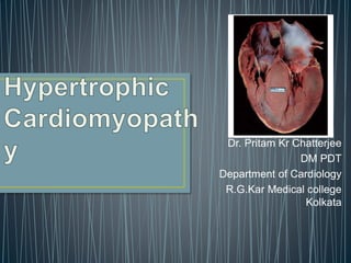
  • 1. Dr. Pritam Kr Chatterjee DM PDT Department of Cardiology R.G.Kar Medical college Kolkata
  • 2. • Definition • Genetics • Pathophysiology • Clinical features • Evaluation • Treatment
  • 3. HCM is a genetic disease state characterized by unexplained LV hypertrophy associated with nondilated ventricular chambers in the absence of another cardiac or systemic disease that itself would be capable of producing the magnitude of hypertrophy evident in given patient. It’s prevalence estimated to be 1:500 IHSS, HOCM, and MSS are older terms
  • 4. • Beta MHC mutations-clinical presentation apparent by late adolescents and develop substantial hypertrophy and more severe diseases. • MyBPC mutations can have delayed clinical presentation until age 50 or older.Less severe symptoms. • cTnT mutations-modest hypertrophy, increased risk of sudden death • cTnI mutations- Greater predisposition of apical hypertrophy • Alpha tropomyosin-relatively good survival. Variable degree of hypertrophy
  • 5.
  • 6.
  • 7. • LV outflow tract obstruction • Diastolic dysfunction • Myocardial ischemia • Mitral regurgitation • Arrhythmias • End stage/ burned out
  • 8. • Produced by SAM of mitral valve • Explanations for the SAM of the mitral valve 1. Mitral valve is drawn toward the septum because of the lower pressure that occurs as blood is ejected at high velocity through a narrowed outflow tract (Venturi effect) 2. Mitral valve is pulled against the septum by contraction of the papillary muscles, which occurs because of the valve's abnormal location and septal hypertrophy altering the orientation of the papillary muscles 3. Hydrodynamic “drag” or the “pushing” force of flow LV OUTFLOW OBSTRUCTION
  • 9. • Increase in contractility VPC Dobutamine,Isoproterenol Exercise • Decrease in afterload/volume Valsalva maneuver Standing Nitroglycerine/amyl nitrite inhalation Blood loss Dehydration
  • 10. Hemodynamic state Conditions Outflow gradient Basal obstruction Rest >30mmHg Non obstructive Rest <30mmHg Physiologically provoked <30mmHg Dynamic obstruction Rest <30mmHg Physiologically provoked >30mmHg
  • 11. • Hyper contractile states • Anomalous papillary muscle insertion • Anteroapical infarction with hyperkinetic basal segments • Elderly women with LVH/sigmoid septum and hyperdynamic ventricular function • After mitral valve repair
  • 12. •Impaired relaxation •Decreased compliance • Hypertrophy • Disorganized cellular architecture • Replacement scarring • Interstitial fibrosis •Accounts for symptoms of exertional dyspnea • Increased filling pressures  increased pulmonary venous pressure
  • 13. •Often occurs without atherosclerotic coronary artery disease •Postulated mechanisms • Abnormally small and partially obliterated intramural coronary arteries as a result of hypertrophy • Inadequate number of capillaries for the degree of LV mass and increased myocardial oxygen consumption-supply demand mismatch • Increased filling pressures resulting in subendocardial ischemia MYOCARDIAL ISCHEMIA
  • 14. • Twice the normal size due to elongation of both leaflets or segmental enlargement of only anterior leaflet or mid portion of posterior leaflet • Congenital and anomalous anterolateral papillary muscle insertion into the anterior leaflet without interposition of chordae tendineae and produce muscular mid cavity outflow obstruction >>SAM>>LVOTO • Variations in leaflet length (posterior/anterior leaflet length mismatch) – restrict the ability of the posterior leaflet to follow the anterior leaflet and to coapt effectively resulting in MR • Severity of MR directly proportional to LV outflow obstruction • Results in symptoms of dyspnea, orthopnea
  • 15. • Adabag AS1 J Am Coll Cardiol. 2005 Mar 1;45(5):697-704- total 178 patients.
  • 16. • Majority are asymptomatic • Dyspnea on exertion (90%), orthopnea, PND • Palpitations (PAC, PVC, sinus pauses, AF, A flutter, SVT and VT) • Congestive heart failure (2o to increased filling pressures and myocardial ischemia) • Sudden cardiac death (<1%) • Angina (70-80%) • Syncope (20%), Presyncope (50%) • Outflow obstruction worsens with increased contractility during exertional activities resulting in decrease in cardiac output • Secondary to arrhythmias
  • 17. • Jugular venous pulse: prominent a- wave • Double carotid arterial pulse: declines in mid systole as gradient develop. • Double apical impulse: • Forceful left atrial contraction against non-compliant ventricle • Triple apical impulse: • Late systolic bulge near isometric contraction • S1: normal • S2: normal or paradoxical split • S3 gallop: decompensated Lt. ventricle • S4: atrial systole against hypertrophic ventricle 17
  • 18.
  • 19. • Holosystolic Murmur of MR: • Retrograde ejection of blood flow into low pressure left atrium • Best heard at apex and axilla • Pt. with SAM* and significant LV outflow gradients • Systolic ejection crescendo-decrescendo murmur: crescendo–decrescendo systolic murmur along the upper left sternal border that increases with the Valsalva manoeuvre, which is indicative of dynamic LVOT obstruction. • Diastolic Decrescendo Murmur of AR: 10% of Pt. * Systolic anterior motion 19
  • 20. • Chronic hypertension • RV hypertrophy • Cardiac amyloidosis • Athlete's heart • Valvular AS Apical hypertrophy - apical cavity obliteration caused by hypereosinophilic syndrome or noncompaction.
  • 21. • Family history of HCM • Asymmetry • Right ventricular hypertrophy • Late gadolinium enhancement at the RV insertion points or localized to segments of maximum LV thickening on CMR • Maximum LV wall thickness ≥15 mm (Caucasian); ≥20 mm (black) • Severe diastolic dysfunction • Marked repolarization abnormalities, conduction disease or Q- waves on 12 lead ECG • ESC Guidelines2014
  • 23. HCM Fixed Obstruction carotid pulse spike and dome parvus murmur radiate to carotids  valsalva, standing  squatting, handgrip  passive leg elevation systolic thrill 4th left ics 2nd right ics systolic click absent present
  • 24. • ECG • IMAGING - ECHOCARDIOGRAPHY - CARDIAC MRI - OTHER MODALITIES • CATH DATA • TESTS TO RISK STRATIFY PATIENTS • GENETIC TESTING • FAMILY SCREENING
  • 25. Abnormal - >90% of pts & >75% of asymptomatic relatives • Increased voltages consistent with LV hypertrophy • ST-T changes - marked T wave inversion in the lateral precordial leads • Left atrial enlargement • Deep and narrow Q waves lateral precordial leads • Diminished R waves in the lateral precordial leads. • 1/3rd patients has delayed his purkinjee conduction in EP study • Yamaguchi disease Normal ECG - 5% of pts • Less severe phenotype and favorable course • Not predictive of future sudden death
  • 26.
  • 27.
  • 28.
  • 29.
  • 30.
  • 31.
  • 32.
  • 33.
  • 34.
  • 35.
  • 36.
  • 37.
  • 38.
  • 39.
  • 40.
  • 41.
  • 42. • Usually normal or increased • Can have small LV end-diastolic volumes and therefore reduced stroke volumes despite having normal EFs • Overt LV systolic dysfunction, termed the ‘‘dilated or progressive phase of HCM,’’ ‘‘end-stage HCM,’’ or ‘‘burnt-out HCM,’’ is usually defined as an LV EF < 50% and occurs in a minority (2%– 5%) of patients • Prognosis is worse in the presence of LV systolic dysfunction
  • 43. • Useful when echocardiography is questionable, particularly with apical hypertrophy • Cines loops typically show obstruction and velocity mapping is useful in the assessment of peak velocities • SAM of the mitral valve is clearly seen on cardiac MRI • Improvement in obstruction after septal ablation or myomectomy can be demonstrated, as can the location and size of the associated infarction, which are useful for planning repeat procedures • Cardiac MRI tagging identifies abnormal patterns of strain, shear, and torsion in cases of HCM, demonstrating significant dysfunction in hypertrophic areas of the ventricle CARDIAC MRI
  • 44. • Gadolinium contrast cardiac MRI - differentiating HCM from other causes of cardiac hypertrophy and other types of cardiomyopathy such as, amyloidosis, athletic heart, and Fabry’s disease • Late gadolinium enhancement occurring in HCM represents myocardial fibrosis • The greater the degree of late gadolinium enhancement, the more likely that the particular HCM patient has 2 or more risk factors for sudden death • More likely the patient has or will develop progression of ventricular dilation toward heart failure, thereby indicating a poorer prognosis CARDIAC MRI
  • 45.
  • 46. • Diagnostic cardiac catheterization is useful to determine the degree of LVOT obstruction, cardiac hemodynamics, the diastolic characteristics of the left ventricle, LV anatomy and coronary anatomy • Reserved for situations when invasive modalities of therapy, such as a pacemaker or surgery, are being considered • Therapeutic cardiac catheterization interventions, include transcatheter septal alcohol ablation • The arterial pressure tracing found on cardiac catheterization may demonstrate a "spike and dome" configuration
  • 47. • Dynamic intraventricular pressure gradient • Swan neck deformity in a banana-shaped ventricle • Spike-and-dome configuration – proximal aorta • Brockenbrough-Braunwald-morrow sign
  • 48.
  • 49.
  • 50.
  • 51. Natural History • Normal life expectancy • Mortality – Adults – 1% / yr – Children - 2% / yr • Subgroups at higher risk for important disease complications – sudden and unexpected death – progressive heart failure – Atrial fibrillation (AF)
  • 52. Heart failure NYHA III - IV [10 - 15 %] • LV outflow obstruction • AF • Diastolic dysfunction • Micro vascular dysfunction
  • 53. • Burnt out HCM - 3% • Systolic dysfunction EF <50% • Associated with AF • Wall thinning and cavity dilation • Diffuse transmural scarring • Progression to refractory heart failure or sudden death • Mortality 10%/year • Most reliable risk marker - a family history of the end stage
  • 54.
  • 55. • Screening all first-degree relatives is recommended • Echocardiography • Children & participating in competitive athletics Every 12 to 18 months • Adults no competitive athletics - every 5 years • Counseled against engaging in competitive athletics • Maintain hydration
  • 56. • Primary ventricular tachycardia and ventricular fibrillation • Adolescents and young adults 30 to 35 years of age • Most common cause of Athletic field deaths • Death most commonly occur at rest
  • 57. Secondary prevention 1. Prior cardiac arrest 2. Sustained ventricular tachycardia Primary prevention one or more of the following 1. Family history of one or more premature HCM-related deaths, particularly if sudden and multiple 2. Unexplained syncope, especially if recent and in the young 3. Hypotensive or attenuated blood pressure response to exercise 4. Multiple, repetitive (or prolonged) NSVT on Holter 5. Massive LVH (wall thickness, ≥30 mm), particularly in young patients
  • 58. • When level of risk judged - ambiguous on the basis of conventional markers 1. CMR - delayed enhancement 2. Thin-walled akinetic LV apical aneurysms 3. End-stage phase 4. Percutaneous alcohol septal ablation • Very limited prognostic significance can be attributed to specific HCM-causing mutations
  • 59. • Prophylactic - empiric treatment with Amiodarone – Obsolete • Amiodarone may be used in pts with AF
  • 60. • Empirical & highly variable Beta blockers • Slowing heart rate • Reducing force of LV contraction • Augmenting ventricular filling and relaxation • Decreasing myocardial oxygen consumption • Long-acting preparations - propranolol, atenolol, metoprolol or nadolol • Blunt LV outflow gradient triggered by physiologic exercise. • Target resting heart rate - 60 beats/min • May require up to 400 mg equivalent of metoprolol
  • 61. Verapamil Improves symptoms and exercise capacity (patients without marked obstruction to LV outflow) Beneficial effect on ventricular relaxation and filling Better angina control than BB Hemodynamic deterioration with CCB agents - lowering of the afterload in the presence of severe outflow tract gradients and high diastolic filling pressures
  • 62. Disopyramide Negative inotropic effect decreases the gradient and improve symptoms. Concomitant beta blockade may be important to prevent rapid atrioventricular node conduction Between 300 and 600 mg/d The corrected QT interval must be monitored Anticholinergic side effects in older patients Diuretic agents may be judiciously administered Either beta blockers or verapamil initially No advantage by combinations of BB & CCB Disopyramide may be combined with BB or CCB
  • 63. Perhexiline Incrs. Calcium sensitivity – Incrs. Contractility – Hypertrophy – Abnl.energy utilisation Improved Energy utilisation Diltiazem Blocks L type Ca channels To prevent Hypertrophy ARB s To block TGF beta – Prevent Fibrosis
  • 64. • Nifedipine, Nitrates, Diuretics -potent vasodilator and hence avoided • Digoxin, dobutamine, noradrenaline, dopamine-positive inotropes • MANAGEMENT OF ACUTE HYPOTENSION IN HOCM • i.v fluids • phenylephrine • Iv beta blockers • MANAGEMENT OF HCM WITH LV SYSTOLIC DYSFUNCTION • ACEI/ARBS, diuretics-standard HF treatment • Discontinue verapamil, diltiazem, disopyramide (class iii)
  • 65. • A/c AF+ hemodynamically unstable= DC • A/C AF + angina /pulmonary edema = BB/A–iv • Hemodynamically stable= 3W OAC >>DC • Rate control with oral BB/ CCB • rhythm control with Amiodarone • Flecainide and propafenone, should be avoided as they may prolong QRS duration and the QT intervaL • AV nodal ablation F/B DDD/VVIR/CRT IN drug refractory • Radiofrequency ablation (II A)
  • 66. 1. Drug-refractory heart failure symptoms 2. NYHA Classes III and IV or exertional syncope 3. LV outflow obstruction • gradient ≥ 50 mm Hg 4. Better surgical candidates 1. Younger age 2. Greater septal thickness (≥30 mm) 3. Concomitant cardiac diseases –valvular/CAD 5. Complications 1. Perioperative Mortality (1-3%) 2. VSD - 3% 3. CHB and PPI (5%) 4. CVA 1-2% 5. LBBB-40%
  • 67.
  • 68. Braunwald, E. N Engl J Med 2002;347:1306-1307 Alcohol-Induced Septal Ablation
  • 69.
  • 70.
  • 71. • Polyvinyl alcohol foam particles, • Microspheres, • Absorbable gelatine sponges, • Septal coils • Gross CM, Schulz-Menger J, Kramer J, Siegel I, Pilz B, Waigand J, Friedrich MG, Uhlich F, Dietz R. Percutaneous transluminal septal artery ablation using polyvinyl alcohol foam particles for septal hypertrophy in patients with hypertrophic obstructive cardiomyopathy: acute and 3-year outcomes. J Endovasc Ther2004;11:705–711. • Llamas-Esperon GA, Sandoval-Navarrete S. Percutaneous septal ablation with absorbable gelatin sponge in hypertrophic obstructive cardiomyopathy. Catheter Cardiovasc Interv 2007;69:231–235. • Lafont A, Durand E, Brasselet C, Mousseaux E, Hagege A, Desnos M. Percutaneous transluminal septal coil embolisation as an alternative to alcohol septal ablation for hypertrophic obstructive cardiomyopathy. Heart2005;91:92
  • 72.
  • 73. • Proposed benefit: • Pacing the RV apex will decrease the outflow tract gradient by decreasing projection of basal septum into LVOT • Several RCTs have found that the improvement in subjective measures provided by dual- chamber pacing is likely a placebo effect • Other treatment • EP • CRT • CARDIAC TRANSPLANTATION/VAD
  • 74.
  • 75.
  • 76. • High risk for- LVOT gradient > 50 mmhg • Class III for those with class III/IV systolic dysfunction • No added risk for patients with controlled symptoms (II A) • Continue drugs in prgnancy (class I)-watch or fetal bradycardia and growth abnormalities in fetus • Guard against post delivery volume loss
  • 77. • Low intensity aerobic exercises • Avoid dehydration • Avoid heavy meals • Risk of syncope in high intensity sports • Unpredictability of SCD  an other reason to avoid high intensity sports
  • 78. • ASA OVERTAKING SURGERY • ALTERNATIVES TO ALCOHOL • INCLUSION OF CARDIAC MRI IN DIAGNOSIS AND RISK STRATIFICATION • PRECLINICAL DIAGNOSIS WITH ECHO