SlideShare uma empresa Scribd logo
1 de 22
07/12/2012   2
Histology of Neuron
 Principle cells of Nervous Tissue
 Consist of 3 parts :
       CELL BODY (perikaryon/soma)
       A single AXON
       Multiple DENDRITES


07/12/2012                             3
CELL BODY (PERIKARYON)
•   Central portion of the cell
•   Generally are polygonal
•   Different shape and size  characteristic regions of nervous system
•   Contain : Nucleus and Perinuclear cytoplasm
   Nucleus :
•   large, spherical to ovoid and centraly located
•   a single prominent nucleolus
•    finely dispersed chromatin
 Cytoplasm : contains :
• R.E.R and S.E.R.
• Polyribosomes
• Basic dyes (a+b) Nissl Bodies
• Golgi complex

     07/12/2012                                                           4
CELL BODY (PERIKARYON)
•   Multiple elongated processes
•   Cytoplasmic~perikaryon (devoid golgi complex)
•   Receiving stimuli


•Single process up to 100 cm
•Originate from axon hillock
•Devoid ribosome
•conducting impulse away from the soma
•It maintains Axonal transport
NEURONS CLASSIFICATION :
According to the size and shape of the processes


07/12/2012                                         7
NEUROGLIAL CELLS
•    Metabolic and mechanical support for neuron
•    Have phagocytic function
•    10 times abundant than neurons
•    Neuroglial cells undergo mitosis
•    CNS neuroglia Includes the following cell types :
      1. Oligodendrocytes
      2. Astrocytes
      3. Ependymal Cells
      4. Microglia
      PNS neuroglia following cell types :
      1- Schwann Cells
      2- Setellite Cells
    07/12/2012                                           8
CNS neuroglia Includes the following cell types :

1- Oligodendrocytes
    – Produce myelin sheath (electrical insulation) in CNS
    – A single cell wrap several axons (40 to 50)
    – Form nodes of Ranvier


2- Astrocytes
    – Pedicles binds to capillaries and to the pia mater form glial
       limitans
    – Controlling the ionic & chemical environment of neurons
    – Energy metabolism
    – Form cellular scar tissue
    – Form the blood-brain barrier
3- Ependymal Cells
     • Low columnar ciliated epithelial cells  line the ventricles of the brain & central
       canal spinal cord
     • Formation of choroid flexus  produce CSF
     • Facilitates the movement of CSF


 4- Microglia
 • Scattered throughout the CNS
 • Clearing debris
 • Act as APC
 • Protect the CNS from viruses and microorganism
PNS neuroglia Includes the following cell types :
1- Schwann cells
• surround all axons of neurons in the PNS creating a neurilemma around them.
Neurilemma allows for potential regeneration of damaged axons
• creates myelin sheath around most axons of PNS


2 - Satellite cells
• support groups of cell bodies of neurons within ganglia of the PNS
•Grey matter consists of
neuronal cell bodies
 neuropil (dendrites and unmyelinated axons)
 glial cells (astroglia and oligodendrocytes)

•Grey matter contains neural cell bodies, in contrast to white matter,
which does not and mostly contains myelinated axon tracts.

•The color difference arises mainly from the whiteness of myelin


•White matter is composed of bundles of myelinated nerve cell processes.

•consists mostly of glial cells and myelinated axons that transmit signals from one region of
the cerebrum to another and between the cerebrum and lower brain centers.

•Its white color is due to its usual preservation in formaldehyde.
NERVE FIBERS
– Consist of axons enveloped by a special sheath
– Group of fibers constitute the peripheral nerve
– Two types :
1- Myelinated fibers
   A single Schwann cell wraps around single axon form myelin sheath 
        nodes of Ranvier
2 - Unmyelinated fibers
   A single Schwann cell envelopes several axon
   Fibers enveloped within simple clefts of Schwann cells
CONNECTIVE TISSUE INVESTMENTS OF NERVES
                     • Epineureum
                       – Dense collagenous Con. Tissue
                         with thick elastic fiber
                       – Prevent damage by
                         overstreching
                     • Perineureum
                       – Dense con. Tissue
                       – Layers of epithelioids
                       – Isolates neural environment
                         (blood-nerve barrier)
                     • Endoneureum
                       – Loose con. Tissue
                       – Regulation of
                         microenvironment of nerve
                         fiber
                                                       16
07/12/2012
GANGLIA
• Ovoid structure containing neuronal cell bodies, glial cells
  supported by connective tissue
• Function : Relay stations to transmit impulses
• Types :
   1- Sensory Ganglia (cell bodies of sensory neuron)
       • Unipolar cell bodies enveloped by cuboidal capsule cells
                – Cranial ganglia : Associated with the cranial nerve
                – Spinal ganglia : Associated with the spinal nerve

   2 -Autonomic Ganglia (cell bodies of postganglionic autonomic nerves)
       • Multipolar neuron enveloped by satellite cells
       • Some are located within certain organ (intramural)


   07/12/2012                                                              17
Sensory Ganglia   Autonomic Ganglia
MENINGES
                  • The dura mater
                     – dense, collagenous connective
                       tissue
                  • The arachnoid layer:
                     – fibroblasts, collagen & elastic
                       fibers
                     – Layer in contact with dura mater
                       & a system of trabeculae
                     – Form arachnoid villi
                  • The pia mater
                     – Loose con. Tissue with blood
                       vessel
                     – There is a physical barrier
                       between pia mater & neuron


07/12/2012                                            19
Cerebellar Cortex
 •Surface of cerebellum show transverse fissures which divide the cerebellum into a
 number of lobules
 .
•Section of cerebellar cortex shows three layers, from out to inside are:

1 - Molecular Layer. :
• Outer most layer mainly consists of cell processes.
•These cell process may be dendrites or unmyelinated axon.
•In this layer make the profuse synapses

2 - Purkinje Cell layer :
•This layer contains the cell bodies of large multipolar neurons called Purkinje cells.
•The purkinje cell consists large flask shaped body.
•cytoplasm contains large number of Nissl granules.

3- Granular Layer :
•This layer consists of thickly populated small cells called granule cells.
•This layer shows irregular scattered lighter staining area called as glomeruli or island.
Cerebellar Cortex
Nervous tissue (Histology)

Mais conteúdo relacionado

Mais procurados

Histology of thyroid and parathyroid, tongue
Histology of thyroid and parathyroid, tongueHistology of thyroid and parathyroid, tongue
Histology of thyroid and parathyroid, tongue
Rohit Paswan
 

Mais procurados (20)

Connective tissue present
Connective tissue presentConnective tissue present
Connective tissue present
 
Skin and its appendages
Skin and its appendagesSkin and its appendages
Skin and its appendages
 
Histology of Muscle Tissues
Histology of Muscle TissuesHistology of Muscle Tissues
Histology of Muscle Tissues
 
Histology of Cartilage.pptx
Histology of Cartilage.pptxHistology of Cartilage.pptx
Histology of Cartilage.pptx
 
5.a.nervous tissue
5.a.nervous tissue5.a.nervous tissue
5.a.nervous tissue
 
Histology of special senses
Histology of special sensesHistology of special senses
Histology of special senses
 
09.22.08: Histology of the Peripheral Nervous System
09.22.08: Histology of the Peripheral Nervous System09.22.08: Histology of the Peripheral Nervous System
09.22.08: Histology of the Peripheral Nervous System
 
Histology of Connective Tissue.pptx
Histology of Connective Tissue.pptxHistology of Connective Tissue.pptx
Histology of Connective Tissue.pptx
 
Epithelium
EpitheliumEpithelium
Epithelium
 
Muscle tissues
Muscle tissuesMuscle tissues
Muscle tissues
 
Muscle histology by dr. armaan singh
Muscle histology by  dr. armaan singhMuscle histology by  dr. armaan singh
Muscle histology by dr. armaan singh
 
Histology of Muscle tissue
Histology of Muscle tissueHistology of Muscle tissue
Histology of Muscle tissue
 
Histology of cns.dk.2014
Histology of cns.dk.2014Histology of cns.dk.2014
Histology of cns.dk.2014
 
Connective tissue
Connective tissue Connective tissue
Connective tissue
 
Connective Tissue
Connective TissueConnective Tissue
Connective Tissue
 
Neuron and neurolgia, nervous system, Anatomy and Physiology
Neuron and neurolgia, nervous system, Anatomy and PhysiologyNeuron and neurolgia, nervous system, Anatomy and Physiology
Neuron and neurolgia, nervous system, Anatomy and Physiology
 
Histology of thyroid and parathyroid, tongue
Histology of thyroid and parathyroid, tongueHistology of thyroid and parathyroid, tongue
Histology of thyroid and parathyroid, tongue
 
Nerve tissue . The Nervous System
Nerve tissue . The Nervous SystemNerve tissue . The Nervous System
Nerve tissue . The Nervous System
 
Bone Histology
Bone HistologyBone Histology
Bone Histology
 
Histology of Endocrine system.
Histology of Endocrine system.Histology of Endocrine system.
Histology of Endocrine system.
 

Destaque

Shunt porto cava
Shunt porto cavaShunt porto cava
Shunt porto cava
Alii Páez
 
VARICEAL HAEMORRHAGE WITH SPECIAL ATTENTION TO PORTAL HYPERTENSION
VARICEAL HAEMORRHAGE WITH SPECIAL ATTENTION TO PORTAL HYPERTENSIONVARICEAL HAEMORRHAGE WITH SPECIAL ATTENTION TO PORTAL HYPERTENSION
VARICEAL HAEMORRHAGE WITH SPECIAL ATTENTION TO PORTAL HYPERTENSION
Arkaprovo Roy
 
ANASTOMOSIS PORTO-CAVA
ANASTOMOSIS PORTO-CAVAANASTOMOSIS PORTO-CAVA
ANASTOMOSIS PORTO-CAVA
Eliecer Zurita
 
anastomosis porto-cava (shunt)
anastomosis porto-cava (shunt)anastomosis porto-cava (shunt)
anastomosis porto-cava (shunt)
anais chang
 

Destaque (13)

hepatic portal system
hepatic portal systemhepatic portal system
hepatic portal system
 
Shunt porto cava
Shunt porto cavaShunt porto cava
Shunt porto cava
 
VARICEAL HAEMORRHAGE WITH SPECIAL ATTENTION TO PORTAL HYPERTENSION
VARICEAL HAEMORRHAGE WITH SPECIAL ATTENTION TO PORTAL HYPERTENSIONVARICEAL HAEMORRHAGE WITH SPECIAL ATTENTION TO PORTAL HYPERTENSION
VARICEAL HAEMORRHAGE WITH SPECIAL ATTENTION TO PORTAL HYPERTENSION
 
Life style diseases
Life style diseasesLife style diseases
Life style diseases
 
Portal
PortalPortal
Portal
 
Portal Hypertension
Portal HypertensionPortal Hypertension
Portal Hypertension
 
ANASTOMOSIS PORTO-CAVA
ANASTOMOSIS PORTO-CAVAANASTOMOSIS PORTO-CAVA
ANASTOMOSIS PORTO-CAVA
 
Ayurvedic management of present life style diseases related to Uttamanga.
Ayurvedic management of present life style diseases related to Uttamanga.Ayurvedic management of present life style diseases related to Uttamanga.
Ayurvedic management of present life style diseases related to Uttamanga.
 
Hepatic Portal vein and portocaval anatomosis
Hepatic Portal vein and portocaval anatomosisHepatic Portal vein and portocaval anatomosis
Hepatic Portal vein and portocaval anatomosis
 
Portacaval Anastomosis
Portacaval AnastomosisPortacaval Anastomosis
Portacaval Anastomosis
 
Principles of vascular anastomosis
Principles of vascular anastomosisPrinciples of vascular anastomosis
Principles of vascular anastomosis
 
Life style diseases
Life style diseasesLife style diseases
Life style diseases
 
anastomosis porto-cava (shunt)
anastomosis porto-cava (shunt)anastomosis porto-cava (shunt)
anastomosis porto-cava (shunt)
 

Semelhante a Nervous tissue (Histology)

dr fareed structure of neuron msk 1 lec 1.pptx
dr fareed structure of neuron msk 1 lec 1.pptxdr fareed structure of neuron msk 1 lec 1.pptx
dr fareed structure of neuron msk 1 lec 1.pptx
WallerianDegenration
 
NEURON SUPPORTIVE CELLS OR ANS
NEURON SUPPORTIVE CELLS OR ANSNEURON SUPPORTIVE CELLS OR ANS
NEURON SUPPORTIVE CELLS OR ANS
optometry student
 
Cellular organization of the nervous system
Cellular organization of the nervous systemCellular organization of the nervous system
Cellular organization of the nervous system
Davis Mburu
 

Semelhante a Nervous tissue (Histology) (20)

LEC 1 ,Excitable tissue nerve and muscle.pptx
LEC 1 ,Excitable tissue nerve and muscle.pptxLEC 1 ,Excitable tissue nerve and muscle.pptx
LEC 1 ,Excitable tissue nerve and muscle.pptx
 
Nervous System 1 (63).pptx
Nervous System 1 (63).pptxNervous System 1 (63).pptx
Nervous System 1 (63).pptx
 
Nervous Tissues.pptx
Nervous Tissues.pptxNervous Tissues.pptx
Nervous Tissues.pptx
 
Nervous tissue 1
Nervous tissue 1Nervous tissue 1
Nervous tissue 1
 
dr fareed structure of neuron msk 1 lec 1.pptx
dr fareed structure of neuron msk 1 lec 1.pptxdr fareed structure of neuron msk 1 lec 1.pptx
dr fareed structure of neuron msk 1 lec 1.pptx
 
Lecture 3.. Dr. Hameed
Lecture 3.. Dr. HameedLecture 3.. Dr. Hameed
Lecture 3.. Dr. Hameed
 
Central nervous system 1
Central nervous system 1Central nervous system 1
Central nervous system 1
 
NSh 2024.pdf histology slides uj university
NSh 2024.pdf histology slides uj universityNSh 2024.pdf histology slides uj university
NSh 2024.pdf histology slides uj university
 
Nueron ppt
Nueron pptNueron ppt
Nueron ppt
 
nervous system
 nervous system nervous system
nervous system
 
Nerve,structure and function
Nerve,structure and functionNerve,structure and function
Nerve,structure and function
 
Introduction to nervous system
Introduction to nervous systemIntroduction to nervous system
Introduction to nervous system
 
Nervous tissue
Nervous tissueNervous tissue
Nervous tissue
 
nervous tissue.pptx
nervous tissue.pptxnervous tissue.pptx
nervous tissue.pptx
 
Neurons
NeuronsNeurons
Neurons
 
Neuron structure
Neuron structureNeuron structure
Neuron structure
 
U4-Neurone System.pdf
U4-Neurone System.pdfU4-Neurone System.pdf
U4-Neurone System.pdf
 
NEURON SUPPORTIVE CELLS OR ANS
NEURON SUPPORTIVE CELLS OR ANSNEURON SUPPORTIVE CELLS OR ANS
NEURON SUPPORTIVE CELLS OR ANS
 
RETINA
RETINA RETINA
RETINA
 
Cellular organization of the nervous system
Cellular organization of the nervous systemCellular organization of the nervous system
Cellular organization of the nervous system
 

Último

Salient Features of India constitution especially power and functions
Salient Features of India constitution especially power and functionsSalient Features of India constitution especially power and functions
Salient Features of India constitution especially power and functions
KarakKing
 

Último (20)

Sociology 101 Demonstration of Learning Exhibit
Sociology 101 Demonstration of Learning ExhibitSociology 101 Demonstration of Learning Exhibit
Sociology 101 Demonstration of Learning Exhibit
 
Food safety_Challenges food safety laboratories_.pdf
Food safety_Challenges food safety laboratories_.pdfFood safety_Challenges food safety laboratories_.pdf
Food safety_Challenges food safety laboratories_.pdf
 
80 ĐỀ THI THỬ TUYỂN SINH TIẾNG ANH VÀO 10 SỞ GD – ĐT THÀNH PHỐ HỒ CHÍ MINH NĂ...
80 ĐỀ THI THỬ TUYỂN SINH TIẾNG ANH VÀO 10 SỞ GD – ĐT THÀNH PHỐ HỒ CHÍ MINH NĂ...80 ĐỀ THI THỬ TUYỂN SINH TIẾNG ANH VÀO 10 SỞ GD – ĐT THÀNH PHỐ HỒ CHÍ MINH NĂ...
80 ĐỀ THI THỬ TUYỂN SINH TIẾNG ANH VÀO 10 SỞ GD – ĐT THÀNH PHỐ HỒ CHÍ MINH NĂ...
 
Jamworks pilot and AI at Jisc (20/03/2024)
Jamworks pilot and AI at Jisc (20/03/2024)Jamworks pilot and AI at Jisc (20/03/2024)
Jamworks pilot and AI at Jisc (20/03/2024)
 
How to setup Pycharm environment for Odoo 17.pptx
How to setup Pycharm environment for Odoo 17.pptxHow to setup Pycharm environment for Odoo 17.pptx
How to setup Pycharm environment for Odoo 17.pptx
 
Single or Multiple melodic lines structure
Single or Multiple melodic lines structureSingle or Multiple melodic lines structure
Single or Multiple melodic lines structure
 
latest AZ-104 Exam Questions and Answers
latest AZ-104 Exam Questions and Answerslatest AZ-104 Exam Questions and Answers
latest AZ-104 Exam Questions and Answers
 
Philosophy of china and it's charactistics
Philosophy of china and it's charactisticsPhilosophy of china and it's charactistics
Philosophy of china and it's charactistics
 
Basic Intentional Injuries Health Education
Basic Intentional Injuries Health EducationBasic Intentional Injuries Health Education
Basic Intentional Injuries Health Education
 
FSB Advising Checklist - Orientation 2024
FSB Advising Checklist - Orientation 2024FSB Advising Checklist - Orientation 2024
FSB Advising Checklist - Orientation 2024
 
Basic Civil Engineering first year Notes- Chapter 4 Building.pptx
Basic Civil Engineering first year Notes- Chapter 4 Building.pptxBasic Civil Engineering first year Notes- Chapter 4 Building.pptx
Basic Civil Engineering first year Notes- Chapter 4 Building.pptx
 
Plant propagation: Sexual and Asexual propapagation.pptx
Plant propagation: Sexual and Asexual propapagation.pptxPlant propagation: Sexual and Asexual propapagation.pptx
Plant propagation: Sexual and Asexual propapagation.pptx
 
ICT role in 21st century education and it's challenges.
ICT role in 21st century education and it's challenges.ICT role in 21st century education and it's challenges.
ICT role in 21st century education and it's challenges.
 
Salient Features of India constitution especially power and functions
Salient Features of India constitution especially power and functionsSalient Features of India constitution especially power and functions
Salient Features of India constitution especially power and functions
 
HMCS Max Bernays Pre-Deployment Brief (May 2024).pptx
HMCS Max Bernays Pre-Deployment Brief (May 2024).pptxHMCS Max Bernays Pre-Deployment Brief (May 2024).pptx
HMCS Max Bernays Pre-Deployment Brief (May 2024).pptx
 
SOC 101 Demonstration of Learning Presentation
SOC 101 Demonstration of Learning PresentationSOC 101 Demonstration of Learning Presentation
SOC 101 Demonstration of Learning Presentation
 
OSCM Unit 2_Operations Processes & Systems
OSCM Unit 2_Operations Processes & SystemsOSCM Unit 2_Operations Processes & Systems
OSCM Unit 2_Operations Processes & Systems
 
Google Gemini An AI Revolution in Education.pptx
Google Gemini An AI Revolution in Education.pptxGoogle Gemini An AI Revolution in Education.pptx
Google Gemini An AI Revolution in Education.pptx
 
How to Add New Custom Addons Path in Odoo 17
How to Add New Custom Addons Path in Odoo 17How to Add New Custom Addons Path in Odoo 17
How to Add New Custom Addons Path in Odoo 17
 
HMCS Vancouver Pre-Deployment Brief - May 2024 (Web Version).pptx
HMCS Vancouver Pre-Deployment Brief - May 2024 (Web Version).pptxHMCS Vancouver Pre-Deployment Brief - May 2024 (Web Version).pptx
HMCS Vancouver Pre-Deployment Brief - May 2024 (Web Version).pptx
 

Nervous tissue (Histology)

  • 1.
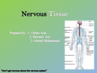
  • 3. Histology of Neuron  Principle cells of Nervous Tissue  Consist of 3 parts :  CELL BODY (perikaryon/soma)  A single AXON  Multiple DENDRITES 07/12/2012 3
  • 4. CELL BODY (PERIKARYON) • Central portion of the cell • Generally are polygonal • Different shape and size  characteristic regions of nervous system • Contain : Nucleus and Perinuclear cytoplasm  Nucleus : • large, spherical to ovoid and centraly located • a single prominent nucleolus • finely dispersed chromatin  Cytoplasm : contains : • R.E.R and S.E.R. • Polyribosomes • Basic dyes (a+b) Nissl Bodies • Golgi complex 07/12/2012 4
  • 6. Multiple elongated processes • Cytoplasmic~perikaryon (devoid golgi complex) • Receiving stimuli •Single process up to 100 cm •Originate from axon hillock •Devoid ribosome •conducting impulse away from the soma •It maintains Axonal transport
  • 7. NEURONS CLASSIFICATION : According to the size and shape of the processes 07/12/2012 7
  • 8. NEUROGLIAL CELLS • Metabolic and mechanical support for neuron • Have phagocytic function • 10 times abundant than neurons • Neuroglial cells undergo mitosis • CNS neuroglia Includes the following cell types : 1. Oligodendrocytes 2. Astrocytes 3. Ependymal Cells 4. Microglia PNS neuroglia following cell types : 1- Schwann Cells 2- Setellite Cells 07/12/2012 8
  • 9. CNS neuroglia Includes the following cell types : 1- Oligodendrocytes – Produce myelin sheath (electrical insulation) in CNS – A single cell wrap several axons (40 to 50) – Form nodes of Ranvier 2- Astrocytes – Pedicles binds to capillaries and to the pia mater form glial limitans – Controlling the ionic & chemical environment of neurons – Energy metabolism – Form cellular scar tissue – Form the blood-brain barrier
  • 10. 3- Ependymal Cells • Low columnar ciliated epithelial cells  line the ventricles of the brain & central canal spinal cord • Formation of choroid flexus  produce CSF • Facilitates the movement of CSF 4- Microglia • Scattered throughout the CNS • Clearing debris • Act as APC • Protect the CNS from viruses and microorganism
  • 11. PNS neuroglia Includes the following cell types : 1- Schwann cells • surround all axons of neurons in the PNS creating a neurilemma around them. Neurilemma allows for potential regeneration of damaged axons • creates myelin sheath around most axons of PNS 2 - Satellite cells • support groups of cell bodies of neurons within ganglia of the PNS
  • 12. •Grey matter consists of neuronal cell bodies  neuropil (dendrites and unmyelinated axons)  glial cells (astroglia and oligodendrocytes) •Grey matter contains neural cell bodies, in contrast to white matter, which does not and mostly contains myelinated axon tracts. •The color difference arises mainly from the whiteness of myelin •White matter is composed of bundles of myelinated nerve cell processes. •consists mostly of glial cells and myelinated axons that transmit signals from one region of the cerebrum to another and between the cerebrum and lower brain centers. •Its white color is due to its usual preservation in formaldehyde.
  • 13.
  • 14. NERVE FIBERS – Consist of axons enveloped by a special sheath – Group of fibers constitute the peripheral nerve – Two types : 1- Myelinated fibers A single Schwann cell wraps around single axon form myelin sheath  nodes of Ranvier 2 - Unmyelinated fibers A single Schwann cell envelopes several axon Fibers enveloped within simple clefts of Schwann cells
  • 15.
  • 16. CONNECTIVE TISSUE INVESTMENTS OF NERVES • Epineureum – Dense collagenous Con. Tissue with thick elastic fiber – Prevent damage by overstreching • Perineureum – Dense con. Tissue – Layers of epithelioids – Isolates neural environment (blood-nerve barrier) • Endoneureum – Loose con. Tissue – Regulation of microenvironment of nerve fiber 16 07/12/2012
  • 17. GANGLIA • Ovoid structure containing neuronal cell bodies, glial cells supported by connective tissue • Function : Relay stations to transmit impulses • Types : 1- Sensory Ganglia (cell bodies of sensory neuron) • Unipolar cell bodies enveloped by cuboidal capsule cells – Cranial ganglia : Associated with the cranial nerve – Spinal ganglia : Associated with the spinal nerve 2 -Autonomic Ganglia (cell bodies of postganglionic autonomic nerves) • Multipolar neuron enveloped by satellite cells • Some are located within certain organ (intramural) 07/12/2012 17
  • 18. Sensory Ganglia Autonomic Ganglia
  • 19. MENINGES • The dura mater – dense, collagenous connective tissue • The arachnoid layer: – fibroblasts, collagen & elastic fibers – Layer in contact with dura mater & a system of trabeculae – Form arachnoid villi • The pia mater – Loose con. Tissue with blood vessel – There is a physical barrier between pia mater & neuron 07/12/2012 19
  • 20. Cerebellar Cortex •Surface of cerebellum show transverse fissures which divide the cerebellum into a number of lobules . •Section of cerebellar cortex shows three layers, from out to inside are: 1 - Molecular Layer. : • Outer most layer mainly consists of cell processes. •These cell process may be dendrites or unmyelinated axon. •In this layer make the profuse synapses 2 - Purkinje Cell layer : •This layer contains the cell bodies of large multipolar neurons called Purkinje cells. •The purkinje cell consists large flask shaped body. •cytoplasm contains large number of Nissl granules. 3- Granular Layer : •This layer consists of thickly populated small cells called granule cells. •This layer shows irregular scattered lighter staining area called as glomeruli or island.