SlideShare uma empresa Scribd logo
1 de 81
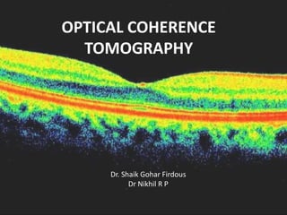
OPTICAL COHERENCE
TOMOGRAPHY
Dr. Shaik Gohar Firdous
Dr Nikhil R P
INTRODUCTION
• OCT is a diagnostic technology,provides a
cross sectional image of the anterior eye and
retina in-vivo with a high resolution, similar to
histological section.
• OCT allows assessment of retinal disease,
understanding of pathology and correlations
between structure and function.
It allows detection and measurement of:
• Morphological changes in retina
• Retinal thickness
• Retinal volume
• Retinal nerve fiber layer thickness (RNFL)
• Various parameters of the optic nerve head
(ONH)
PRINCIPLE
• Low coherence interferometry.
Michelson Interferometer
• A beam of light passes through
semitransparent mirror that splits the beam
into two.
• These two beams are then thrown on two
equidistant mirrors; reflected light from these
mirrors is then picked up and summed up by a
detector.
• The equidistant mirrors reflect the light wave in
same phase; however, if one of the mirrors is
moved by a distance less than the wavelength of
the incident light, the reflected lights from the
two mirrors will then possess a phase difference.
• This phase difference then produces an
interference pattern at the level of the detector.
• The resulting interference patterns are used to
reconstruct an axial A-scan, which represents
the scattering properties of the tissue along
the beam path.
• Moving the beam of light along the tissue in a
line results in a compilation of A-scans with
each A-scan having a different incidence point.
• From all these A-scans, a two-dimensional
crosssectional image of the target tissue can be
reconstructed and this is known as a B-scan.
• OCT operates like a fundus camera but resolves
like a USG machine.
USG OCT
• Source Sound waves Infrared light
• Resolution 150 μ 10 μ
• Patient contact Needed Non-invasive
TYPES OF OCT
• Time Domain OCT
• Spectral Domain OCT
• In TD OCT, image resolution and acquisition
speed are inversely related.
• Simultaneous increase in imaging speed and
resolution can be brought about by spectral
domain OCT (SD OCT).
DIFFERENCES
TD-OCT
• LIGHT SOURCE: 840nm
• AXIAL RESOLUTION: 6-7µ
• TRANSVERSE : 10µ
• SCANNING SPEED : 28000
• SCAN DEPTH : 2mm
• MAXIMUM ASCANS: 8000
SD-OCT
• 820nm
• 10µ
• 20µ
• 400
• 2mm
• 512
Technique
• In the presence of clear media and cooperative
patients quality images can be taken even with a 3
mm pupil; otherwise dilatation is recommended.
• The patient is seated comfortably in front of the
OCT machine with chin positioned on the chin rest.
He is asked to fixate on the fixation target. The
internal fixation (green color light) target is the
commonly used fixation target. Those patients who
are unable to fixate with macula can focus with the
opposite eye on an external target. After Fixation
the operator selects the desired scan and aligns the
instrument so that fundus image and scan beam is
displayed on the screen.
Scanning protocols
• The OCT software offers a variety of retinal
scanning protocols including linear, circular,
radial and parallel line scans
• The duration of scan acquisition can be
shortened by using fast scan protocols, which
acquire three fast scans and averages them to
give the final interpretation.
• The alignment algorithm reduces artifacts caused by
axial movement of the eye during scan acquisition.
• The normalization algorithm allows comparison of
scans with varying signals. Apart from this the machine
has Gaussian smoothing, proportional evaluation and
profile algorithm scan for further refinement of the
scan image
• Application of gray scale image allows better
sensitivity for detection of minute difference in the
contrast.
• A normative database is also available for comparison
of peripapillary retinal nerve fiber layer as well as the
macular thickness in the latest software.
Specific scanning protocols
• Retina scanning protocols: Line scan, Raster lines,
Cross hair, Radial lines, X-line and Circle.
• Macular scanning protocols: Macular thickness scan,
Fast macular thickness, Raster lines and Single line
scan
• RNFL scanning protocols: RNFL thickness protocol
(3.4 mm), Fast RNFL thickness protocol (3.4 mm),
Proportional circle, Concentric 3 rings, RNFL
thickness (2.27Xdisc), RNFL map.
• ONH scanning protocols: Optical disc scan and Fast
Optical disc scan.
NORMAL RETINAL SCAN
• Posterior hyaloid ,visible as very faint, fine and
slightly reflective line.
• Internal limiting membrane is clearly defined
in the OCT scans due to contrast between the
reflective retina and non-reflective vitreous.
• The nerve fiber layer is highly reflective and
more visible on the nasal side due to the
density of papillomacular bundle.
• The fovea shows a characteristic depression
on the macular scan.
• The plexiform layers with reflectivity that is
slightly greater than the reflectivity of the
nuclear layers.
• The outer retina is bounded by a highly
reflective band (70 microns thick) that
represents the retinal pigment epithelium.
• This band can be divided into three layers.
• The first is thin and hyperreflective
representing the junction of inner and outer
photoreceptors.
• The second of hyporeflective,outer segment
• The third one is the thickest and most
hyperreflective
• The Bruchs’ membrane and the
choriocapillaris are seen as a single less
reflective structure but in some scans the
choriocapillaris may be visible separate from
the RPE and the Bruchs’ membrane.
• The larger retinal vessels are located
indirectly by the shadow cone that they form
on the posterior layers
• The OCT image can be displayed on a gray
scale where more highly reflected light is
brighter than less highly reflected light.
• Alternatively, it can be displayed in color,
colors correspond to different degrees of
reflectivity.
• Highly reflective:bright colors(red and yellow)
• Low reflectivity:darker colors(black and blue).
• Intermediate reflectivity -green.
• First generation OCT became available in 1996
as Humphrey Optical Coherence Tomography
Scanner.
• Infrared light source with wavelength of 850
nm,with a resolution of 10 to 17 μm.
• Commercial 3rd generation OCT (StratusOCT, Carl
Zeiss Meditec, Dublin, CA) was introduced in
2002.
Light source - super luminescent diode (SLD)
Wavelength - 820 nm
STRATUS OCT:
Scanning speed - 400 A-scan/seconds (4х 1st
gen).
Axial resolution of 10 μm
Transverse resolution of 20 μm.
Various newer OCT systems are:
• Ultra-high resolution OCT
• Combined OCT/SLO
• Doppler-OCT
• High Speed UHR-OCT
• CAS OCT-Visante™ OCT
• Polarization sensitive OCT
• Combined FFA and en-face OCT
• Intraoperative OCT
ULTRA HIGH RESOLUTION OCT
• Axial resolution - 3 μm.
• Transverse resolution - 15-20 μm.
• Uses femto-second titanium sapphire laser that
generates light with bandwidth of 125 nm
centered at 815 nm.
• Time for image acquisition is 4.3 sec(1.3 sec with
Stratus OCT).
• Hence, UHR OCT images need correction for axial
motion.
• With OCT (Stratus OCT); GCL,ELM and
photoreceptor details are not well visualized.
• UHR OCT may be useful in evaluation of these
retinal layers.
1.Showing photoreceptor integrity in patients with
macular hole. Foveal photoreceptor degeneration
is represented by outer hyporeflective
disruptions of the junction between the inner
and outer segments of the photoreceptors
2.Detection of milder forms of ERM and
vitreomacular traction.
3.Earlier detection of RNFL thinning and
treatment of glaucoma patients.
• UHR OCT can not only demonstrate focal RNFL
changes before appearance of field defect but
also detect progression of disease in an
established case.
HIGH SPEED UHR OCT
High Speed UHR-OCT uses SD OCT technology.
• It allows simultaneous ultra-high speed and
ultra-high resolution.
• Imaging speed is 100 times faster than time
domain UHR OCT.
• 40 times faster than the standard- resolution
OCT.
• It not only gives structural information but
also functional retinal blood flow similar to
Doppler ultrasound.
• It may reduce the need for FFA.
• Raster scan to obtain 3-dimensional (3-D)
images of ocular structure.
• Quantitative mapping of retina layers,
including measurements of the retinal
thickness, RNFL photoreceptor layer and other
intraretinal layers can be performed.
• Its applications would be similar to that
indicated for UHR-OCT.
COLOR DOPPLER OCT
• CD OCT is the technique that combines laser
Doppler velocimetry and OCT for imaging the
depth, diameter, flow rate and retinal
hemodynamic characteristics.
• Color coded velocity data are superimposed
on the conventional OCT image for CD OCT
display. The direction and magnitude of blood
flow are designated by red and blue color and
intensity respectively.
• As it can measure blood flow profile in a few
milliseconds it is able to show vascular
autoregulation and response to changes in
perfusion pressure, oxygen contents and
following laser photocoagulation.
• Presently only larger vessels near the optic
disc have been mostly studied.
CORNEA ANTERIOR SEGMENT OCT
• ASOCT with 1300 nm wavelength ASOCT was
first reported by Radhakrishnan.
• The CAS OCT image is a gray scale or false
color two-dimensional representation of
backscattered light intensity in a cross-
sectional plane.
• The scanning speed of the system is 4000 axial
scans per second
• CAS OCT with a wavelength of 1.3-μm
(present model) provides adequate resolution
of both the cornea and the AC angle.
• Detailed image of the cornea, iris root,angle
recess, anterior ciliary body, scleral spur,
and,in some eyes, the canal of Schlemm is
possible.
ADVANTAGE OVER OTHERS
• Being noncontact, the patient comfort, cooperation and
safety is increased (pediatric) and there is no mechanical
distortion of the tissue.
• Provides more accurate biometry of anterior segment than
Orbscan or Scheimpflug photography.
• Though confocal scanning microscopy gives higher
resolution than CAS OCT, it can scan only a small area of the
eye at a time.
• Unlike UBM, CAS OCT can perform measurements without
any need of anesthesia or coupling medium. CAS OCT can
also perform scanning of all 4 quadrants at a time.
OTHER USES
• Measure corneal thickness, flap and residual posterior
stromal bed following refractive surgery.
• Important landmarks such as the scleral spur are more
distinct in CAS OCT images.
• Traumatic angle recession can be easily picked up.
• Detecting gonioscopically occludable angle.
• Also being used for imaging ocular surface and iris
neoplasia.
LIMITATIONS
• Speed and depth of penetration.
• It cannot obtain clear images through opaque
media.
• Is obstructed by the eyelids making imaging of
the superior and inferior angles difficult.
• It provides limited visualization of the ciliary
body.
INTERPRETATION
EPIRETINAL MEMBRANE:
• ERMs can be classified as idiopathic or secondary.
• Idiopathic ERMs -fibroglial proliferation on the
inner surface of the retina,secondary to a break
in ILM,during posterior vitreous detachment.
• Secondary ERMs result from an already-existing
ocular pathology such as central or branch retinal
vein occlusion, diabetic retinopathy, uveitis,and
retinal breaks with or without detachment.
• ERMs are seen as a highly reflective layer on
the inner retinal surface.
• In most eyes, the membrane is globally
adherent to the retina but,in some cases, it
can be separated from the inner aspect of the
retina, which enhances its visibility by OCT.
MACULAR HOLE
• Macular hole is partial or full thickness
dissolution of retinal tissue at the foveal region.
• It may occur following blunt trauma, long-
standing macular edema or as an idiopathic
condition.
• Pseudoholes are seen in dense sheet of ERM
with a central defect that overlies the foveal
center, giving the ophthalmoscopic appearance of
a true macular hole.
OCT STAGING OF MACULAR HOLE
• Stage 1
• Stage 1a: Foveolar detachment with yellow spot. OCT
shows a cystoid space occupying the inner part of the
foveal tissue.
• Stage 1b: Foveolar detachment with yellow halo. OCT
shows impending hole with extension of cystoid space
posteriorly, disrupting the outer retinal layers.
• Stage 2: Formation of minute eccentric holes. OCT
shows eccentric opening of the roof of the hole with
presence of an operculum (Figs 23.6 to 23.8).
• Stage 3: Full thickness macular hole with or
without operculum. OCT shows a central full
thickness macular hole with detached
posterior vitreous.
• Stage 4: Full thickness macular hole with
posterior vitreous detachment. OCT shows a
central full thickness macular hole with a cuff
of subretinal fluid and completely detached
posterior vitreous.
BERLINS EDEMA
• Acute retinal opacification (macula or elsewhere)
following closed globe injury.
• Mild cases resolve spontaneously without any
sequelae; severe cases result in permanent vision loss.
• Histopathological features:
• Disruption of photoreceptor outer segments
• Phagocytosis of retinal pigment epithelial cells (RPE)
• Intraretinal migration of RPE
• Multilayered, disorganized RPE
• OCT findings depend on the severity and the
duration of commotio retinae .
• Photoreceptor disruption is seen as optically
clear spaces in the area corresponding to the
photoreceptors.
RETINAL ARTERY OCCLUSIONS
• The clinical presentation depends on the type
of vessel that is occluded. It may be
asymptomatic or sudden rapid loss of vision or
no PL.
• The ophthalmoscopic include cotton wool
exudates, opacification of retina and a cherry
red spot.
OCT FINDINGS
• Diffuse thickening of the neurosensory retina.
• Increased reflectivity in the inner retinal
layers, decreased reflectivity of the
photoreceptor layers and the retinal pigment
epithelium secondary to the shadowing effect.
• Involvement of macula,of cystoid changes in
the macular area with loss of the macular
contour.
• In old cases decrease in macular thickness.
RETINAL VENOUS OCCLUSIONS
• Retinal thickening and cystoid macular edema (CME):
Increase in retinal thickness is seen as loss of macular
contour on OCT. In the area of retinal edema,
presence of cystoid spaces with reduced reflectivity
depicts CME.
• Intraretinal and subretinal hemorrhages are seen as
focal areas with bright and high reflectivity with back
scattering. Area of shadowing appears as black
spaces in the RPE and choriocapillaris layer
• Cotton wool spots are seen as highly reflective
well demarcated areas with reduced
reflectivity from outer retinal layers.
• Optic disc edema is a common feature in
CRVO. OCT can demonstrate disc edema and
help in monitoring it in a quantitative manner.
AGE RELATED MACULAR
DEGENERATION
• Age related macular degeneration is the most
common cause of blindness.
• Early grades of the disease more frequent
than the advanced grade.
• Early stage – drusen
• Advanced stage by geographic atrophy (non-
exudative or dry form)
choroidal neovascularization (exudative or
wet form).
• Geographic atrophy - large area of
irregular,well-defined chorioretinal atrophy
involving the macula.
• Exudative form-choroidal neovascular
membrane and its sequlae like serous
detachment, hemorrhagic
detachment,exudation and distortion of the
retinal photoreceptors.
• Drusen - hyper-reflective discrete protrusions
within the RPE complex.
• Active choroidal neovascular membrane -
multi-layered, highly hyper-reflective fusiform
or irregular mass with loss of retinal contour
in the overlying region.
• It may be located in the pre choriocapillaris
region or in front of the pigment epithelial
layer or in both these spaces.
CENTRAL SEROUS
CHORIORETINOPATHY
• Central serous chorioretinopathy is a
noninflammatory, idiopathic serous
detachment of the macula with or without
associated RPE detachment.
• Spontaneous resolution occurs - majority.
Concurrent pigment epithelial detachments
persist for a longer time despite resolution of
the serous detachment.
• OCT to detect subtle serous detachments of
the macula, either in the first episode, during
recurrences or following treatment.
• Serous detachment - hypo-reflective, shallow
separation of the neurosensory retina from
the RPE.
CSR
CSR with PED
CSR with PED

Mais conteúdo relacionado

Mais procurados

Corneal Topography
Corneal TopographyCorneal Topography
Corneal TopographyRaman Gupta
 
FUNDUS FLUORESCEIN ANGIOGRAPHY
FUNDUS FLUORESCEIN ANGIOGRAPHYFUNDUS FLUORESCEIN ANGIOGRAPHY
FUNDUS FLUORESCEIN ANGIOGRAPHYAnuraag Singh
 
Optical coherence tomography(OCT) --macula
Optical coherence tomography(OCT) --maculaOptical coherence tomography(OCT) --macula
Optical coherence tomography(OCT) --maculaAkshay Nayak
 
Fundus Fluoroscein Angiography
Fundus Fluoroscein AngiographyFundus Fluoroscein Angiography
Fundus Fluoroscein AngiographyRashmi Ranjan
 
Specular microscopy
Specular microscopySpecular microscopy
Specular microscopyRuchi sood
 
Slit lamp (methods of illumination)
Slit lamp (methods of illumination)Slit lamp (methods of illumination)
Slit lamp (methods of illumination)maclester manahan
 
Humphrey visual field analyser (HVFA)
Humphrey visual field analyser (HVFA)Humphrey visual field analyser (HVFA)
Humphrey visual field analyser (HVFA)Azizul Islam
 
Ultrasound of eye - B scan
Ultrasound of eye - B scan Ultrasound of eye - B scan
Ultrasound of eye - B scan Shruti Laddha
 
Accommodation/ Accommodation of Eye, Measurement of Accommodation of Eye (hea...
Accommodation/ Accommodation of Eye, Measurement of Accommodation of Eye (hea...Accommodation/ Accommodation of Eye, Measurement of Accommodation of Eye (hea...
Accommodation/ Accommodation of Eye, Measurement of Accommodation of Eye (hea...Bikash Sapkota
 
Slit Lamp Illumination Techniques
Slit Lamp Illumination TechniquesSlit Lamp Illumination Techniques
Slit Lamp Illumination TechniquesIrina Kezik
 
Hess chart, diplopia chart, cover tests
Hess chart, diplopia chart, cover testsHess chart, diplopia chart, cover tests
Hess chart, diplopia chart, cover testsNikhil Rp
 

Mais procurados (20)

corneal Pachymetry
 corneal Pachymetry corneal Pachymetry
corneal Pachymetry
 
Corneal Topography
Corneal TopographyCorneal Topography
Corneal Topography
 
A scan biometry
A scan biometryA scan biometry
A scan biometry
 
FUNDUS FLUORESCEIN ANGIOGRAPHY
FUNDUS FLUORESCEIN ANGIOGRAPHYFUNDUS FLUORESCEIN ANGIOGRAPHY
FUNDUS FLUORESCEIN ANGIOGRAPHY
 
Contrast sensitivity
Contrast sensitivityContrast sensitivity
Contrast sensitivity
 
Biometry
BiometryBiometry
Biometry
 
Optical coherence tomography(OCT) --macula
Optical coherence tomography(OCT) --maculaOptical coherence tomography(OCT) --macula
Optical coherence tomography(OCT) --macula
 
B scan
B scanB scan
B scan
 
Pachymetry
PachymetryPachymetry
Pachymetry
 
Fundus Fluoroscein Angiography
Fundus Fluoroscein AngiographyFundus Fluoroscein Angiography
Fundus Fluoroscein Angiography
 
Specular microscopy
Specular microscopySpecular microscopy
Specular microscopy
 
Slit lamp (methods of illumination)
Slit lamp (methods of illumination)Slit lamp (methods of illumination)
Slit lamp (methods of illumination)
 
Humphrey visual field analyser (HVFA)
Humphrey visual field analyser (HVFA)Humphrey visual field analyser (HVFA)
Humphrey visual field analyser (HVFA)
 
Ultrasound of eye - B scan
Ultrasound of eye - B scan Ultrasound of eye - B scan
Ultrasound of eye - B scan
 
Perimetry
PerimetryPerimetry
Perimetry
 
Accommodation/ Accommodation of Eye, Measurement of Accommodation of Eye (hea...
Accommodation/ Accommodation of Eye, Measurement of Accommodation of Eye (hea...Accommodation/ Accommodation of Eye, Measurement of Accommodation of Eye (hea...
Accommodation/ Accommodation of Eye, Measurement of Accommodation of Eye (hea...
 
Slit Lamp Illumination Techniques
Slit Lamp Illumination TechniquesSlit Lamp Illumination Techniques
Slit Lamp Illumination Techniques
 
Test for stereopsis
Test for stereopsisTest for stereopsis
Test for stereopsis
 
Synaptophore
SynaptophoreSynaptophore
Synaptophore
 
Hess chart, diplopia chart, cover tests
Hess chart, diplopia chart, cover testsHess chart, diplopia chart, cover tests
Hess chart, diplopia chart, cover tests
 

Semelhante a Oct

Semelhante a Oct (20)

Oct introduction
Oct  introductionOct  introduction
Oct introduction
 
OPTICAL COHERENCE DEMYSTIFIED
OPTICAL COHERENCE DEMYSTIFIED OPTICAL COHERENCE DEMYSTIFIED
OPTICAL COHERENCE DEMYSTIFIED
 
OCT MACULA INTERPRETATION.
OCT MACULA  INTERPRETATION.OCT MACULA  INTERPRETATION.
OCT MACULA INTERPRETATION.
 
Oct in post seg disorders
Oct in post seg disordersOct in post seg disorders
Oct in post seg disorders
 
Final oct
Final octFinal oct
Final oct
 
Recent advances in ophthalmology - Dr. Parag Apte
Recent advances in ophthalmology - Dr. Parag ApteRecent advances in ophthalmology - Dr. Parag Apte
Recent advances in ophthalmology - Dr. Parag Apte
 
Oct
Oct Oct
Oct
 
OCT For Everyone..
OCT For Everyone..OCT For Everyone..
OCT For Everyone..
 
Glaucoma oct (optical coherence tomography)
Glaucoma oct (optical coherence tomography)Glaucoma oct (optical coherence tomography)
Glaucoma oct (optical coherence tomography)
 
Optical Coherence Tomography
Optical Coherence TomographyOptical Coherence Tomography
Optical Coherence Tomography
 
OCT Machines
OCT Machines OCT Machines
OCT Machines
 
Imaging in Glaucoma
Imaging in Glaucoma Imaging in Glaucoma
Imaging in Glaucoma
 
Optical coherence tomography
Optical coherence tomographyOptical coherence tomography
Optical coherence tomography
 
Optical Coherence Tomography
Optical Coherence Tomography Optical Coherence Tomography
Optical Coherence Tomography
 
Oct demystified
Oct demystified  Oct demystified
Oct demystified
 
OCT
OCTOCT
OCT
 
OCT
OCTOCT
OCT
 
Role of oct in glaucoma
Role of oct in glaucomaRole of oct in glaucoma
Role of oct in glaucoma
 
LASER IN OPHTHALMOLOGY
LASER IN OPHTHALMOLOGYLASER IN OPHTHALMOLOGY
LASER IN OPHTHALMOLOGY
 
Optical coherence tomography)(OCT)
Optical coherence tomography)(OCT)Optical coherence tomography)(OCT)
Optical coherence tomography)(OCT)
 

Mais de Nikhil Rp

Pentacam analysis
Pentacam analysisPentacam analysis
Pentacam analysisNikhil Rp
 
Ocular blood flow in glaucoma
Ocular  blood flow in glaucomaOcular  blood flow in glaucoma
Ocular blood flow in glaucomaNikhil Rp
 
Malignant glaucoma
Malignant glaucomaMalignant glaucoma
Malignant glaucomaNikhil Rp
 
Choroidal neovascularisation(cnv)
Choroidal neovascularisation(cnv)Choroidal neovascularisation(cnv)
Choroidal neovascularisation(cnv)Nikhil Rp
 
Keratoprsthesis
KeratoprsthesisKeratoprsthesis
KeratoprsthesisNikhil Rp
 
Tractional RD
Tractional RD Tractional RD
Tractional RD Nikhil Rp
 
Retinal detachment
Retinal detachment Retinal detachment
Retinal detachment Nikhil Rp
 
Pneumatic retinopexy
Pneumatic retinopexyPneumatic retinopexy
Pneumatic retinopexyNikhil Rp
 
White dot syndromes
White dot syndromesWhite dot syndromes
White dot syndromesNikhil Rp
 
General slides
General slidesGeneral slides
General slidesNikhil Rp
 
Ocular viscosurgical devices
Ocular viscosurgical devicesOcular viscosurgical devices
Ocular viscosurgical devicesNikhil Rp
 

Mais de Nikhil Rp (11)

Pentacam analysis
Pentacam analysisPentacam analysis
Pentacam analysis
 
Ocular blood flow in glaucoma
Ocular  blood flow in glaucomaOcular  blood flow in glaucoma
Ocular blood flow in glaucoma
 
Malignant glaucoma
Malignant glaucomaMalignant glaucoma
Malignant glaucoma
 
Choroidal neovascularisation(cnv)
Choroidal neovascularisation(cnv)Choroidal neovascularisation(cnv)
Choroidal neovascularisation(cnv)
 
Keratoprsthesis
KeratoprsthesisKeratoprsthesis
Keratoprsthesis
 
Tractional RD
Tractional RD Tractional RD
Tractional RD
 
Retinal detachment
Retinal detachment Retinal detachment
Retinal detachment
 
Pneumatic retinopexy
Pneumatic retinopexyPneumatic retinopexy
Pneumatic retinopexy
 
White dot syndromes
White dot syndromesWhite dot syndromes
White dot syndromes
 
General slides
General slidesGeneral slides
General slides
 
Ocular viscosurgical devices
Ocular viscosurgical devicesOcular viscosurgical devices
Ocular viscosurgical devices
 

Último

High Profile Call Girls Coimbatore Saanvi☎️ 8250192130 Independent Escort Se...
High Profile Call Girls Coimbatore Saanvi☎️  8250192130 Independent Escort Se...High Profile Call Girls Coimbatore Saanvi☎️  8250192130 Independent Escort Se...
High Profile Call Girls Coimbatore Saanvi☎️ 8250192130 Independent Escort Se...narwatsonia7
 
VIP Call Girls Tirunelveli Aaradhya 8250192130 Independent Escort Service Tir...
VIP Call Girls Tirunelveli Aaradhya 8250192130 Independent Escort Service Tir...VIP Call Girls Tirunelveli Aaradhya 8250192130 Independent Escort Service Tir...
VIP Call Girls Tirunelveli Aaradhya 8250192130 Independent Escort Service Tir...narwatsonia7
 
Bangalore Call Girl Whatsapp Number 100% Complete Your Sexual Needs
Bangalore Call Girl Whatsapp Number 100% Complete Your Sexual NeedsBangalore Call Girl Whatsapp Number 100% Complete Your Sexual Needs
Bangalore Call Girl Whatsapp Number 100% Complete Your Sexual NeedsGfnyt
 
VIP Russian Call Girls in Varanasi Samaira 8250192130 Independent Escort Serv...
VIP Russian Call Girls in Varanasi Samaira 8250192130 Independent Escort Serv...VIP Russian Call Girls in Varanasi Samaira 8250192130 Independent Escort Serv...
VIP Russian Call Girls in Varanasi Samaira 8250192130 Independent Escort Serv...Neha Kaur
 
Vip Call Girls Anna Salai Chennai 👉 8250192130 ❣️💯 Top Class Girls Available
Vip Call Girls Anna Salai Chennai 👉 8250192130 ❣️💯 Top Class Girls AvailableVip Call Girls Anna Salai Chennai 👉 8250192130 ❣️💯 Top Class Girls Available
Vip Call Girls Anna Salai Chennai 👉 8250192130 ❣️💯 Top Class Girls AvailableNehru place Escorts
 
Call Girls Cuttack Just Call 9907093804 Top Class Call Girl Service Available
Call Girls Cuttack Just Call 9907093804 Top Class Call Girl Service AvailableCall Girls Cuttack Just Call 9907093804 Top Class Call Girl Service Available
Call Girls Cuttack Just Call 9907093804 Top Class Call Girl Service AvailableDipal Arora
 
Book Paid Powai Call Girls Mumbai 𖠋 9930245274 𖠋Low Budget Full Independent H...
Book Paid Powai Call Girls Mumbai 𖠋 9930245274 𖠋Low Budget Full Independent H...Book Paid Powai Call Girls Mumbai 𖠋 9930245274 𖠋Low Budget Full Independent H...
Book Paid Powai Call Girls Mumbai 𖠋 9930245274 𖠋Low Budget Full Independent H...Call Girls in Nagpur High Profile
 
Night 7k to 12k Chennai City Center Call Girls 👉👉 7427069034⭐⭐ 100% Genuine E...
Night 7k to 12k Chennai City Center Call Girls 👉👉 7427069034⭐⭐ 100% Genuine E...Night 7k to 12k Chennai City Center Call Girls 👉👉 7427069034⭐⭐ 100% Genuine E...
Night 7k to 12k Chennai City Center Call Girls 👉👉 7427069034⭐⭐ 100% Genuine E...hotbabesbook
 
(👑VVIP ISHAAN ) Russian Call Girls Service Navi Mumbai🖕9920874524🖕Independent...
(👑VVIP ISHAAN ) Russian Call Girls Service Navi Mumbai🖕9920874524🖕Independent...(👑VVIP ISHAAN ) Russian Call Girls Service Navi Mumbai🖕9920874524🖕Independent...
(👑VVIP ISHAAN ) Russian Call Girls Service Navi Mumbai🖕9920874524🖕Independent...Taniya Sharma
 
Call Girls Ludhiana Just Call 9907093804 Top Class Call Girl Service Available
Call Girls Ludhiana Just Call 9907093804 Top Class Call Girl Service AvailableCall Girls Ludhiana Just Call 9907093804 Top Class Call Girl Service Available
Call Girls Ludhiana Just Call 9907093804 Top Class Call Girl Service AvailableDipal Arora
 
Call Girls Varanasi Just Call 9907093804 Top Class Call Girl Service Available
Call Girls Varanasi Just Call 9907093804 Top Class Call Girl Service AvailableCall Girls Varanasi Just Call 9907093804 Top Class Call Girl Service Available
Call Girls Varanasi Just Call 9907093804 Top Class Call Girl Service AvailableDipal Arora
 
Call Girls Ooty Just Call 9907093804 Top Class Call Girl Service Available
Call Girls Ooty Just Call 9907093804 Top Class Call Girl Service AvailableCall Girls Ooty Just Call 9907093804 Top Class Call Girl Service Available
Call Girls Ooty Just Call 9907093804 Top Class Call Girl Service AvailableDipal Arora
 
Kesar Bagh Call Girl Price 9548273370 , Lucknow Call Girls Service
Kesar Bagh Call Girl Price 9548273370 , Lucknow Call Girls ServiceKesar Bagh Call Girl Price 9548273370 , Lucknow Call Girls Service
Kesar Bagh Call Girl Price 9548273370 , Lucknow Call Girls Servicemakika9823
 
Call Girls Aurangabad Just Call 9907093804 Top Class Call Girl Service Available
Call Girls Aurangabad Just Call 9907093804 Top Class Call Girl Service AvailableCall Girls Aurangabad Just Call 9907093804 Top Class Call Girl Service Available
Call Girls Aurangabad Just Call 9907093804 Top Class Call Girl Service AvailableDipal Arora
 
Bangalore Call Girls Hebbal Kempapura Number 7001035870 Meetin With Bangalor...
Bangalore Call Girls Hebbal Kempapura Number 7001035870  Meetin With Bangalor...Bangalore Call Girls Hebbal Kempapura Number 7001035870  Meetin With Bangalor...
Bangalore Call Girls Hebbal Kempapura Number 7001035870 Meetin With Bangalor...narwatsonia7
 
Call Girls Coimbatore Just Call 9907093804 Top Class Call Girl Service Available
Call Girls Coimbatore Just Call 9907093804 Top Class Call Girl Service AvailableCall Girls Coimbatore Just Call 9907093804 Top Class Call Girl Service Available
Call Girls Coimbatore Just Call 9907093804 Top Class Call Girl Service AvailableDipal Arora
 
Russian Escorts Girls Nehru Place ZINATHI 🔝9711199012 ☪ 24/7 Call Girls Delhi
Russian Escorts Girls  Nehru Place ZINATHI 🔝9711199012 ☪ 24/7 Call Girls DelhiRussian Escorts Girls  Nehru Place ZINATHI 🔝9711199012 ☪ 24/7 Call Girls Delhi
Russian Escorts Girls Nehru Place ZINATHI 🔝9711199012 ☪ 24/7 Call Girls DelhiAlinaDevecerski
 
Low Rate Call Girls Kochi Anika 8250192130 Independent Escort Service Kochi
Low Rate Call Girls Kochi Anika 8250192130 Independent Escort Service KochiLow Rate Call Girls Kochi Anika 8250192130 Independent Escort Service Kochi
Low Rate Call Girls Kochi Anika 8250192130 Independent Escort Service KochiSuhani Kapoor
 
Call Girls Service Jaipur Grishma WhatsApp ❤8445551418 VIP Call Girls Jaipur
Call Girls Service Jaipur Grishma WhatsApp ❤8445551418 VIP Call Girls JaipurCall Girls Service Jaipur Grishma WhatsApp ❤8445551418 VIP Call Girls Jaipur
Call Girls Service Jaipur Grishma WhatsApp ❤8445551418 VIP Call Girls Jaipurparulsinha
 

Último (20)

High Profile Call Girls Coimbatore Saanvi☎️ 8250192130 Independent Escort Se...
High Profile Call Girls Coimbatore Saanvi☎️  8250192130 Independent Escort Se...High Profile Call Girls Coimbatore Saanvi☎️  8250192130 Independent Escort Se...
High Profile Call Girls Coimbatore Saanvi☎️ 8250192130 Independent Escort Se...
 
VIP Call Girls Tirunelveli Aaradhya 8250192130 Independent Escort Service Tir...
VIP Call Girls Tirunelveli Aaradhya 8250192130 Independent Escort Service Tir...VIP Call Girls Tirunelveli Aaradhya 8250192130 Independent Escort Service Tir...
VIP Call Girls Tirunelveli Aaradhya 8250192130 Independent Escort Service Tir...
 
Bangalore Call Girl Whatsapp Number 100% Complete Your Sexual Needs
Bangalore Call Girl Whatsapp Number 100% Complete Your Sexual NeedsBangalore Call Girl Whatsapp Number 100% Complete Your Sexual Needs
Bangalore Call Girl Whatsapp Number 100% Complete Your Sexual Needs
 
VIP Russian Call Girls in Varanasi Samaira 8250192130 Independent Escort Serv...
VIP Russian Call Girls in Varanasi Samaira 8250192130 Independent Escort Serv...VIP Russian Call Girls in Varanasi Samaira 8250192130 Independent Escort Serv...
VIP Russian Call Girls in Varanasi Samaira 8250192130 Independent Escort Serv...
 
Vip Call Girls Anna Salai Chennai 👉 8250192130 ❣️💯 Top Class Girls Available
Vip Call Girls Anna Salai Chennai 👉 8250192130 ❣️💯 Top Class Girls AvailableVip Call Girls Anna Salai Chennai 👉 8250192130 ❣️💯 Top Class Girls Available
Vip Call Girls Anna Salai Chennai 👉 8250192130 ❣️💯 Top Class Girls Available
 
Call Girls Cuttack Just Call 9907093804 Top Class Call Girl Service Available
Call Girls Cuttack Just Call 9907093804 Top Class Call Girl Service AvailableCall Girls Cuttack Just Call 9907093804 Top Class Call Girl Service Available
Call Girls Cuttack Just Call 9907093804 Top Class Call Girl Service Available
 
Book Paid Powai Call Girls Mumbai 𖠋 9930245274 𖠋Low Budget Full Independent H...
Book Paid Powai Call Girls Mumbai 𖠋 9930245274 𖠋Low Budget Full Independent H...Book Paid Powai Call Girls Mumbai 𖠋 9930245274 𖠋Low Budget Full Independent H...
Book Paid Powai Call Girls Mumbai 𖠋 9930245274 𖠋Low Budget Full Independent H...
 
Night 7k to 12k Chennai City Center Call Girls 👉👉 7427069034⭐⭐ 100% Genuine E...
Night 7k to 12k Chennai City Center Call Girls 👉👉 7427069034⭐⭐ 100% Genuine E...Night 7k to 12k Chennai City Center Call Girls 👉👉 7427069034⭐⭐ 100% Genuine E...
Night 7k to 12k Chennai City Center Call Girls 👉👉 7427069034⭐⭐ 100% Genuine E...
 
(👑VVIP ISHAAN ) Russian Call Girls Service Navi Mumbai🖕9920874524🖕Independent...
(👑VVIP ISHAAN ) Russian Call Girls Service Navi Mumbai🖕9920874524🖕Independent...(👑VVIP ISHAAN ) Russian Call Girls Service Navi Mumbai🖕9920874524🖕Independent...
(👑VVIP ISHAAN ) Russian Call Girls Service Navi Mumbai🖕9920874524🖕Independent...
 
Call Girls Ludhiana Just Call 9907093804 Top Class Call Girl Service Available
Call Girls Ludhiana Just Call 9907093804 Top Class Call Girl Service AvailableCall Girls Ludhiana Just Call 9907093804 Top Class Call Girl Service Available
Call Girls Ludhiana Just Call 9907093804 Top Class Call Girl Service Available
 
Call Girls Varanasi Just Call 9907093804 Top Class Call Girl Service Available
Call Girls Varanasi Just Call 9907093804 Top Class Call Girl Service AvailableCall Girls Varanasi Just Call 9907093804 Top Class Call Girl Service Available
Call Girls Varanasi Just Call 9907093804 Top Class Call Girl Service Available
 
Call Girls Ooty Just Call 9907093804 Top Class Call Girl Service Available
Call Girls Ooty Just Call 9907093804 Top Class Call Girl Service AvailableCall Girls Ooty Just Call 9907093804 Top Class Call Girl Service Available
Call Girls Ooty Just Call 9907093804 Top Class Call Girl Service Available
 
Kesar Bagh Call Girl Price 9548273370 , Lucknow Call Girls Service
Kesar Bagh Call Girl Price 9548273370 , Lucknow Call Girls ServiceKesar Bagh Call Girl Price 9548273370 , Lucknow Call Girls Service
Kesar Bagh Call Girl Price 9548273370 , Lucknow Call Girls Service
 
Call Girls Aurangabad Just Call 9907093804 Top Class Call Girl Service Available
Call Girls Aurangabad Just Call 9907093804 Top Class Call Girl Service AvailableCall Girls Aurangabad Just Call 9907093804 Top Class Call Girl Service Available
Call Girls Aurangabad Just Call 9907093804 Top Class Call Girl Service Available
 
Bangalore Call Girls Hebbal Kempapura Number 7001035870 Meetin With Bangalor...
Bangalore Call Girls Hebbal Kempapura Number 7001035870  Meetin With Bangalor...Bangalore Call Girls Hebbal Kempapura Number 7001035870  Meetin With Bangalor...
Bangalore Call Girls Hebbal Kempapura Number 7001035870 Meetin With Bangalor...
 
Escort Service Call Girls In Sarita Vihar,, 99530°56974 Delhi NCR
Escort Service Call Girls In Sarita Vihar,, 99530°56974 Delhi NCREscort Service Call Girls In Sarita Vihar,, 99530°56974 Delhi NCR
Escort Service Call Girls In Sarita Vihar,, 99530°56974 Delhi NCR
 
Call Girls Coimbatore Just Call 9907093804 Top Class Call Girl Service Available
Call Girls Coimbatore Just Call 9907093804 Top Class Call Girl Service AvailableCall Girls Coimbatore Just Call 9907093804 Top Class Call Girl Service Available
Call Girls Coimbatore Just Call 9907093804 Top Class Call Girl Service Available
 
Russian Escorts Girls Nehru Place ZINATHI 🔝9711199012 ☪ 24/7 Call Girls Delhi
Russian Escorts Girls  Nehru Place ZINATHI 🔝9711199012 ☪ 24/7 Call Girls DelhiRussian Escorts Girls  Nehru Place ZINATHI 🔝9711199012 ☪ 24/7 Call Girls Delhi
Russian Escorts Girls Nehru Place ZINATHI 🔝9711199012 ☪ 24/7 Call Girls Delhi
 
Low Rate Call Girls Kochi Anika 8250192130 Independent Escort Service Kochi
Low Rate Call Girls Kochi Anika 8250192130 Independent Escort Service KochiLow Rate Call Girls Kochi Anika 8250192130 Independent Escort Service Kochi
Low Rate Call Girls Kochi Anika 8250192130 Independent Escort Service Kochi
 
Call Girls Service Jaipur Grishma WhatsApp ❤8445551418 VIP Call Girls Jaipur
Call Girls Service Jaipur Grishma WhatsApp ❤8445551418 VIP Call Girls JaipurCall Girls Service Jaipur Grishma WhatsApp ❤8445551418 VIP Call Girls Jaipur
Call Girls Service Jaipur Grishma WhatsApp ❤8445551418 VIP Call Girls Jaipur
 

Oct

  • 1. OPTICAL COHERENCE TOMOGRAPHY Dr. Shaik Gohar Firdous Dr Nikhil R P
  • 2. INTRODUCTION • OCT is a diagnostic technology,provides a cross sectional image of the anterior eye and retina in-vivo with a high resolution, similar to histological section. • OCT allows assessment of retinal disease, understanding of pathology and correlations between structure and function.
  • 3. It allows detection and measurement of: • Morphological changes in retina • Retinal thickness • Retinal volume • Retinal nerve fiber layer thickness (RNFL) • Various parameters of the optic nerve head (ONH)
  • 4. PRINCIPLE • Low coherence interferometry. Michelson Interferometer • A beam of light passes through semitransparent mirror that splits the beam into two. • These two beams are then thrown on two equidistant mirrors; reflected light from these mirrors is then picked up and summed up by a detector.
  • 5. • The equidistant mirrors reflect the light wave in same phase; however, if one of the mirrors is moved by a distance less than the wavelength of the incident light, the reflected lights from the two mirrors will then possess a phase difference. • This phase difference then produces an interference pattern at the level of the detector.
  • 6.
  • 7.
  • 8. • The resulting interference patterns are used to reconstruct an axial A-scan, which represents the scattering properties of the tissue along the beam path. • Moving the beam of light along the tissue in a line results in a compilation of A-scans with each A-scan having a different incidence point.
  • 9. • From all these A-scans, a two-dimensional crosssectional image of the target tissue can be reconstructed and this is known as a B-scan. • OCT operates like a fundus camera but resolves like a USG machine. USG OCT • Source Sound waves Infrared light • Resolution 150 μ 10 μ • Patient contact Needed Non-invasive
  • 10. TYPES OF OCT • Time Domain OCT • Spectral Domain OCT
  • 11. • In TD OCT, image resolution and acquisition speed are inversely related. • Simultaneous increase in imaging speed and resolution can be brought about by spectral domain OCT (SD OCT).
  • 12. DIFFERENCES TD-OCT • LIGHT SOURCE: 840nm • AXIAL RESOLUTION: 6-7µ • TRANSVERSE : 10µ • SCANNING SPEED : 28000 • SCAN DEPTH : 2mm • MAXIMUM ASCANS: 8000 SD-OCT • 820nm • 10µ • 20µ • 400 • 2mm • 512
  • 13. Technique • In the presence of clear media and cooperative patients quality images can be taken even with a 3 mm pupil; otherwise dilatation is recommended. • The patient is seated comfortably in front of the OCT machine with chin positioned on the chin rest. He is asked to fixate on the fixation target. The internal fixation (green color light) target is the commonly used fixation target. Those patients who are unable to fixate with macula can focus with the opposite eye on an external target. After Fixation the operator selects the desired scan and aligns the instrument so that fundus image and scan beam is displayed on the screen.
  • 14. Scanning protocols • The OCT software offers a variety of retinal scanning protocols including linear, circular, radial and parallel line scans • The duration of scan acquisition can be shortened by using fast scan protocols, which acquire three fast scans and averages them to give the final interpretation.
  • 15. • The alignment algorithm reduces artifacts caused by axial movement of the eye during scan acquisition. • The normalization algorithm allows comparison of scans with varying signals. Apart from this the machine has Gaussian smoothing, proportional evaluation and profile algorithm scan for further refinement of the scan image • Application of gray scale image allows better sensitivity for detection of minute difference in the contrast. • A normative database is also available for comparison of peripapillary retinal nerve fiber layer as well as the macular thickness in the latest software.
  • 16. Specific scanning protocols • Retina scanning protocols: Line scan, Raster lines, Cross hair, Radial lines, X-line and Circle. • Macular scanning protocols: Macular thickness scan, Fast macular thickness, Raster lines and Single line scan • RNFL scanning protocols: RNFL thickness protocol (3.4 mm), Fast RNFL thickness protocol (3.4 mm), Proportional circle, Concentric 3 rings, RNFL thickness (2.27Xdisc), RNFL map. • ONH scanning protocols: Optical disc scan and Fast Optical disc scan.
  • 17.
  • 18.
  • 19.
  • 20.
  • 21.
  • 22. NORMAL RETINAL SCAN • Posterior hyaloid ,visible as very faint, fine and slightly reflective line. • Internal limiting membrane is clearly defined in the OCT scans due to contrast between the reflective retina and non-reflective vitreous. • The nerve fiber layer is highly reflective and more visible on the nasal side due to the density of papillomacular bundle.
  • 23. • The fovea shows a characteristic depression on the macular scan. • The plexiform layers with reflectivity that is slightly greater than the reflectivity of the nuclear layers. • The outer retina is bounded by a highly reflective band (70 microns thick) that represents the retinal pigment epithelium.
  • 24. • This band can be divided into three layers. • The first is thin and hyperreflective representing the junction of inner and outer photoreceptors. • The second of hyporeflective,outer segment • The third one is the thickest and most hyperreflective
  • 25. • The Bruchs’ membrane and the choriocapillaris are seen as a single less reflective structure but in some scans the choriocapillaris may be visible separate from the RPE and the Bruchs’ membrane. • The larger retinal vessels are located indirectly by the shadow cone that they form on the posterior layers
  • 26. • The OCT image can be displayed on a gray scale where more highly reflected light is brighter than less highly reflected light. • Alternatively, it can be displayed in color, colors correspond to different degrees of reflectivity. • Highly reflective:bright colors(red and yellow) • Low reflectivity:darker colors(black and blue). • Intermediate reflectivity -green.
  • 27.
  • 28.
  • 29. • First generation OCT became available in 1996 as Humphrey Optical Coherence Tomography Scanner. • Infrared light source with wavelength of 850 nm,with a resolution of 10 to 17 μm.
  • 30. • Commercial 3rd generation OCT (StratusOCT, Carl Zeiss Meditec, Dublin, CA) was introduced in 2002. Light source - super luminescent diode (SLD) Wavelength - 820 nm STRATUS OCT: Scanning speed - 400 A-scan/seconds (4х 1st gen). Axial resolution of 10 μm Transverse resolution of 20 μm.
  • 31. Various newer OCT systems are: • Ultra-high resolution OCT • Combined OCT/SLO • Doppler-OCT • High Speed UHR-OCT • CAS OCT-Visante™ OCT • Polarization sensitive OCT • Combined FFA and en-face OCT • Intraoperative OCT
  • 32. ULTRA HIGH RESOLUTION OCT • Axial resolution - 3 μm. • Transverse resolution - 15-20 μm. • Uses femto-second titanium sapphire laser that generates light with bandwidth of 125 nm centered at 815 nm. • Time for image acquisition is 4.3 sec(1.3 sec with Stratus OCT). • Hence, UHR OCT images need correction for axial motion.
  • 33. • With OCT (Stratus OCT); GCL,ELM and photoreceptor details are not well visualized. • UHR OCT may be useful in evaluation of these retinal layers. 1.Showing photoreceptor integrity in patients with macular hole. Foveal photoreceptor degeneration is represented by outer hyporeflective disruptions of the junction between the inner and outer segments of the photoreceptors
  • 34. 2.Detection of milder forms of ERM and vitreomacular traction. 3.Earlier detection of RNFL thinning and treatment of glaucoma patients. • UHR OCT can not only demonstrate focal RNFL changes before appearance of field defect but also detect progression of disease in an established case.
  • 35. HIGH SPEED UHR OCT High Speed UHR-OCT uses SD OCT technology. • It allows simultaneous ultra-high speed and ultra-high resolution. • Imaging speed is 100 times faster than time domain UHR OCT. • 40 times faster than the standard- resolution OCT.
  • 36. • It not only gives structural information but also functional retinal blood flow similar to Doppler ultrasound. • It may reduce the need for FFA.
  • 37. • Raster scan to obtain 3-dimensional (3-D) images of ocular structure. • Quantitative mapping of retina layers, including measurements of the retinal thickness, RNFL photoreceptor layer and other intraretinal layers can be performed. • Its applications would be similar to that indicated for UHR-OCT.
  • 38.
  • 39.
  • 40. COLOR DOPPLER OCT • CD OCT is the technique that combines laser Doppler velocimetry and OCT for imaging the depth, diameter, flow rate and retinal hemodynamic characteristics. • Color coded velocity data are superimposed on the conventional OCT image for CD OCT display. The direction and magnitude of blood flow are designated by red and blue color and intensity respectively.
  • 41. • As it can measure blood flow profile in a few milliseconds it is able to show vascular autoregulation and response to changes in perfusion pressure, oxygen contents and following laser photocoagulation. • Presently only larger vessels near the optic disc have been mostly studied.
  • 42. CORNEA ANTERIOR SEGMENT OCT • ASOCT with 1300 nm wavelength ASOCT was first reported by Radhakrishnan. • The CAS OCT image is a gray scale or false color two-dimensional representation of backscattered light intensity in a cross- sectional plane. • The scanning speed of the system is 4000 axial scans per second
  • 43. • CAS OCT with a wavelength of 1.3-μm (present model) provides adequate resolution of both the cornea and the AC angle. • Detailed image of the cornea, iris root,angle recess, anterior ciliary body, scleral spur, and,in some eyes, the canal of Schlemm is possible.
  • 44. ADVANTAGE OVER OTHERS • Being noncontact, the patient comfort, cooperation and safety is increased (pediatric) and there is no mechanical distortion of the tissue. • Provides more accurate biometry of anterior segment than Orbscan or Scheimpflug photography. • Though confocal scanning microscopy gives higher resolution than CAS OCT, it can scan only a small area of the eye at a time. • Unlike UBM, CAS OCT can perform measurements without any need of anesthesia or coupling medium. CAS OCT can also perform scanning of all 4 quadrants at a time.
  • 45. OTHER USES • Measure corneal thickness, flap and residual posterior stromal bed following refractive surgery. • Important landmarks such as the scleral spur are more distinct in CAS OCT images. • Traumatic angle recession can be easily picked up. • Detecting gonioscopically occludable angle. • Also being used for imaging ocular surface and iris neoplasia.
  • 46. LIMITATIONS • Speed and depth of penetration. • It cannot obtain clear images through opaque media. • Is obstructed by the eyelids making imaging of the superior and inferior angles difficult. • It provides limited visualization of the ciliary body.
  • 47. INTERPRETATION EPIRETINAL MEMBRANE: • ERMs can be classified as idiopathic or secondary. • Idiopathic ERMs -fibroglial proliferation on the inner surface of the retina,secondary to a break in ILM,during posterior vitreous detachment. • Secondary ERMs result from an already-existing ocular pathology such as central or branch retinal vein occlusion, diabetic retinopathy, uveitis,and retinal breaks with or without detachment.
  • 48. • ERMs are seen as a highly reflective layer on the inner retinal surface. • In most eyes, the membrane is globally adherent to the retina but,in some cases, it can be separated from the inner aspect of the retina, which enhances its visibility by OCT.
  • 49.
  • 50.
  • 51.
  • 52. MACULAR HOLE • Macular hole is partial or full thickness dissolution of retinal tissue at the foveal region. • It may occur following blunt trauma, long- standing macular edema or as an idiopathic condition. • Pseudoholes are seen in dense sheet of ERM with a central defect that overlies the foveal center, giving the ophthalmoscopic appearance of a true macular hole.
  • 53. OCT STAGING OF MACULAR HOLE • Stage 1 • Stage 1a: Foveolar detachment with yellow spot. OCT shows a cystoid space occupying the inner part of the foveal tissue. • Stage 1b: Foveolar detachment with yellow halo. OCT shows impending hole with extension of cystoid space posteriorly, disrupting the outer retinal layers. • Stage 2: Formation of minute eccentric holes. OCT shows eccentric opening of the roof of the hole with presence of an operculum (Figs 23.6 to 23.8).
  • 54. • Stage 3: Full thickness macular hole with or without operculum. OCT shows a central full thickness macular hole with detached posterior vitreous. • Stage 4: Full thickness macular hole with posterior vitreous detachment. OCT shows a central full thickness macular hole with a cuff of subretinal fluid and completely detached posterior vitreous.
  • 55.
  • 56.
  • 57.
  • 58.
  • 59.
  • 60.
  • 61. BERLINS EDEMA • Acute retinal opacification (macula or elsewhere) following closed globe injury. • Mild cases resolve spontaneously without any sequelae; severe cases result in permanent vision loss. • Histopathological features: • Disruption of photoreceptor outer segments • Phagocytosis of retinal pigment epithelial cells (RPE) • Intraretinal migration of RPE • Multilayered, disorganized RPE
  • 62. • OCT findings depend on the severity and the duration of commotio retinae . • Photoreceptor disruption is seen as optically clear spaces in the area corresponding to the photoreceptors.
  • 63.
  • 64.
  • 65. RETINAL ARTERY OCCLUSIONS • The clinical presentation depends on the type of vessel that is occluded. It may be asymptomatic or sudden rapid loss of vision or no PL. • The ophthalmoscopic include cotton wool exudates, opacification of retina and a cherry red spot.
  • 66. OCT FINDINGS • Diffuse thickening of the neurosensory retina. • Increased reflectivity in the inner retinal layers, decreased reflectivity of the photoreceptor layers and the retinal pigment epithelium secondary to the shadowing effect. • Involvement of macula,of cystoid changes in the macular area with loss of the macular contour. • In old cases decrease in macular thickness.
  • 67.
  • 68. RETINAL VENOUS OCCLUSIONS • Retinal thickening and cystoid macular edema (CME): Increase in retinal thickness is seen as loss of macular contour on OCT. In the area of retinal edema, presence of cystoid spaces with reduced reflectivity depicts CME. • Intraretinal and subretinal hemorrhages are seen as focal areas with bright and high reflectivity with back scattering. Area of shadowing appears as black spaces in the RPE and choriocapillaris layer
  • 69. • Cotton wool spots are seen as highly reflective well demarcated areas with reduced reflectivity from outer retinal layers. • Optic disc edema is a common feature in CRVO. OCT can demonstrate disc edema and help in monitoring it in a quantitative manner.
  • 70.
  • 71. AGE RELATED MACULAR DEGENERATION • Age related macular degeneration is the most common cause of blindness. • Early grades of the disease more frequent than the advanced grade. • Early stage – drusen • Advanced stage by geographic atrophy (non- exudative or dry form) choroidal neovascularization (exudative or wet form).
  • 72. • Geographic atrophy - large area of irregular,well-defined chorioretinal atrophy involving the macula. • Exudative form-choroidal neovascular membrane and its sequlae like serous detachment, hemorrhagic detachment,exudation and distortion of the retinal photoreceptors.
  • 73. • Drusen - hyper-reflective discrete protrusions within the RPE complex. • Active choroidal neovascular membrane - multi-layered, highly hyper-reflective fusiform or irregular mass with loss of retinal contour in the overlying region. • It may be located in the pre choriocapillaris region or in front of the pigment epithelial layer or in both these spaces.
  • 74.
  • 75.
  • 76.
  • 77. CENTRAL SEROUS CHORIORETINOPATHY • Central serous chorioretinopathy is a noninflammatory, idiopathic serous detachment of the macula with or without associated RPE detachment. • Spontaneous resolution occurs - majority. Concurrent pigment epithelial detachments persist for a longer time despite resolution of the serous detachment.
  • 78. • OCT to detect subtle serous detachments of the macula, either in the first episode, during recurrences or following treatment. • Serous detachment - hypo-reflective, shallow separation of the neurosensory retina from the RPE.
  • 79. CSR

Notas do Editor

  1. A) Stage 1b macular hole and vitreofoveal traction; (B and C) Evolving into stage 2 macular hole (full thickness eccentric defect with operculum
  2. Stage 2 macular hole (full thickness eccentric defect with operculum
  3. full thickness defect with cystoid changes at the edge and pseudo-operculum
  4. nd yellow pigment deposit at the base of macular hol
  5. central area of retinal elevation and subneurosensory collection of moderate reflectivity material suggestive of fibrin along with retinal pigment epithelium detachment.