
Recomendados
Mais conteúdo relacionado
Mais procurados
Mais procurados (20)
Semelhante a Surgical complication 3
Semelhante a Surgical complication 3 (20)
Último
Último (20)
Surgical complication 3
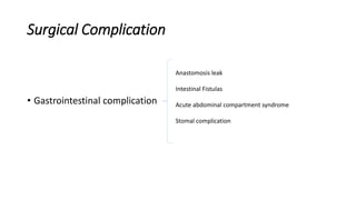
- 1. Surgical Complication • Gastrointestinal complication Anastomosis leak Intestinal Fistulas Acute abdominal compartment syndrome Stomal complication
- 2. Anastomotic Leak • Causes Numerous factors can cause or are associated with an increased risk for anastomotic leak .
- 3. • The level of the anastomosis in the GI tract is important. • Although small bowel, ileocolic, and ileorectal anastomoses are considered safe, esophageal, pancreaticoenteric, and colorectal anastomoses are considered high risk for leakage.
- 4. • In the esophagus, lack of serosa appears to be a significant contributing factor. • In the pancreas, the texture of the gland and size of the pancreatic duct, the presence of pancreatic duct obstructive lesions, the experience of the operating surgeon, and probably the type of enteric anastomosis are implicated. • In the rectum, the highest leak rate is found in anastomoses in the distal rectum, 6 to 8 cm from the anal verge.
- 5. • Adequate microcirculation at the resection margins is crucial for the healing of any anastomosis. • Factors interfering with the perianastomotic microcirculation include: 1. smoking, 2. hypertension, 3. locally enhanced coagulation activity as a result of surgical trauma, 4. perianastomotic hematoma, and 5. presence of macrovascular disease.
- 6. • In colorectal anastomoses, relative ischemia in the rectal remnant is a factor because its blood supply is derived from the internal iliac artery via the inferior hemorrhoidal vessels; contribution from the middle hemorrhoidal artery is minimal and, at best, variable because the vessels are mostly absent and, when present, are unilateral. • Total mesorectal excision, neoadjuvant therapy, and extended lymphadenectomy with high ligation of the inferior mesenteric artery are additional contributing factors.
- 7. • Intraluminal distention is believed to be responsible for rupture of an anastomosis. • The mechanical strength of the anastomosis is important and, in the early period, is dependent on sutures or staples, with endothelial cells and fibrin-fibrinonectin complex additionally contributing to the tension force. • Construction of a watertight and airtight anastomosis is essential.
- 8. • Intra-abdominally placed open rubber drains are not helpful and are associated with an increased risk of infection if left for more than 24 to 48 hours. • In the pelvis, drains have been shown in some studies to be associated with a higher leak rate.
- 9. • Conversely, drains may remove blood, cellular debris, and serum that act as good culture media for perianastomotic sepsis or abscess formation.
- 10. • Local sepsis affects the integrity of the anastomosis negatively as it reduces collagen synthesis and increases collagenase activity, which results in increased lysis of collagen at the anastomosis.
- 11. • Defunctioning or protective stomas do not decrease the overall leak rate, but rather minimize the severity and sequelae of perianastomotic contamination and decrease the reoperation rate. • However, defunctioning stomas deprive the colon of shortchain fatty acids, resulting in exclusion colitis and delay in epithelialization of the anastomosis, and are associated with altered collagen metabolism observed in left-sided anastomoses.
- 12. • Bevacizumab, an angiogenesis inhibitor, is associated with increased risk for surgical site complications. This agent is a humanized monoclonal antibody that targets vascular endothelial growth factor (VEGF). VEGF is a critical factor for the survival of endothelial cells and is selectively present in the neovasculature of growing tumors
- 13. • Bevacizumab binds with high specificity and affinity to VEGF, inhibiting the binding of VEGF to its receptors and negatively affecting angiogenesis or the remodeling of the existing network of blood vessels • Bevacizumab is used in combination with standard chemotherapy (irinotecan, 5-fluorouracil, and leucovorin) in the treatment of patients with metastatic colorectal cancer.
- 14. • In patients with metastatic colorectal cancer, it increases the risk of surgical site complications—spontaneous dehiscence of primary anastomosis and colocutaneous fistula formation from an anastomosis. • Such complications may occur 2 years after surgery.
- 15. The mechanism is probably related to : • microthromboembolic disease leading to bowel ischemia, • inhibition of angiogenesis in the microvascular bed of the new anastomosis, • inhibition of neoangiogenesis in postradiated tissue, and • reduction in the number of newly formed vessels in granulation tissue surrounding anastomotic sites.
- 16. Emergency bowel surgery is associated with high morbidity and mortality, in part because of sepsis and anastomotic leakage; this is related to : • the poor nutritional status of the patient, • presence of underlying malignancy, • immunocompromised state, • presence of intra-abdominal contamination or sepsis, and • hemodynamic instability.
- 17. • Transfusion : • on the one hand, causes impaired cellmediated immunity and predisposes to infection and, • on the other hand, alleviates anemia and improves the oxygen- carrying capacity of red blood cells that may have a positive impact on healing.
- 18. • Obesity : • increases the difficulty and complexity of the surgery; has been shown to be associated with increased postoperative complications; and is an independent risk factor for an increasing leakage rate, especially after a low colorectal anastomosis.
- 19. • Steroids : • affect healing by decreasing collagen synthesis, • delaying the appearance of the inflammatory reaction, • and reducing the production of transforming growth factor-β and insulin-like growth factor in wounds, which are essential for wound healing.
- 21. Presentation and Diagnosis • Anastomotic leak results in sepsis and enteric fistula formation, leads to reoperation and a possible permanent stoma, and is associated with decreased survival and increased local recurrence rate after curative resection of cancer and possibly leads to death.
- 22. • The clinical manifestations are the result of a cascade of events that start with loss of integrity of the anastomosis and leakage of intestinal contents. • The leakage may be diffuse throughout the peritoneal cavity (uncontrolled leak) or become walled off by omentum; abdominal wall; and contiguous loops of bowel, pelvic wall, or adhesions from prior operations. If a surgical drain is present, intestinal contents are discharged onto the skin.
- 23. Intraabdominal fluid collections may contain: • intestinal contents, • frank pus, or • pus mixed with intestinal contents. • If the fluid collection is drained surgically or percutaneously, there is an initial discharge of purulent material followed by feculent material heralding the formation of an enterocutaneous fistula (controlled fistula).
- 24. • If allowed to drain through the surgical incision or abdominal wall, surgical wound infection and dehiscence with evisceration or an abdominal wall abscess may occur. • If the fluid collection burrows into a contiguous structure such as the urinary bladder or vagina, spontaneous drainage occurs, with the formation of an enterovesical or enterovaginal fistula.
- 25. • After the index surgery, a patient may have an initial normal postoperative course or may not have been progressing as expected. • The early warning signs of anastomotic leak are malaise, fever, abdominal pain, ileus, localized erythema around the surgical incision, and leukocytosis. • Patients may also develop bowel obstruction, induration and erythema in the abdominal wall, rectal bleeding, or suprapubic pain.
- 26. • There may be initial excessive drainage from the surgical wound or surgical wound dehiscence or evisceration or both. • An intra-abdominal fluid collection or abdominal wall abscess may be identified and drained surgically or percutaneously. • Patients may also experience pneumaturia, fecaluria, and pyuria.
- 27. • Sepsis is a prominent feature of anastomotic leakage and results from diffuse peritonitis or localized abscess, abdominal wall infection, or contamination of a sterile site with intestinal contents. • Abdominal wall infection develops as a result of contact of purulent material with the muscle and subcutaneous tissue; or contact of corrosive intestinal juices with the abdominal wall, resulting in chemical erosion and extension of the infectious process.
- 28. • Contamination of the urinary bladder with intestinal contents (enterovesical fistula) results in urosepsis.
- 29. Treatment • Treatment of anastomotic leakage starts with prevention. • In elective cases, nutritional support for 5 to 7 days is appropriate for patients who are malnourished or have lost significant amounts of weight. • Mechanical and chemical bowel preparations are still recommended by many surgeons before colorectal resection.
- 30. • In patients receiving or who have received bevacizumab, the appropriate interval between the last dose administered and the surgery is unknown. • The terminal half-life of the medication is long—20 days—so wound- healing complications are documented 56 days after treatment. It is advisable to delay elective surgery for at least 4 to 8 weeks or, preferably, three half-lives (60 days) after treatment.
- 31. • In emergencies, especially in hemodynamically unstable, immunocompromised, and nutritionally depleted patients, in the presence of fecal peritonitis, significant bowel dilation, and edema, an anastomosis is best avoided because a leak may prove fatal.
- 32. • Construction of an anastomosis that is at low risk for disruption requires the following: • 1. Adequate exposure, gentle handling of tissues, aseptic precaution, and meticulous, careful dissection • 2. Adequate mobilization so that the two attached organs have a tension-free anastomosis • 3. Correct technical placement of sutures or staples with little variance • 4. Matching of the lumens of the two organs to be connected, which can be done by various techniques • 5. Preservation of the blood supply to the ends of structures to be anastomosed
- 33. • Sufficient microcirculation is essential for healing of the anastomosis. • In intestinal anastomoses, the marginal artery of the colon and last vascular arcade of small bowel mesentery must be preserved.
- 34. • The small bowel serosa must not be denuded of mesentery more than 3 to 4 cm for hand-sewn anastomoses.
- 35. • In the distal colon, the following maneuvers may be required to ensure a tension-free anastomosis: • inferior mesenteric artery divided at its origin, • windows created in the mesentery of the small bowel up to the third portion of the duodenum, and small branches interrupted between the arcades creating mesenteric windows and dividing the ileocolic vessels at their origin.
- 36. • For intestinal and colorectal anastomoses, there is no difference in the rate of anastomotic leakage between hand-sewn and stapled anastomoses and among various stapling techniques, • The decision to construct a one-layer or two-layer intestinal anastomosis is a matter of preference. A colorectal anastomosis is easier to perform in one layer.
- 37. • However, since the advent of stapling devices, an anastomosis deep in the pelvis has most commonly been stapled. • The technique is not only faster but also improves asepsis because the anastomosis is performed in a closed fashion compared with a hand- sewn anastomosis, which is considered an “open anastomosis” and allows for more contamination
- 38. • In low anterior resection, the omentum may be advanced to the pelvis and placed around the colorectal anastomosis. This maneuver may reduce the rate of anastomotic leak or disruption but mostly appears to decrease the severity of the complication.
- 39. • Drainage of a colorectal anastomosis is advisable in difficult cases and when technical problems are encountered or when neoadjuvant therapy has been used. • Defunctioning stomas are used for extraperitoneal anastomoses, when technical difficulties are encountered, or after neoadjuvant therapy.
- 40. • The routine placement of drains in proximity to pancreatic anastomoses is controversial. • Drains and octreotide can be used when an anastomosis is performed to a soft pancreas with a small duct and in centers with lower surgical volume or centers with a high leak rate (>10%).
- 41. • Pancreatic duct stents (placed intraoperatively) continue to be used, despite the lack of data to suggest that they decrease the leak rate. • A pancreatic stent placed before a distal pancreatectomy decompresses the pancreatic duct by abolishing the pressure gradient between the pancreatic duct and duodenum and may decrease the risk of fistula formation, allowing the site of a leak to seal.
- 42. • When an anastomotic leak is suspected or diagnosed, resuscitation is started immediately because patients are in the postoperative period and have been without nutrition. Furthermore, they have a contracted intravascular volume because of third spacing and lost intestinal contents and may have an electrolyte imbalance.
- 43. • Intravascular volume is restored with crystalloid fluids and a blood transfusion if anemia is present, and electrolyte imbalances are corrected. • Oral intake is stopped, and the bowel is put at rest to decrease luminal contents and GI stimulation and secretion. • An NG tube is placed if obstructive symptoms are present • Infected surgical wounds are opened, and any abdominal wall abscesses are incised and drained.
- 44. • Reoperation is indicated if there is : 1. diffuse peritonitis, 2. intra-abdominal hemorrhage, 3. suspected intestinal ischemia, 4. major wound disruption, or 5. evisceration. • Reoperation is a major undertaking and is associated with significant mortality and morbidity. • Primary closure of the leaking point only is avoided because failure is certain.
- 45. • The management of duodenal and proximal jejunal leaks is challenging. • In these situations, transgastric placement of a jejunal tube helps divert gastric and biliopancreatic secretions, and placement of drains in close proximity to the leak allows external drainage of the intestinal contents. • Pyloric exclusion and gastrojejunostomy should be used judiciously in these situations.
- 46. . Management of jejunal, ileal, and colorectal leaking anastomoses depends on : • the severity and duration of contamination, • condition of the bowel, and • hemodynamic stability of the patient.
- 47. • In a critically ill and unstable patient, especially a patient with fecal peritonitis, a damage control type of procedure is performed : 1. the anastomosis is taken down, 2. the ends of the bowel are stapled, 3. peritoneal lavage is performed, and 4. the incision is left open. 5. A second-look laparotomy with stomal formation is performed in 24 to 48 hours or when the patient is more stable.
- 48. • Otherwise: • in the small bowel, an anastomosis may be performed or the ends of the bowel are delivered as stomas; • in the colon, the proximal end of the colon is brought out as a colostomy, and the distal end is closed or brought out as a mucous fistula; and • in the rectum, the distal end is closed, and the proximal end of the colon is delivered as a stoma.
- 49. • In the absence of diffuse peritonitis and evisceration, a CT scan may identify single or multiple abscesses, pneumoperitoneum, ascites, and sometimes extravasation of oral contrast agent into the peritoneal cavity. • Multiple abscesses require open drainage, • a single intra-abdominal abscess can be drained percutaneously, and • a pelvic abscess can be drained transrectally or transvaginally
- 50. • An external fistula may develop after drainage. • If percutaneous drainage fails to control sepsis, reoperation is indicated. • At the time of open drainage of a pelvic abscess, if there is any doubt about the origin of the abscess (de novo abscess versus abscess secondary to a small anastomotic leak that has sealed), a defunctioning stoma is constructed, unless there is complete disruption of the anastomosis. In that case, the ends of the bowel are exteriorized as a stoma.
- 51. • A pancreaticojejunostomy leak, if small, can be treated by placing a drain next to the leak. However, for an anastomosis that has almost fallen apart, the patient would probably require completion pancreatectomy. • A patient who has a bile duct leak requires drainage of the infection and placement of a drain next to the leak or, in the case of a large leak, may require bile duct reconstruction.