SlideShare uma empresa Scribd logo
1 de 49
DIGESTIVE SYSTEM
Digestive System
Organs of GI Tract
• Oral cavity
• Pharynx
• Esophagus
• Stomach
• Small intestine
• Large intestine
Accessory Organs of GI Tract
• Teeth
• Tongue
• Salivary glands
• Liver
• Gall bladder
• Pancreas
Functions of digestive system
• Ingestion
• Mastication
• Deglutition
• Digestion
• Absorption
• Peristalsis
• Defecation
LAYERS OF GASTROINTESTINAL TRACT
1. MUCOSA
2. SUB MUCOSA
3. TUNICA MUSCULARIS
4. SEROSA
LAYERS OF GASTROINTESTINAL TRACT
Innervation of the Gastrointestinal Tract
 The GI tract is innervated by the sympathetic and parasympathetic divisions of
the autonomic nervous system
 The vagus nerves are the source of parasympathetic activity in the esophagus,
stomach, pancreas, gallbladder, small intestine, and upper portion of the large
intestine.
 The lower portion of the large intestine receives parasympathetic innervation
from spinal nerves in the sacral region.
 The submucosal plexus and myenteric plexus are the sites where preganglionic
neurons synapse with postganglionic neurons that innervate the smooth muscle of
the GI tract.
 Stimulation of the parasympathetic neurons increases peristalsis and the
secretions of the GI tract.
 Postganglionic sympathetic fibers pass through the submucosal and myenteric
plexuses and innervate the GI tract.
 The effects of sympathetic nerve stimulation are antagonistic to those of
parasympathetic nerve stimulation.
 Sympathetic impulses inhibit peristalsis, reduce secretions, and constrict muscle
sphincters along the GI tract.
MOUTH
• Mouth also known as oral cavity is formed by
cheeks, lips, hard palate & soft palate.
• Vestibule of oral cavity is the depression
between the cheeks & lips externally and
gums & teeth internally.
• Opening of oral cavity is referred to as
oral orifice.
• Opening between oral cavity & pharynx is
called fauces.
CHEEKS
• Cheeks form the lateral walls of the oral cavity
• They consist of outer layer of skin,
subcutaneous fats, facial muscles that assist in
manipulating food in oral cavity & inner lining
of stratified squamous epithelium.
• Anterior portion of lips terminate in superior
& inferior lips that surrounds oral orifice.
LIPS
• Lips are fleshy highly mobile organs whose
principle function in human is associated with
speech.
• Lips also serve for suckling, manipulating food
& keeping food between upper & lower teeth.
• Each lip is attached from its inner surface to
the gum by a midline fold of mucus
membrane called labial frenulum.
Between outer skin &
mucous membrane of oral
cavity is a transition zone
called the Vermilion
PALATE
• Palate which forms the roof of oral cavity
consists of bony hard palate anteriorly & soft
palate posteriorly.
Hard Palate
• Hard palate formed palatine process of
maxillae & horizontal plates of palatine bones
& is covered with mucous membrane.
• Transverse palatine folds or Palatal Rugae are
located along the mucous membrane of the
hard palate.
• Soft Palate is a muscular arch covered mucous
membrane & is continuous with the hard
palate anteriorly.
• Suspended from the middle lower border of
the soft palate is a cone shaped projection
called Palatine Uvula.
Soft Palate
• Two muscular folds extend
downward from both
sides of the base of
palatine uvula.
• The anterior fold is called
glossopalatine arch &
posterior fold is called
pharyngopalatine arch.
• Between these two arches
is the palatine tonsil.
TONGUE
• Tongue is a mass of skeletal muscle covered with
mucous membrane.
• Tongue function to move food around the mouth
during mastication & to assist in swallowing food
• Essential in producing speech.
• Extrinsic tongue muscles move tongue from side
to side & in and out.
• Only anterior 2/3 of tongue lies in oral cavity &
remaining 1/3 lies in pharynx and is attached to
hyoid bone.
• Rounded masses of lingual tonsils are located
on the superior surface of the base of the
tongue.
• The inferior surface of the tongue is
connected along midline anteriorly to the
floor of mouth by vertically positioned
Lingual frenulum .
• Papillae are numerous small elevations on the surface
of tongue.
• They give tongue a distinct roughened surface that aids
in handling of food.
• They also contain taste buds that respond to sweet,
salty, sour & bitter chemical stimuli.
• 4 types of papillae are present on surface of tongue
1. Filiform
2. Fungiform
3. Foliate
4. Circumvallate
Surface of tongue
• Filiform papillae Filiform papillae are sensitive to touch,
have tapered tips, and are by far the most numerous. These
papillae lack taste buds and are not involved in the
perception of taste.
• Fungiform papillae -The fungiform papillae are involved in
the sensations of taste and have taste buds on their upper
surface which can distinguish the
tastes: sweet, sour, bitter, salty
• Circumvallate papillae - there are only about 10 to 14 of
these papillae on most people, and are arranged in a V-
shaped formation directed toward the throat. The taste
buds of the vallate papillae are sensitive to bitter flavors.
• Foliate papillae - these are ridges and grooves towards the
posterior part of the tongue found at the lateral borders
TEETH
• Humans & other mammals have heterodont
dentition means they have various types of
teeth.
• The four pairs (upper & lower jaws) of anterior
most teeth are the incisors adapted for
cutting & shearing food.
• Two pairs of cone shaped canines (cuspids)
are located at the anterior corners of the
mouth they are adapted for holding & tearing.
• Incisors & canines are characterized by singly
root on each tooth
TEETH
• Located behind the canines are the premolars
& molars
• They have 2 or 3 roots & somewhat rounded,
irregular surfaces called dental cusps for
crushing & grinding food.
The buccal surface
of the premolars &
molars is adjacent
to cheek.
The labial surface
of the incisors &
canines is adjacent
to the lip.
The lingual surface
of all teeth is
adjacent to tongue.
TEETH
• Humans are diphyodont that is normally 2 sets of teeth
develop in person’s lifetime.
• Twenty deciduous (milk) teeth begin to erupt at about
6 months of age, beginning with the incisors.
• All erupt at the age of 2.5 yrs
• 32 permanent teeth replace the deciduous teeth in
predictable sequence.
• Process begins at age of 6 and continues until about
age of 17yrs.
• Third molars (wisdom teeth ) are the last to erupt at
age 17-25
• A dental formula is a graphic representation of
the types, number & position of teeth in the oral
cavity
• Formula for deciduous dentition
I 2/2, C 1/1 , DM 2/2 = 10*2 =20 teeth
• Formula for permanent dentition
I 2/2, C1/1, P 2/2, M 3/3 = 16*2=32 teeth
Where I = incisor
C= canine
P = premolar
DM = deciduous molar
M = molar
Structure of tooth
• A tooth consists of exposed crown, which is
supported by neck that is anchored firmly into
the jaw by one or more roots.
• Root of teeth fits into sockets called dental
alveoli, in the alveolar processes of mandible &
maxillae.
• Each socket is lined by connective tissue
periosteum, specifically called periodontal
membrane.
• Root of tooth is covered with a bone like material
called cementum.
• The gingiva(gum) is the mucous membrane
surrounding the alveolar processes in oral
cavity.
• Bulk of tooth consists of dentin a substance
similar to bone but harder.
• Covering the dentin on the outside & forming
the crown is a tough, durable layer of enamel .
• Enamel is composed of calcium phosphate &
is the hardest substance in body.
• Central region of tooth contains the pulp
cavity
• It contains pulp which is composed of
connective tissue with blood vessels, lymph
vessels & nerves.
• A root canal continuous with the pulp cavity
opens to the connective tissue surrounding
the root through an apical foramen.
SALIVARY GLANDS
SALIVARY GLANDS
• The salivary glands are accessory digestive glands
that produce a fluid secretion called saliva.
• The amount of saliva secreted daily ranges from
1.0 to 1.5 L.
• Saliva also contains starch-digesting enzymes and
lubricating mucus, which aids swallowing.
• PAROTID GLANDS
• SUBMANDIBULAR GLANDS
• SUBLINGUAL GLANDS
Two types of secretory cells, serous and mucous cells, are
found in all salivary glands in various proportions .
Serous cells produce a watery fluid containing digestive enzymes;
mucous cells secrete a thick, stringy mucus. Cuboidal epithelial
cells line the lumina of the salivary ducts.
The salivary glands are innervated by both divisions of the
autonomic nervous system. Sympathetic impulses stimulate the
secretion of small amounts of viscous saliva. Parasympathetic
stimulation causes the secretion of large volumes of watery saliva.
Physiological responses of this type occur whenever a person
sees, smells, tastes, or even thinks about desirable food
HISTOLOGY OF SALIVARY GLANDS
Acini:
 Secretory cells are found in a group, or acinus (plural, acini).
 Each acinus is located at the terminal part of the gland connected to the ductal system, with
many acini within each lobule of the gland.
 Each acinus consists of a single layer of cuboidal epithelial cells surrounding a lumen, a
central opening where the saliva is deposited after being produced by the secretory cells.
 The three forms of acini are classified in terms of the type of epithelial cell present and the
secretory product being produced: serous, mucoserous and mucous.
 Two types of secretory cells, serous and mucous cells, arefound in all salivary glands in
various proportions .Serous cells produce a watery fluid containing digestive enzymes;
mucous cells secrete a thick, stringy mucus.
Ducts:
 In the duct system, the lumina are formed by intercalated ducts, which in turn join
to form striated ducts. These drain into ducts situated between the lobes of the
gland (called interlobar ducts or secretory ducts). These are found on most major
and minor glands (exception may be the sublingual gland).
Acinus ……….Intercalated Duct ……. Striated Duct (Intralobular)
………. Excretory Duct (Interlobular)
PAROTID GLANDS
• Largest of salivary glands
• Positioned below & in front of the auricle of
ear,between skin & masseter muscle.
• Saliva produced drain through parotid
( Stensen’s) duct.
• Secretes watery serous fluids, salts &
enzymes.
• It become infected & swollen with the
mumps.
HISTOLOGY OF PAROTID GLANDS
• The acini of glands secrete enzymes
• The acinus is rounded & is lined by pyramidal cells
surrounding a very small lumen
• Nuclie are rounded & basal in position.
SUBMANDIBULAR GLAND
• Lies inferior to the body of the mandible,
about midway along the inner side of jaw.
• Saliva produced in the submandibular gland
drains through the submandibular (Wharton’s
duct) & empties into the floor of the mouth
on lateral side of lingual frenulum.
HISTOLOGY OF SUBMANDIBULAR
GLANDS
• Consists of both serous & mucous acini.
• Mucous acini is lined by columnar cells.
• Size of mucous acini is larger than the serous
one & shows bigger lumen.
• Between the mucous cells & the basement
membrane are half moon shaped polyhedral
granular serous cells.
• These cells are known as demilunes of
Gianuzzi.
SUBMANDIBULAR GLAND
SUBLINGUAL GLANDS
• Lies under the mucous membrane of the floor
of the mouth.
• Contains small sublingual ducts (Rivinus ducts)
that empty into the floor of the mouth in as
area posterior to the papilla of submandibular
duct
HISTOLOGY OF SUBLINGUAL GLANDS
• Sublingual gland is predominantly mucous.
• The acinus is lined by truncated columnar
cells.
• Size of acinus is bigger than serous acinus with
wider lumen
• Nucleus is flattened and rest against basement
membrane.
SUBLINGUAL GLAND

Mais conteúdo relacionado

Mais procurados

Mais procurados (20)

Stomach
StomachStomach
Stomach
 
Synovial joints
Synovial jointsSynovial joints
Synovial joints
 
Digestive System
Digestive SystemDigestive System
Digestive System
 
Lymphatic system
Lymphatic systemLymphatic system
Lymphatic system
 
Appendicular skeleton
Appendicular skeletonAppendicular skeleton
Appendicular skeleton
 
Structure and functions of liver
Structure and functions of liverStructure and functions of liver
Structure and functions of liver
 
Renal system
Renal systemRenal system
Renal system
 
Anatomy and physiology of the respiratory system
Anatomy and physiology of the respiratory systemAnatomy and physiology of the respiratory system
Anatomy and physiology of the respiratory system
 
The urinary system
The urinary systemThe urinary system
The urinary system
 
Anatomy and Physiology of Digestive System
Anatomy and Physiology of Digestive System Anatomy and Physiology of Digestive System
Anatomy and Physiology of Digestive System
 
Connective tissue
Connective tissue Connective tissue
Connective tissue
 
Anatomy and physiology of the digestive system
Anatomy and physiology of the digestive systemAnatomy and physiology of the digestive system
Anatomy and physiology of the digestive system
 
Anatomy and physiology of Lymphatic System
Anatomy and physiology of Lymphatic SystemAnatomy and physiology of Lymphatic System
Anatomy and physiology of Lymphatic System
 
Stomach
StomachStomach
Stomach
 
Erythropoiesis notes
Erythropoiesis notesErythropoiesis notes
Erythropoiesis notes
 
Gastrointestinal Tract: Anatomy & Physiology
Gastrointestinal Tract: Anatomy & PhysiologyGastrointestinal Tract: Anatomy & Physiology
Gastrointestinal Tract: Anatomy & Physiology
 
Anatomy and Physiology Cell organelles
 Anatomy and Physiology Cell organelles Anatomy and Physiology Cell organelles
Anatomy and Physiology Cell organelles
 
Villi and Absorption
Villi  and  AbsorptionVilli  and  Absorption
Villi and Absorption
 
Anatomical position.ppt (1)
Anatomical position.ppt (1)Anatomical position.ppt (1)
Anatomical position.ppt (1)
 
Cranial and spinal nerve
Cranial and spinal nerveCranial and spinal nerve
Cranial and spinal nerve
 

Semelhante a Digestive system part 1

Histology Of The Oral Cavity
Histology Of The Oral CavityHistology Of The Oral Cavity
Histology Of The Oral Cavity
MBBS IMS MSU
 
1st Semester Anatomy - Digestive System - GIT - By thirumurugan.pptx
1st Semester Anatomy - Digestive System - GIT - By thirumurugan.pptx1st Semester Anatomy - Digestive System - GIT - By thirumurugan.pptx
1st Semester Anatomy - Digestive System - GIT - By thirumurugan.pptx
thiru murugan
 
Histology of gastrointestinal tract
Histology of gastrointestinal tract Histology of gastrointestinal tract
Histology of gastrointestinal tract
2015101
 
Lect. 13 digestive system
Lect. 13   digestive systemLect. 13   digestive system
Lect. 13 digestive system
Hara O.
 

Semelhante a Digestive system part 1 (20)

Histology Of The Oral Cavity
Histology Of The Oral CavityHistology Of The Oral Cavity
Histology Of The Oral Cavity
 
1st Semester Anatomy - Digestive System - GIT - By thirumurugan.pptx
1st Semester Anatomy - Digestive System - GIT - By thirumurugan.pptx1st Semester Anatomy - Digestive System - GIT - By thirumurugan.pptx
1st Semester Anatomy - Digestive System - GIT - By thirumurugan.pptx
 
Digestive System
Digestive SystemDigestive System
Digestive System
 
development & growth of tongue
development & growth of tonguedevelopment & growth of tongue
development & growth of tongue
 
GItract.pptx
GItract.pptxGItract.pptx
GItract.pptx
 
Digestive system
Digestive system Digestive system
Digestive system
 
Digestive system-1.pptx
Digestive system-1.pptxDigestive system-1.pptx
Digestive system-1.pptx
 
Histology of gastrointestinal tract
Histology of gastrointestinal tract Histology of gastrointestinal tract
Histology of gastrointestinal tract
 
digestive system nursing [Autosaved].pptx
digestive system nursing [Autosaved].pptxdigestive system nursing [Autosaved].pptx
digestive system nursing [Autosaved].pptx
 
digestion system.pptx
digestion system.pptxdigestion system.pptx
digestion system.pptx
 
Anatomyandphysiologyofdigestivesystem
AnatomyandphysiologyofdigestivesystemAnatomyandphysiologyofdigestivesystem
Anatomyandphysiologyofdigestivesystem
 
Tongue anatomy & diseases
Tongue anatomy & diseasesTongue anatomy & diseases
Tongue anatomy & diseases
 
Tongue, papillae and some special structures.
Tongue, papillae and some special structures.Tongue, papillae and some special structures.
Tongue, papillae and some special structures.
 
Lect. 13 digestive system
Lect. 13   digestive systemLect. 13   digestive system
Lect. 13 digestive system
 
1.anatomy of Oral cavity & Salivary glands.pdf
1.anatomy of  Oral cavity & Salivary glands.pdf1.anatomy of  Oral cavity & Salivary glands.pdf
1.anatomy of Oral cavity & Salivary glands.pdf
 
Head and neck chapter 13 copy.pptx
Head and neck chapter 13 copy.pptxHead and neck chapter 13 copy.pptx
Head and neck chapter 13 copy.pptx
 
COMPARITIVE ANATOMY OF DIGESTIVE SYSTEM IN VERTEBRATES.pdf
COMPARITIVE ANATOMY OF DIGESTIVE SYSTEM IN VERTEBRATES.pdfCOMPARITIVE ANATOMY OF DIGESTIVE SYSTEM IN VERTEBRATES.pdf
COMPARITIVE ANATOMY OF DIGESTIVE SYSTEM IN VERTEBRATES.pdf
 
Histology digestive-system
Histology digestive-system Histology digestive-system
Histology digestive-system
 
Anatomy and physiology of Digestive system
Anatomy and physiology of Digestive systemAnatomy and physiology of Digestive system
Anatomy and physiology of Digestive system
 
Chapter 1-oral cavity
Chapter 1-oral cavityChapter 1-oral cavity
Chapter 1-oral cavity
 

Mais de Zainab&Sons (20)

Proteins lecture 3
Proteins lecture 3Proteins lecture 3
Proteins lecture 3
 
Strutural organisation of proteins
Strutural organisation of proteinsStrutural organisation of proteins
Strutural organisation of proteins
 
Lipoproteins
LipoproteinsLipoproteins
Lipoproteins
 
Lipids
LipidsLipids
Lipids
 
Chemistry of lipids
Chemistry of lipidsChemistry of lipids
Chemistry of lipids
 
Lipids digestion
Lipids digestionLipids digestion
Lipids digestion
 
Chemistry of lipids ii
Chemistry of lipids iiChemistry of lipids ii
Chemistry of lipids ii
 
Hormones
HormonesHormones
Hormones
 
Enzymes
EnzymesEnzymes
Enzymes
 
Urea cycle
Urea cycleUrea cycle
Urea cycle
 
Lipid catabolism (fatty acid oxidation)
Lipid catabolism (fatty acid oxidation)Lipid catabolism (fatty acid oxidation)
Lipid catabolism (fatty acid oxidation)
 
Glycogenolysis
GlycogenolysisGlycogenolysis
Glycogenolysis
 
Glycogen metabolism
Glycogen metabolismGlycogen metabolism
Glycogen metabolism
 
Gluconeogenesis
GluconeogenesisGluconeogenesis
Gluconeogenesis
 
Citric acid cycle (2)
Citric acid cycle (2)Citric acid cycle (2)
Citric acid cycle (2)
 
Cholesterol biosynthesis
Cholesterol biosynthesisCholesterol biosynthesis
Cholesterol biosynthesis
 
Carbohydrate
CarbohydrateCarbohydrate
Carbohydrate
 
Carb lec 2 & 3 slides
Carb lec 2 & 3 slidesCarb lec 2 & 3 slides
Carb lec 2 & 3 slides
 
Transcription translation
Transcription translationTranscription translation
Transcription translation
 
Nucleic acids
Nucleic acidsNucleic acids
Nucleic acids
 

Último

1029-Danh muc Sach Giao Khoa khoi 6.pdf
1029-Danh muc Sach Giao Khoa khoi  6.pdf1029-Danh muc Sach Giao Khoa khoi  6.pdf
1029-Danh muc Sach Giao Khoa khoi 6.pdf
QucHHunhnh
 
Seal of Good Local Governance (SGLG) 2024Final.pptx
Seal of Good Local Governance (SGLG) 2024Final.pptxSeal of Good Local Governance (SGLG) 2024Final.pptx
Seal of Good Local Governance (SGLG) 2024Final.pptx
negromaestrong
 
Russian Escort Service in Delhi 11k Hotel Foreigner Russian Call Girls in Delhi
Russian Escort Service in Delhi 11k Hotel Foreigner Russian Call Girls in DelhiRussian Escort Service in Delhi 11k Hotel Foreigner Russian Call Girls in Delhi
Russian Escort Service in Delhi 11k Hotel Foreigner Russian Call Girls in Delhi
kauryashika82
 

Último (20)

General Principles of Intellectual Property: Concepts of Intellectual Proper...
General Principles of Intellectual Property: Concepts of Intellectual  Proper...General Principles of Intellectual Property: Concepts of Intellectual  Proper...
General Principles of Intellectual Property: Concepts of Intellectual Proper...
 
How to Manage Global Discount in Odoo 17 POS
How to Manage Global Discount in Odoo 17 POSHow to Manage Global Discount in Odoo 17 POS
How to Manage Global Discount in Odoo 17 POS
 
This PowerPoint helps students to consider the concept of infinity.
This PowerPoint helps students to consider the concept of infinity.This PowerPoint helps students to consider the concept of infinity.
This PowerPoint helps students to consider the concept of infinity.
 
1029-Danh muc Sach Giao Khoa khoi 6.pdf
1029-Danh muc Sach Giao Khoa khoi  6.pdf1029-Danh muc Sach Giao Khoa khoi  6.pdf
1029-Danh muc Sach Giao Khoa khoi 6.pdf
 
Python Notes for mca i year students osmania university.docx
Python Notes for mca i year students osmania university.docxPython Notes for mca i year students osmania university.docx
Python Notes for mca i year students osmania university.docx
 
Dyslexia AI Workshop for Slideshare.pptx
Dyslexia AI Workshop for Slideshare.pptxDyslexia AI Workshop for Slideshare.pptx
Dyslexia AI Workshop for Slideshare.pptx
 
Unit-IV; Professional Sales Representative (PSR).pptx
Unit-IV; Professional Sales Representative (PSR).pptxUnit-IV; Professional Sales Representative (PSR).pptx
Unit-IV; Professional Sales Representative (PSR).pptx
 
Introduction to Nonprofit Accounting: The Basics
Introduction to Nonprofit Accounting: The BasicsIntroduction to Nonprofit Accounting: The Basics
Introduction to Nonprofit Accounting: The Basics
 
Seal of Good Local Governance (SGLG) 2024Final.pptx
Seal of Good Local Governance (SGLG) 2024Final.pptxSeal of Good Local Governance (SGLG) 2024Final.pptx
Seal of Good Local Governance (SGLG) 2024Final.pptx
 
Making communications land - Are they received and understood as intended? we...
Making communications land - Are they received and understood as intended? we...Making communications land - Are they received and understood as intended? we...
Making communications land - Are they received and understood as intended? we...
 
psychiatric nursing HISTORY COLLECTION .docx
psychiatric  nursing HISTORY  COLLECTION  .docxpsychiatric  nursing HISTORY  COLLECTION  .docx
psychiatric nursing HISTORY COLLECTION .docx
 
Accessible Digital Futures project (20/03/2024)
Accessible Digital Futures project (20/03/2024)Accessible Digital Futures project (20/03/2024)
Accessible Digital Futures project (20/03/2024)
 
Russian Escort Service in Delhi 11k Hotel Foreigner Russian Call Girls in Delhi
Russian Escort Service in Delhi 11k Hotel Foreigner Russian Call Girls in DelhiRussian Escort Service in Delhi 11k Hotel Foreigner Russian Call Girls in Delhi
Russian Escort Service in Delhi 11k Hotel Foreigner Russian Call Girls in Delhi
 
Magic bus Group work1and 2 (Team 3).pptx
Magic bus Group work1and 2 (Team 3).pptxMagic bus Group work1and 2 (Team 3).pptx
Magic bus Group work1and 2 (Team 3).pptx
 
Application orientated numerical on hev.ppt
Application orientated numerical on hev.pptApplication orientated numerical on hev.ppt
Application orientated numerical on hev.ppt
 
Spatium Project Simulation student brief
Spatium Project Simulation student briefSpatium Project Simulation student brief
Spatium Project Simulation student brief
 
PROCESS RECORDING FORMAT.docx
PROCESS      RECORDING        FORMAT.docxPROCESS      RECORDING        FORMAT.docx
PROCESS RECORDING FORMAT.docx
 
Unit-IV- Pharma. Marketing Channels.pptx
Unit-IV- Pharma. Marketing Channels.pptxUnit-IV- Pharma. Marketing Channels.pptx
Unit-IV- Pharma. Marketing Channels.pptx
 
How to Give a Domain for a Field in Odoo 17
How to Give a Domain for a Field in Odoo 17How to Give a Domain for a Field in Odoo 17
How to Give a Domain for a Field in Odoo 17
 
TỔNG ÔN TẬP THI VÀO LỚP 10 MÔN TIẾNG ANH NĂM HỌC 2023 - 2024 CÓ ĐÁP ÁN (NGỮ Â...
TỔNG ÔN TẬP THI VÀO LỚP 10 MÔN TIẾNG ANH NĂM HỌC 2023 - 2024 CÓ ĐÁP ÁN (NGỮ Â...TỔNG ÔN TẬP THI VÀO LỚP 10 MÔN TIẾNG ANH NĂM HỌC 2023 - 2024 CÓ ĐÁP ÁN (NGỮ Â...
TỔNG ÔN TẬP THI VÀO LỚP 10 MÔN TIẾNG ANH NĂM HỌC 2023 - 2024 CÓ ĐÁP ÁN (NGỮ Â...
 

Digestive system part 1

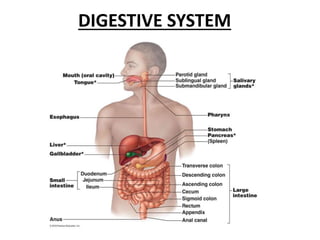
  • 2. Digestive System Organs of GI Tract • Oral cavity • Pharynx • Esophagus • Stomach • Small intestine • Large intestine Accessory Organs of GI Tract • Teeth • Tongue • Salivary glands • Liver • Gall bladder • Pancreas
  • 3. Functions of digestive system • Ingestion • Mastication • Deglutition • Digestion • Absorption • Peristalsis • Defecation
  • 4.
  • 5. LAYERS OF GASTROINTESTINAL TRACT 1. MUCOSA 2. SUB MUCOSA 3. TUNICA MUSCULARIS 4. SEROSA
  • 7.
  • 8. Innervation of the Gastrointestinal Tract  The GI tract is innervated by the sympathetic and parasympathetic divisions of the autonomic nervous system  The vagus nerves are the source of parasympathetic activity in the esophagus, stomach, pancreas, gallbladder, small intestine, and upper portion of the large intestine.  The lower portion of the large intestine receives parasympathetic innervation from spinal nerves in the sacral region.  The submucosal plexus and myenteric plexus are the sites where preganglionic neurons synapse with postganglionic neurons that innervate the smooth muscle of the GI tract.  Stimulation of the parasympathetic neurons increases peristalsis and the secretions of the GI tract.  Postganglionic sympathetic fibers pass through the submucosal and myenteric plexuses and innervate the GI tract.  The effects of sympathetic nerve stimulation are antagonistic to those of parasympathetic nerve stimulation.  Sympathetic impulses inhibit peristalsis, reduce secretions, and constrict muscle sphincters along the GI tract.
  • 9.
  • 10. MOUTH • Mouth also known as oral cavity is formed by cheeks, lips, hard palate & soft palate. • Vestibule of oral cavity is the depression between the cheeks & lips externally and gums & teeth internally. • Opening of oral cavity is referred to as oral orifice. • Opening between oral cavity & pharynx is called fauces.
  • 11.
  • 12. CHEEKS • Cheeks form the lateral walls of the oral cavity • They consist of outer layer of skin, subcutaneous fats, facial muscles that assist in manipulating food in oral cavity & inner lining of stratified squamous epithelium. • Anterior portion of lips terminate in superior & inferior lips that surrounds oral orifice.
  • 13. LIPS • Lips are fleshy highly mobile organs whose principle function in human is associated with speech. • Lips also serve for suckling, manipulating food & keeping food between upper & lower teeth. • Each lip is attached from its inner surface to the gum by a midline fold of mucus membrane called labial frenulum.
  • 14. Between outer skin & mucous membrane of oral cavity is a transition zone called the Vermilion
  • 15. PALATE • Palate which forms the roof of oral cavity consists of bony hard palate anteriorly & soft palate posteriorly.
  • 16. Hard Palate • Hard palate formed palatine process of maxillae & horizontal plates of palatine bones & is covered with mucous membrane. • Transverse palatine folds or Palatal Rugae are located along the mucous membrane of the hard palate.
  • 17.
  • 18. • Soft Palate is a muscular arch covered mucous membrane & is continuous with the hard palate anteriorly. • Suspended from the middle lower border of the soft palate is a cone shaped projection called Palatine Uvula. Soft Palate
  • 19. • Two muscular folds extend downward from both sides of the base of palatine uvula. • The anterior fold is called glossopalatine arch & posterior fold is called pharyngopalatine arch. • Between these two arches is the palatine tonsil.
  • 20. TONGUE • Tongue is a mass of skeletal muscle covered with mucous membrane. • Tongue function to move food around the mouth during mastication & to assist in swallowing food • Essential in producing speech. • Extrinsic tongue muscles move tongue from side to side & in and out. • Only anterior 2/3 of tongue lies in oral cavity & remaining 1/3 lies in pharynx and is attached to hyoid bone.
  • 21. • Rounded masses of lingual tonsils are located on the superior surface of the base of the tongue. • The inferior surface of the tongue is connected along midline anteriorly to the floor of mouth by vertically positioned Lingual frenulum .
  • 22. • Papillae are numerous small elevations on the surface of tongue. • They give tongue a distinct roughened surface that aids in handling of food. • They also contain taste buds that respond to sweet, salty, sour & bitter chemical stimuli. • 4 types of papillae are present on surface of tongue 1. Filiform 2. Fungiform 3. Foliate 4. Circumvallate
  • 24. • Filiform papillae Filiform papillae are sensitive to touch, have tapered tips, and are by far the most numerous. These papillae lack taste buds and are not involved in the perception of taste. • Fungiform papillae -The fungiform papillae are involved in the sensations of taste and have taste buds on their upper surface which can distinguish the tastes: sweet, sour, bitter, salty • Circumvallate papillae - there are only about 10 to 14 of these papillae on most people, and are arranged in a V- shaped formation directed toward the throat. The taste buds of the vallate papillae are sensitive to bitter flavors. • Foliate papillae - these are ridges and grooves towards the posterior part of the tongue found at the lateral borders
  • 25. TEETH • Humans & other mammals have heterodont dentition means they have various types of teeth. • The four pairs (upper & lower jaws) of anterior most teeth are the incisors adapted for cutting & shearing food. • Two pairs of cone shaped canines (cuspids) are located at the anterior corners of the mouth they are adapted for holding & tearing. • Incisors & canines are characterized by singly root on each tooth
  • 26. TEETH
  • 27. • Located behind the canines are the premolars & molars • They have 2 or 3 roots & somewhat rounded, irregular surfaces called dental cusps for crushing & grinding food.
  • 28. The buccal surface of the premolars & molars is adjacent to cheek. The labial surface of the incisors & canines is adjacent to the lip. The lingual surface of all teeth is adjacent to tongue.
  • 29. TEETH • Humans are diphyodont that is normally 2 sets of teeth develop in person’s lifetime. • Twenty deciduous (milk) teeth begin to erupt at about 6 months of age, beginning with the incisors. • All erupt at the age of 2.5 yrs • 32 permanent teeth replace the deciduous teeth in predictable sequence. • Process begins at age of 6 and continues until about age of 17yrs. • Third molars (wisdom teeth ) are the last to erupt at age 17-25
  • 30. • A dental formula is a graphic representation of the types, number & position of teeth in the oral cavity • Formula for deciduous dentition I 2/2, C 1/1 , DM 2/2 = 10*2 =20 teeth • Formula for permanent dentition I 2/2, C1/1, P 2/2, M 3/3 = 16*2=32 teeth Where I = incisor C= canine P = premolar DM = deciduous molar M = molar
  • 31. Structure of tooth • A tooth consists of exposed crown, which is supported by neck that is anchored firmly into the jaw by one or more roots. • Root of teeth fits into sockets called dental alveoli, in the alveolar processes of mandible & maxillae. • Each socket is lined by connective tissue periosteum, specifically called periodontal membrane. • Root of tooth is covered with a bone like material called cementum.
  • 32.
  • 33. • The gingiva(gum) is the mucous membrane surrounding the alveolar processes in oral cavity. • Bulk of tooth consists of dentin a substance similar to bone but harder. • Covering the dentin on the outside & forming the crown is a tough, durable layer of enamel . • Enamel is composed of calcium phosphate & is the hardest substance in body. • Central region of tooth contains the pulp cavity
  • 34. • It contains pulp which is composed of connective tissue with blood vessels, lymph vessels & nerves. • A root canal continuous with the pulp cavity opens to the connective tissue surrounding the root through an apical foramen.
  • 36. SALIVARY GLANDS • The salivary glands are accessory digestive glands that produce a fluid secretion called saliva. • The amount of saliva secreted daily ranges from 1.0 to 1.5 L. • Saliva also contains starch-digesting enzymes and lubricating mucus, which aids swallowing. • PAROTID GLANDS • SUBMANDIBULAR GLANDS • SUBLINGUAL GLANDS
  • 37. Two types of secretory cells, serous and mucous cells, are found in all salivary glands in various proportions . Serous cells produce a watery fluid containing digestive enzymes; mucous cells secrete a thick, stringy mucus. Cuboidal epithelial cells line the lumina of the salivary ducts. The salivary glands are innervated by both divisions of the autonomic nervous system. Sympathetic impulses stimulate the secretion of small amounts of viscous saliva. Parasympathetic stimulation causes the secretion of large volumes of watery saliva. Physiological responses of this type occur whenever a person sees, smells, tastes, or even thinks about desirable food
  • 38. HISTOLOGY OF SALIVARY GLANDS Acini:  Secretory cells are found in a group, or acinus (plural, acini).  Each acinus is located at the terminal part of the gland connected to the ductal system, with many acini within each lobule of the gland.  Each acinus consists of a single layer of cuboidal epithelial cells surrounding a lumen, a central opening where the saliva is deposited after being produced by the secretory cells.  The three forms of acini are classified in terms of the type of epithelial cell present and the secretory product being produced: serous, mucoserous and mucous.  Two types of secretory cells, serous and mucous cells, arefound in all salivary glands in various proportions .Serous cells produce a watery fluid containing digestive enzymes; mucous cells secrete a thick, stringy mucus. Ducts:  In the duct system, the lumina are formed by intercalated ducts, which in turn join to form striated ducts. These drain into ducts situated between the lobes of the gland (called interlobar ducts or secretory ducts). These are found on most major and minor glands (exception may be the sublingual gland).
  • 39. Acinus ……….Intercalated Duct ……. Striated Duct (Intralobular) ………. Excretory Duct (Interlobular)
  • 40.
  • 41.
  • 42. PAROTID GLANDS • Largest of salivary glands • Positioned below & in front of the auricle of ear,between skin & masseter muscle. • Saliva produced drain through parotid ( Stensen’s) duct. • Secretes watery serous fluids, salts & enzymes. • It become infected & swollen with the mumps.
  • 43. HISTOLOGY OF PAROTID GLANDS • The acini of glands secrete enzymes • The acinus is rounded & is lined by pyramidal cells surrounding a very small lumen • Nuclie are rounded & basal in position.
  • 44. SUBMANDIBULAR GLAND • Lies inferior to the body of the mandible, about midway along the inner side of jaw. • Saliva produced in the submandibular gland drains through the submandibular (Wharton’s duct) & empties into the floor of the mouth on lateral side of lingual frenulum.
  • 45. HISTOLOGY OF SUBMANDIBULAR GLANDS • Consists of both serous & mucous acini. • Mucous acini is lined by columnar cells. • Size of mucous acini is larger than the serous one & shows bigger lumen. • Between the mucous cells & the basement membrane are half moon shaped polyhedral granular serous cells. • These cells are known as demilunes of Gianuzzi.
  • 47. SUBLINGUAL GLANDS • Lies under the mucous membrane of the floor of the mouth. • Contains small sublingual ducts (Rivinus ducts) that empty into the floor of the mouth in as area posterior to the papilla of submandibular duct
  • 48. HISTOLOGY OF SUBLINGUAL GLANDS • Sublingual gland is predominantly mucous. • The acinus is lined by truncated columnar cells. • Size of acinus is bigger than serous acinus with wider lumen • Nucleus is flattened and rest against basement membrane.