facial nerve (7th cranial nerve)
•Transferir como DOCX, PDF•
21 gostaram•7,760 visualizações

• The Facial nerve is the 7th of twelve paired cranial nerves. • It is a mixed nerve with motor and sensory roots. • It also supplies pre-ganglionic parasympathetic fibres to several head and neck ganglia Branches 1. Greater superficial petrosal – arises from the geniculate ganglion. 2. Branches within the facial canal: • i) nerve to stapedius • ii) Chorda tympani 3. After exit from stylomastoid foramen: • i) Posterior auricular • ii) Nerve to posterior belly of digastric • iii) Nerve to stylohyoid. 4. On the face - Five major branches: • i) Temporal • ii) Zygomatic • iii) Buccal • iv) Marginal mandibular • v) Cervical

Recomendados
Mais conteúdo relacionado
Mais procurados
Mais procurados (20)
Destaque
Destaque (16)
Semelhante a facial nerve (7th cranial nerve)
Semelhante a facial nerve (7th cranial nerve) (20)
Mais de D.A.B.M
Mais de D.A.B.M (20)
Último
Último (20)
facial nerve (7th cranial nerve)
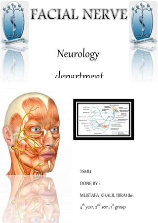
- 1. Neurology department TSMU DONE BY : MUSTAFA KHALIL IBRAHIm 4th year, 2nd sem, 1st group
- 2. CONTENTS 1.Introduction 2. Nuclei of origin 3. Course & Relations 4. Branches of facial nerve 5. Ganglia associated with facial nerve 6. Testing of facial nerve 7. Identification of facial nerve 8. Nerve injury
- 3. • The Facial nerve is the 7th of twelve paired cranial nerves. • It is a mixed nerve with motor and sensory roots. • It also supplies pre-ganglionic parasympathetic fibres to several head and neck ganglia Introduction Nuclei of Origin 1.Motor nucleus 2. Superior salivatory nucleus 3.Nucleus of tractus solitaries This facial nuclei in pons is divided into : 1- Lower parts which innervate the upper half of The face & it has double cortical innervation. 2- upper parts which innervate the lower half of The face & it has single cortical innervation.
- 4. The facial nerve is formed mainly of two parts: • 1- Facial nerve proper (motor): arising from facial motor nucleus in pons. • 2- Nervus intermedius: it is the sensory root of facial lies position between the facial proper and vestibulcochlear nerve in the pontocerebellar angle. Carrying para-sympathetic fibers (from superior salivary nucleus) and taste fibers ( to the solitary nucleus). Course & Relations • I- Intracranial (intrapetrosal) course • II- Extracranial course
- 7. Branches 1. Greater superficial petrosal – arises from the geniculate ganglion. 2. Branches within the facial canal: • i) nerve to stapedius • ii) Chorda tympani 3. After exit from stylomastoid foramen: • i) Posterior auricular • ii) Nerve to posterior belly of digastric • iii) Nerve to stylohyoid. 4. On the face - Five major branches: • i) Temporal • ii) Zygomatic • iii) Buccal • iv) Marginal mandibular • v) Cervical
- 8. Ganglia associated with facial nerve • Geniculate ganglion • Submandibular ganglion • Pterygopalatine ganglion Geniculate ganglion • The geniculate ganglion (from Latin genu, for "knee") is an L-shaped collectionof fibers and sensoryneurons of the facial nerve located in the facial canal of the head. • It receives fibers from the motor, sensory, and parasympathetic components of the facial nerve Submandibular ganglion • The submandibular ganglion is small and fusiform in shape. It is situated above the deep portion of the submandibular gland, on the hyoglossus muscle, near the posterior border of the mylohyoid muscle Pterygopalatine ganglion • The pterygopalatine ganglion (meckel's ganglion, nasal ganglion or sphenopalatine ganglion) is a parasympathetic ganglion found in the pterygopalatine fossa. • It's largely innervated by the greater petrosal nerve (a branch of the facial nerve); and its axons project to the lacrimal glands and nasal mucosa Facial Nerve blood supply • The facial nerve gets it’s blood supply from 5 vessels: Anterior inferior cerebellar artery – at the cerebellopontine angle Labyrinthine artery (branch of anterior inferior cerebellar artery) – within internal acoustic meatus Superficial petrosal artery (branch of middle meningeal artery) – geniculate ganglion and nearby parts Stylomastoid artery (branch of posterior auricular artery) – mastoid segment Posterior auricular artery supplies the facial nerve at & distal to stylomastoid foramen • Venous drainage parallels the arterial blood supply
- 9. Testing of Facial Nerve Branches • Temporal branches -patient is asked to frown and wrinkle his or her forehead. • Zygomatic branches -the patient is asked to close their eyes tightly • Buccal branches -puff up cheeks (buccinator) smile and show teeth (orbicularis oris) tap with finger over each cheek to detect ease of air expulsion on the affected side Temporal branch • It exits the parotid gland anterior to superficial temporal artery During an open approach to the TMJ, violation of this branch is possible Zygomatic Branch • Its course is antero superior crossing the zygomatic bone Inadvertent damage may occur to this nerve during open reduction of zygomatic arch or with the use of zygomatic hook during closed approaches Buccal Branch: • It runs almost horizontally and will often divide into separate branch above and below parotid duct as it runs anteriorly Injury is possible in association with soft tissue trauma to the cheek region Marginal mandibular branch • It extends anteriorly and inferiorly within the substance of parotid gland, there may be two or three branches of this nerve. These branches run anteriorly parallel to inferior border of mandible and in some cases the course of the nerve is above the inferior border. In essentially all cases the nerve is located above the inferior border of mandible beyond the facial artery. The marginal mandibular branch is an important structure encountered at the inferior border of the mandible just beneath the platysma muscle fibres during an open approach to the mandibular angle and body area. • For this reason, an initial incision made approximately 1 to 1.5cm below the inferior border which prevents direct exposure or trauma to the nerve
- 10. Cervical Branch: • The cervical branch exits the parotid gland above its inferior pole and runs downwards underneath the platysma muscle 3 surgicalmaneuvers used to identify nerve trunk A. Bloodfree plane in front of external acoustic meatus B. Exposure of anterior border of SCM below insertion into mastoid process C. Peripheral identification of terminal branch of facialnerve (marginal mandibular branch) Identification of Facial Nerve
- 12. Duus'_Topical_Diagnosis_in_Neurology Neuroanatomy atlas ginsberg_l_lecture_notes_neurology Dan Kirshenbaum BUSM Class of 2011 - Gross Anatomy 2007 Dr.Hassan Elwan Neurology Also Some internet photos References