SlideShare uma empresa Scribd logo
1 de 90
SEGMENTAL ANALYSIS OF
CONGENITAL HEART DISEASE
Murtaza Kamal M.D., D.N.B.
Fellow Pediatric Cardiology
murtaza.vmmc@gmail.com (Dt: 12-14/12/2017) 1
MAUDE ELIZABETH SEYMOUR ABBOTT
1936
McGill University, Canada
1st
systematic study
Based on study of 1000 heart specimen
2
Richard Van praagh
• 1964
• Pediatric Cardiologist
• Boston Children
Hospital, Massachusetts
• Segmental approach to
CHD
3
History Cont…
• Kirklin(1973): Modified the segmental approach
using concordance/discordance to describe
connections between 3 main cardiac segments
• Shinebourne/Anderson/Tynan(1976): Popularized
the sequential segmental approach to CHD
4
5
6
THORACO-ABDOMINAL SITUS
Situs: Topology/ spatial position of structureSitus: Topology/ spatial position of structure
1st
segment evaluated is visceral situs
Atrial and visceral situs considered together
because they usually are concordant ( atrial and
visceral situs are same)
7
Pulmonary Situs (Sidedness)
• Determined by positions of morphologic rt & lt lungs
• Pulmonary morphology, in turn, defined by
relationship of PAs to their adjacent bronchi, and not by
the number of lobes
• PA of a morphologic right lung travels anterior to its
upper and intermediate bronchi, whereas that of a
morphologic left lung travels over its main bronchus
and posterior to the upper lobe bronchus
8
Pulmonary Situs (Sidedness)
Cont…
• Rt bronchus branches, then RPA crosses it
• LPA crosses lt bronchus, then it branches
• Clinically, pulmonary situs may be inferred by
comparing relative lengths of 2 main bronchi, as
measured on a CXR that shows an air bronchogram
• Distance from carina to origin of upper lobe
bronchus is 1.5- 2.5 X greater for morphologic left
lung than for right lung, and this ratio holds true
regardless of the sidedness of the aortic arch
9
10
The morphologically left bronchus is long, and it branches only after it has been crossed by its
accompanying pulmonary artery, making the bronchus hyparterial. In contrast, the
morphologically right bronchus is short, and is crossed by its pulmonary artery only after it
has branched, giving an eparterial pattern of branching
LR
11
2D ECHO: STRUCTURE??
12
2D ECHO: STRUCTURE??
13
14
Cardiac malposition
• Often an integral part of complex associated
anomalies of visceral atrial situs
• Prevalence 1 in 10, 000 live births
Cardiac malpositions:
Dextrocardia
Mesocardia
Levocardia (isolated)
Ectopia cordis
15
Cardiac Malposition
16
17
DEXTROCARDIA
• Detro position
• Dextro version
• Mirror image dextrocardia
18
Dextroposition: Heart pushed/ pulled into right
chest
Push:
Lt sided tension pneumothoax
Lt congenital lobar emphysema
Lt diaphragmatic hernia
Pull:
Hypoplasia/ agenesis of rt. lung
DEXTROCARDIA
19
DEXTROCARDIA Cont…
Dextroversion: Failure of pivoting of cardiac apex to
left, frequently associated with A-V discordance
Mirror-image dextrocardia with atrial situs inversus
There may be major and complex pathology
associated with this type of dextrocardia
20
21
MESOCARDIA
Location of heart with cardiac base apex axis
directed to midline of thorax or with ventricular
apices equally directed to both right and left
sides
This is often ignored as being infrequent and
atypical
22
Isolated levocardia
Occurring in conjunction with situs inverses
and situs ambiguous
 An abnormal atrial and visceral situs, the
heart is in its normal location in the left hemi
thorax with the apex pointing to left
23
Ectopia Cordis
• Partially/ completely
exteriorized
• Pentalogy of Cantrell:
– 1. Deficiency of ant. Diaphragm
– 2. Midline supra umbilical
abdominal wall defect
– 3. Defect in diaphragmatic
pericardium
– 4. CHD
– 5. Defect of lower sternum
24
BASIC CARDIAC POSITIONS
95% CHD 100% CHD
3-5%CHD<1%CHD
25
26
SEGMENT BY SEGMENT
ANALYSIS
27
SEGMENT CONNECTION
Great veins
Atria
Ventricles
Great arteries
Venoatrial
Atrioventricular
Ventriculoarterial
28
29
30
VENOUS SEGMENT
Systemic veins: IVC, hepatic
veins, SVC, CS
Pulmonary veins
HEPATIC VEINS: SC view,
drains into RA
CS – RA
IVC – RA
31
Venous Segment Cont…
ABNORMALITY OF IVC: Subcostal view
Interrupted IVC (Polysplenia):
Large azygous vein entering SVC seen along the spine in
abdomen, can be either right or left sided
Even then there is another vein draining into RA called:
Suprahepatic IVC
Suprahepatic IVC always drains into RA
Suprahepatic IVC differentiated from IVC in being small and
can’t be traced below liver
32
SVC
• SVC: Single right sided
• Subcostal view, high parasternal or suprasternal view
• LSVC:
• Connected to CS posteriorly/ laterally
• Border of LA (dilated CS)
• Directly visualized in a long axis plane
• Parasternal short axis images: Circular vessel anterior to
LPA near bifurcation
• SVC less reliable than IVC for situs
33
CS & LSVC
34
Pulmonary veins
• Subcostal, suprasternal, apical
• Color Doppler: Distinguishes from atrial
appendage
• 4 in number
• Left 2 veins may join: 3 entries into LA
35
ATRIAL SITUS
The identification of the morphologic right
atrium is important for establishing atrial
situs
Anatomic hallmark is the limbus of the
fossa ovalis
36
Atrial Situs
• Describes location of atria
• Solitus: Morphological RA on rt
• Inverses: Morphological RA on lt
• Ambiguous: Undifferentiated atria
37
Right atrium Left atrium
SEPTUM:
Limbus of fossa ovalis Flap valve of fossa ovalis
APPENDAGES:
Broad based, triangular, anterior Long and narrow, finger like,
posterior
MYOCARDIAL FEATURES:
Crista terminalis, tinea sagittalis,
extension of pectinate muscles towards
AV valve
Pectinate muscle confined to
appendage
VEINS:
Receives IVC, suprahepatic IVC, SVC,
coronary sinus
Receives pulmonary veins
38
Rightatrium Left atrium
39
40
Subcostal sagittal views demonstrating eustachian valve (arrow in A) and flap
valve (arrow in B).
41
ATRIAL APPENDAGE
42
43
Viscero atrial situs and its abnormality
Isomerism of Lt atrial
appendage:
•B/l lt atrial appendage:
Smooth pectinate muscles
•Interrupted IVC: 85%
Isomerism of Rt atrial
appendage:
• B/l rt atrial appendage:
Coarse pectinate muscles
•Abscent coronary sinus:
100%
•Abnormal relation of aorta
and IVC
44
ATRIAL ABNORMALITIES
azygous
45
46
VENTRICULAR SITUS AND
MORPHOLOGY
Right ventricle Left ventricle
Large apical trabeculations Small apical trabeculations
Coarse septal surface Smooth upper surface
Moderator band No moderator band
Receives tricuspid valve Receives mitral valve
Tricuspid-pulmonary discontinuity Mitral-aortic continuity
Crescentic in cross section Circular in cross section
Thin free wall (3-5 mm) Thick free wall (12-15 mm)
47
Apical four-chamber view in a patient with ventricular inversion
with L-TGA
48
VENTRICULAR LOOPING
After morphology Determine looping
Bulboventricular loop: Describes location of ventricles
d-Loop: Morphologic RV on right
l-Loop: Morphologic RV on left
49
VENTRICULAR LOOPING Cont…
 Performed by imagining one is standing on the right
ventricle side facing the right ventricular face of the
interventricular septum
 The palm of one hand is placed against the septum
 The looping is determined by establishing which of the two
hands allow the thumb to point into the atrioventricular
valve and the fingers to point into the outflow tract
 If rt. hand meets criteria: d-looped, if lt hand meets criteria:
l-looped
50
The palmar surface of the right hand can
be placed on the septal surface of the normal morphologically right ventricle
with the thumb in the inlet component and the fingers extending into the
ventricular outlet. (d-ventricular loop.)
51
The mirror-imaged normal heart. In this setting,
it is the palmar surface of the left hand that can be placed on the septal
surface of the morphologically right ventricle with the thumb in the inlet
and the fingers in the outlet. (l-ventricular loop)
52
VENTRICULAR LOOPING Cont…
 In general, convexity of aorta points to the position of the
right ventricle and thus helps indicate bulboventricular loop
 Definitive indicator of bulboventricular loop, however, is
relative positioning of ventricular inlets/ AV valves
 Thus, in a d-loop the tricuspid valve is to the right of the
mitral valve, and in an l-loop the tricuspid valve is to the left
of the mitral valve
 In a normal d-loop the apex pivots to the left hemithorax;
in a "normal" l-loop (i.e., one in the setting of situs inversus)
the apex pivots to the right hemithorax
53
SINGLE VENTRICLE
• Single ventricle: Determined by morphology
of ventricle
• Single vent of RV morphology: Hypo plastic LV
remnant, posterior to main ventricular
chamber
• LV morphology: Rudimentary RV, anterior 54
55
56
AV Connections and AV Valve
Morphology
First see: Bi/uni ventricular connection
If double see: AV concordance/discordance
If single ventricle see: Double inlet, single inlet,
common inlet
57
50% rule
 Anderson et al: Rule of 50% for determining
whether a cardiac chamber is a ventricle
 For the assessment of AV connections, an atrium is
considered to join the ventricle into which >50% of
the valve orifice empties
 This rule states that a chamber is a ventricle if it
receives 50% more of an inlet
 The inlet consists of the fibrous ring of the AV valve
and need not always include a patent AV valve with
well formed valve leaflets 58
50% RULE Cont…
 Eg: In HLHS with aortic and mitral atresia, the
fibrous ring of the MV contains an imperforate
membrane and is situated over the small LV
 Thus, this small left sided chamber is a ventricle
because it receives 100% of an inlet
 A chamber need not have an outlet to be a ventricle
 The rule of 50% has also been used to define VA
connections
 Thus, if 50% or more of a great artery arises above a
chamber, the great artery is defined as being
59
50% RULE IN AV, VA
• A great vessel should be related to a
ventricle by 50% of its dimension to be
considered committed to it
• Av valve is committed to that ventricle to
which >50% of that valve is connected
60
SINGLE VENTRICLE
61
ATRIOVENTRICULAR VALVE
MORPHOLOGY
Tricuspid valve Mitral valve
Septal chordal attachments No septal chordal
attachments
Low septal annular
attachment
High septal annular
attachment
Triangular orifice
(midleaflet)
Elliptical orifice (midleaflet)
Three leaflets Two leaflets
Several papillary muscles Two large papillary muscles
Empties into right ventricle Empties into left ventricle
62
ATRIOVENTRICULAR CONNECTIONS
63
OVERRIDING AV VALVE
Abnormality of AV valve
alignment or connection in
which one valve annulus
opens into another chamber
through a VSD
64
STRADDLING
A feature of the tensor apparatus (chordae tendineae
and papillary muscles) of an AV valve and indicates
anomalous insertion into the contralateral ventricle,
either along its septum or its free wall
65
66
67
VENTRICULOARTERIAL CONNECTION
If Ao from LV and PA from RV: Concordant
If Ao from RV and PA from LV: Discordant
Origins of Ao and PA are evident on PLAX view, sweeping the
transducer inferiorly from the basal short-axis view, A5CV and
SC coronal and sagittal views
3rd
type of VA connection: Double outlet, almost always from
RV
Final type of VA connection: Single outlet (truncus
arteriosus)
68
VA Connection
Aorta:
•Coronary artery
•Carotids
•Absence of proximal
division
PA:
•Bifurcation
69
Parasternal short-axis images demonstrating different relationships between
the great vessels.
An echocardiogram from an infant with tetralogy of Fallot and pulmonary
atresia demonstrates the identifying features of a pulmonary artery (MPA) with
its bifurcation into right and left pulmonary arteries (RPA, LPA). The great
vessels are normally related
70
71
72
CONAL MORPHOLOGY
Conus/ infundibulum: Cavitary space formed by muscular
segment of heart that connects ventricles with great arteries
and separates the AV and semilunar valves
Abnormalities in conal development consist of variations in
the presence, length and diameters of subpulmonary and
subaortic conus
These variations can lead to (or be associated with) complex
malformations, such as TOF, IAA,TGA and DORV
73
Subpulmonary conus - Best
identified in subcostal views
In normal heart: Conus- Nearly
vertical tubular outflow portion of
RV
Separated from nearly horizontal
right ventricular inflow portion by
distinct muscle bands
These muscle bands form a near-
circular rim formed by parietal band
anteriorly, crista supraventricularis
posteriorly, and septal band
medially and prohibit PV to AV
valve continuity 74
Conus Cont…
 Subaortic conus is evident on
subcostal coronal and sagittal views
 Persistence of subaortic conus and
involution of subpulmonic conus is
the usual conal relationship in d- (or
l-) TGA
 Persistence of subaortic conus
prohibits continuity of the aortic
valve to either AV valve, and
involution of subpulmonary conus
allows continuity between PV and
both AV valves in TGA 75
BILATERAL CONUS
 B/l persistence of the subarterial
conus usually results in DORV
 Because the main goal of surgical
correction of DORV is to connect the
aorta with the morphologic left
ventricle through VSD, it is important
to determine the conal relationships
with each other and with the great
vessels 76
BILATERAL CONUS
When 2 coni are present, their relationship may be
classified as either anterior/posterior or side-by-side
With the anterior/posterior conal relationship, the
ventricular septal defect is usually subaortic; with the
side-by-side relationship, the defect is usually
subpulmonic
 The conal relationship can be determined by subcostal
coronal and sagittal imaging with anterior/posterior
and left/right sweeping, respectively 77
ABSENT CONUS
 A rare type of d-transposition
can exist in the context of
bilaterally deficient
subarterial conus
 This results in an unusual
heart in which d-TGA exists
with a doubly committed
VSD and a posterior aorta
 Associated with DOLV 78
79
SEMILUNAR VALVES
80
Great Artery Relations
8 basic types of great artery relationships are possible
based on the aortic and pulmonary valve positions at the
level of the semilunar valves
• Right posterior aorta (normally related)
Right lateral aorta (side by side)
Right anterior aorta (d-malposed)
Directly anterior aorta
Left anterior aorta (l-malposed)
Left lateral aorta (left side by side)
Left posterior aorta (inverted normal)
Directly posterior aorta.
81
82
83
ARCH OF AORTA
 Unexpected position of aortic arch: Well recognized
associated anomaly of conditions such eg. TOF/ Truncus
 Distinction should be made b/w position of arch and side of
descending aorta, particularly in describing vascular rings
 Side of aortic arch depends on whether it passes to right or
left of the trachea
 Position of descending aorta is defined relative to vertebral
column
84
ARCH OF AORTA Cont…
 Aorta lies more posterior in center of
 Visualized in PLAX,PSAX, A5CV,SC , and suprasternal
notch views
 Arch best seen in subcostal oblique view and
suprasternal views
 Side of arch diagnosed by sweeping transducer in
suprasternal long-axis view and noting the
relationship of the arch to the trachea, the rings of
which resemble a stack of coins
85
This transducer position allows visualization of the ascending aorta
(Asc), aortic arch (Arch), origin of the brachiocephalic vessels
(arrows), descending thoracic aorta (Dsc), and right pulmonary
artery (*).
ARCH OF AORTA LONG-AXIS
86
The short-axis view of the aortic arch is obtained by rotating the
transducer clockwise, which also allows visualization of the right
pulmonary artery (RPA) in its long-axis format, located inferiorly to
the aortic arch (Arch). Inferior to RPA is the left atrial (LA) cavity
with connections of the four pulmonary veins (arrows
SHORT-AXIS VIEW
87
88
FINAL DIAGNOSIS
• SS, LEVOCARDIA, NORMAL SYSTEMIC &
PULMONARY VENOUS COMMUNICATIONS, NRGAs,
AV & VA CONCORDANCE, INTACT IA & IV SEPTUM,
NO PDA, GOOD SIZED CONFLUENT BPAs, NORMAL
ARCH, NO COA, NORMAL CORONARIES, NORMAL
SIZED CHAMBERS, GOOD BV FUNCTION, NO
VEGETATION/ CLOT OR PE 89
THANKS FOR UR PATIENCE
90

Mais conteúdo relacionado

Mais procurados

Echo assessment of lv systolic function and swma
Echo assessment of lv systolic function and swmaEcho assessment of lv systolic function and swma
Echo assessment of lv systolic function and swma
Fuad Farooq
 
Tte and tee assessment for asd closure 2
Tte and tee assessment for asd closure 2Tte and tee assessment for asd closure 2
Tte and tee assessment for asd closure 2
Rahul Chalwade
 

Mais procurados (20)

Aortic stenosis Echo
Aortic stenosis Echo Aortic stenosis Echo
Aortic stenosis Echo
 
Echo assesment of rv function
Echo assesment of rv function Echo assesment of rv function
Echo assesment of rv function
 
Cath hemodynamics vir
Cath hemodynamics virCath hemodynamics vir
Cath hemodynamics vir
 
Echo assessment of RV function
Echo assessment of RV functionEcho assessment of RV function
Echo assessment of RV function
 
Echo assessment of mitral regurgitation
Echo assessment of mitral regurgitationEcho assessment of mitral regurgitation
Echo assessment of mitral regurgitation
 
Echo Mitral Stenosis
Echo Mitral StenosisEcho Mitral Stenosis
Echo Mitral Stenosis
 
Echocardiographic Evaluation of Hypertrophic Cardiomyopathy
Echocardiographic Evaluation of Hypertrophic CardiomyopathyEchocardiographic Evaluation of Hypertrophic Cardiomyopathy
Echocardiographic Evaluation of Hypertrophic Cardiomyopathy
 
Echocardiography of Mitral regurgitation
Echocardiography of Mitral regurgitationEchocardiography of Mitral regurgitation
Echocardiography of Mitral regurgitation
 
Lv systolic function
Lv systolic functionLv systolic function
Lv systolic function
 
Echocardiography of CHD in Adults
Echocardiography of CHD in AdultsEchocardiography of CHD in Adults
Echocardiography of CHD in Adults
 
Collection of cath tracings by navin
Collection of cath tracings by navinCollection of cath tracings by navin
Collection of cath tracings by navin
 
Natural history of Pre tricuspid shunts
Natural history of Pre tricuspid shuntsNatural history of Pre tricuspid shunts
Natural history of Pre tricuspid shunts
 
MVP Mitral Valve Prolapse - Echocardiographic Evaluation
 MVP Mitral Valve  Prolapse - Echocardiographic Evaluation MVP Mitral Valve  Prolapse - Echocardiographic Evaluation
MVP Mitral Valve Prolapse - Echocardiographic Evaluation
 
Cardiac dyssynchrony ppt by dr awadhesh
Cardiac dyssynchrony ppt   by dr awadheshCardiac dyssynchrony ppt   by dr awadhesh
Cardiac dyssynchrony ppt by dr awadhesh
 
Echo assessment of lv systolic function and swma
Echo assessment of lv systolic function and swmaEcho assessment of lv systolic function and swma
Echo assessment of lv systolic function and swma
 
M mode echocardiography
M mode echocardiographyM mode echocardiography
M mode echocardiography
 
Echocardiography assessment of Aortic Regurgitation severity
Echocardiography assessment of Aortic Regurgitation severityEchocardiography assessment of Aortic Regurgitation severity
Echocardiography assessment of Aortic Regurgitation severity
 
TEE VIEWS
TEE VIEWSTEE VIEWS
TEE VIEWS
 
Ebstein's anomaly echocardiogram
Ebstein's anomaly echocardiogramEbstein's anomaly echocardiogram
Ebstein's anomaly echocardiogram
 
Tte and tee assessment for asd closure 2
Tte and tee assessment for asd closure 2Tte and tee assessment for asd closure 2
Tte and tee assessment for asd closure 2
 

Semelhante a SEGMENTAL ANALYSIS OF CONGENITAL HEART DISEASE

Congenitally corrected transposition of great arteries
Congenitally corrected transposition of great arteriesCongenitally corrected transposition of great arteries
Congenitally corrected transposition of great arteries
Dheeraj Sharma
 
Complete transposition of great arteries
Complete transposition of great arteriesComplete transposition of great arteries
Complete transposition of great arteries
Dheeraj Sharma
 

Semelhante a SEGMENTAL ANALYSIS OF CONGENITAL HEART DISEASE (20)

ECHO SEGMENTAL APPROACH- DR BINJO.ppsx
ECHO SEGMENTAL APPROACH- DR BINJO.ppsxECHO SEGMENTAL APPROACH- DR BINJO.ppsx
ECHO SEGMENTAL APPROACH- DR BINJO.ppsx
 
Segmental analysis of heart anatomy
Segmental analysis of heart anatomySegmental analysis of heart anatomy
Segmental analysis of heart anatomy
 
L tga anatomy, management-
L tga anatomy, management-L tga anatomy, management-
L tga anatomy, management-
 
Cc tga
Cc tgaCc tga
Cc tga
 
Transposition of Great Arteries
Transposition of Great ArteriesTransposition of Great Arteries
Transposition of Great Arteries
 
avsd
avsdavsd
avsd
 
Segmental analysis in congenital heart diseases m.gibreel
Segmental analysis in congenital heart diseases  m.gibreelSegmental analysis in congenital heart diseases  m.gibreel
Segmental analysis in congenital heart diseases m.gibreel
 
Double outlet right ventricle
Double outlet right ventricleDouble outlet right ventricle
Double outlet right ventricle
 
The anatomy of the cardiac valves and their abnormalities
The anatomy of the cardiac valves and their abnormalitiesThe anatomy of the cardiac valves and their abnormalities
The anatomy of the cardiac valves and their abnormalities
 
Congenital LV and RV inflow anomalies by Echocardiography
Congenital LV and RV inflow anomalies by EchocardiographyCongenital LV and RV inflow anomalies by Echocardiography
Congenital LV and RV inflow anomalies by Echocardiography
 
Congenitally corrected transposition of great arteries
Congenitally corrected transposition of great arteriesCongenitally corrected transposition of great arteries
Congenitally corrected transposition of great arteries
 
VASCULAR RINGS AND SLINGS TYPES HAEMODYNAMICS PRESENTATION AND DIAGNOSIS.pptx
VASCULAR RINGS AND SLINGS TYPES HAEMODYNAMICS PRESENTATION AND DIAGNOSIS.pptxVASCULAR RINGS AND SLINGS TYPES HAEMODYNAMICS PRESENTATION AND DIAGNOSIS.pptx
VASCULAR RINGS AND SLINGS TYPES HAEMODYNAMICS PRESENTATION AND DIAGNOSIS.pptx
 
Transposition of great arteries
Transposition of great arteriesTransposition of great arteries
Transposition of great arteries
 
Complete transposition of great arteries
Complete transposition of great arteriesComplete transposition of great arteries
Complete transposition of great arteries
 
Single ventricle
Single ventricleSingle ventricle
Single ventricle
 
Single ventricle
Single ventricleSingle ventricle
Single ventricle
 
Single ventricle
Single ventricleSingle ventricle
Single ventricle
 
Anomalies of fetal cardiac system
Anomalies of fetal cardiac systemAnomalies of fetal cardiac system
Anomalies of fetal cardiac system
 
Double outlet right ventricle
Double outlet right ventricleDouble outlet right ventricle
Double outlet right ventricle
 
Surgical Management Of TAPVR - surgical options.pdf
Surgical Management Of TAPVR - surgical options.pdfSurgical Management Of TAPVR - surgical options.pdf
Surgical Management Of TAPVR - surgical options.pdf
 

Mais de Dr. Murtaza Kamal MD,DNB,DrNB Ped Cardiology

Mais de Dr. Murtaza Kamal MD,DNB,DrNB Ped Cardiology (20)

PEDIATRIC SUDDEN CARDIAC DEATH, SYNCOPE, INHERITABLE ARRHYTHMIAS
PEDIATRIC SUDDEN CARDIAC DEATH, SYNCOPE, INHERITABLE ARRHYTHMIASPEDIATRIC SUDDEN CARDIAC DEATH, SYNCOPE, INHERITABLE ARRHYTHMIAS
PEDIATRIC SUDDEN CARDIAC DEATH, SYNCOPE, INHERITABLE ARRHYTHMIAS
 
FETAL CARDIAC SCREENING
FETAL CARDIAC SCREENINGFETAL CARDIAC SCREENING
FETAL CARDIAC SCREENING
 
SYNCOPE, SUDDEN CARDIAC DEATH AND INHERITED ARRHYTHMIAS
SYNCOPE, SUDDEN CARDIAC DEATH AND INHERITED ARRHYTHMIASSYNCOPE, SUDDEN CARDIAC DEATH AND INHERITED ARRHYTHMIAS
SYNCOPE, SUDDEN CARDIAC DEATH AND INHERITED ARRHYTHMIAS
 
PEDIATRIC CARDIOLOGY CASE SCENARIOS
PEDIATRIC CARDIOLOGY CASE SCENARIOSPEDIATRIC CARDIOLOGY CASE SCENARIOS
PEDIATRIC CARDIOLOGY CASE SCENARIOS
 
PERCUTANEOUS DEVICE CLOSURE OF AORTO- PULMONARY WINDOW (RESIDUAL)
PERCUTANEOUS DEVICE CLOSURE OF AORTO- PULMONARY WINDOW (RESIDUAL) PERCUTANEOUS DEVICE CLOSURE OF AORTO- PULMONARY WINDOW (RESIDUAL)
PERCUTANEOUS DEVICE CLOSURE OF AORTO- PULMONARY WINDOW (RESIDUAL)
 
LONG TERM OUTCOMES OF POST OPERATIVE CHILD WITH CONGENITAL HEART DISEASES
LONG TERM OUTCOMES OF POST OPERATIVE CHILD WITH CONGENITAL HEART DISEASESLONG TERM OUTCOMES OF POST OPERATIVE CHILD WITH CONGENITAL HEART DISEASES
LONG TERM OUTCOMES OF POST OPERATIVE CHILD WITH CONGENITAL HEART DISEASES
 
WHEN TO REFER TO A PEDIATRIC CARDIOLOGIST
WHEN TO REFER TO A PEDIATRIC CARDIOLOGISTWHEN TO REFER TO A PEDIATRIC CARDIOLOGIST
WHEN TO REFER TO A PEDIATRIC CARDIOLOGIST
 
PEDIATRIC ECHOCARDIOGRAPHY: APICAL AND PARASTERNAL VIEWS
PEDIATRIC ECHOCARDIOGRAPHY: APICAL AND PARASTERNAL VIEWSPEDIATRIC ECHOCARDIOGRAPHY: APICAL AND PARASTERNAL VIEWS
PEDIATRIC ECHOCARDIOGRAPHY: APICAL AND PARASTERNAL VIEWS
 
WHEN TO REFER A CHILD TO A PEDIATRIC CARDIOLOGIST FOR INTERVENTION
WHEN TO REFER A CHILD TO A PEDIATRIC CARDIOLOGIST FOR INTERVENTIONWHEN TO REFER A CHILD TO A PEDIATRIC CARDIOLOGIST FOR INTERVENTION
WHEN TO REFER A CHILD TO A PEDIATRIC CARDIOLOGIST FOR INTERVENTION
 
PEDAITRIC OBESITY AND HYPERLIPEDEMIA
PEDAITRIC OBESITY AND HYPERLIPEDEMIAPEDAITRIC OBESITY AND HYPERLIPEDEMIA
PEDAITRIC OBESITY AND HYPERLIPEDEMIA
 
Micronutrient deficiency In Children
Micronutrient deficiency In ChildrenMicronutrient deficiency In Children
Micronutrient deficiency In Children
 
DYSBIOSIS IN CHILDREN BORN BY CAESAREAN SECTION
DYSBIOSIS IN CHILDREN BORN BY CAESAREAN SECTIONDYSBIOSIS IN CHILDREN BORN BY CAESAREAN SECTION
DYSBIOSIS IN CHILDREN BORN BY CAESAREAN SECTION
 
PEDIATRIC CARDIAC SERVICES IN INDIA: WHERE DO WE ACTUALLY STAND?
PEDIATRIC CARDIAC SERVICES IN INDIA: WHERE DO WE ACTUALLY STAND?PEDIATRIC CARDIAC SERVICES IN INDIA: WHERE DO WE ACTUALLY STAND?
PEDIATRIC CARDIAC SERVICES IN INDIA: WHERE DO WE ACTUALLY STAND?
 
Heart diseases in children
Heart diseases in childrenHeart diseases in children
Heart diseases in children
 
ICCU ECGs
ICCU ECGsICCU ECGs
ICCU ECGs
 
CONGENITAL HEART DISEASES: A SIMPLIFIED APPROACH
CONGENITAL HEART DISEASES: A SIMPLIFIED APPROACHCONGENITAL HEART DISEASES: A SIMPLIFIED APPROACH
CONGENITAL HEART DISEASES: A SIMPLIFIED APPROACH
 
Examination of Cardio Vascular System (CVS): Pediatrics+ APPROACH TO A CHILD ...
Examination of Cardio Vascular System (CVS): Pediatrics+ APPROACH TO A CHILD ...Examination of Cardio Vascular System (CVS): Pediatrics+ APPROACH TO A CHILD ...
Examination of Cardio Vascular System (CVS): Pediatrics+ APPROACH TO A CHILD ...
 
TACHYPNIC NEOANTE: IS IS A CHD: APPROACH TO A CHILD WITH CONGENITAL HEART DIS...
TACHYPNIC NEOANTE: IS IS A CHD: APPROACH TO A CHILD WITH CONGENITAL HEART DIS...TACHYPNIC NEOANTE: IS IS A CHD: APPROACH TO A CHILD WITH CONGENITAL HEART DIS...
TACHYPNIC NEOANTE: IS IS A CHD: APPROACH TO A CHILD WITH CONGENITAL HEART DIS...
 
Cath meet 25020202 (TGA, VSD, PS FOR PA PRESSURES)
Cath meet   25020202 (TGA, VSD, PS FOR PA PRESSURES)Cath meet   25020202 (TGA, VSD, PS FOR PA PRESSURES)
Cath meet 25020202 (TGA, VSD, PS FOR PA PRESSURES)
 
Cath meet 03022020 (VSD PAH FOR REVERSIBILITY, PVR)
Cath meet 03022020 (VSD PAH FOR REVERSIBILITY, PVR)Cath meet 03022020 (VSD PAH FOR REVERSIBILITY, PVR)
Cath meet 03022020 (VSD PAH FOR REVERSIBILITY, PVR)
 

Último

Call Girls in Gagan Vihar (delhi) call me [🔝 9953056974 🔝] escort service 24X7
Call Girls in Gagan Vihar (delhi) call me [🔝  9953056974 🔝] escort service 24X7Call Girls in Gagan Vihar (delhi) call me [🔝  9953056974 🔝] escort service 24X7
Call Girls in Gagan Vihar (delhi) call me [🔝 9953056974 🔝] escort service 24X7
9953056974 Low Rate Call Girls In Saket, Delhi NCR
 

Último (20)

Call Girls Tirupati Just Call 8250077686 Top Class Call Girl Service Available
Call Girls Tirupati Just Call 8250077686 Top Class Call Girl Service AvailableCall Girls Tirupati Just Call 8250077686 Top Class Call Girl Service Available
Call Girls Tirupati Just Call 8250077686 Top Class Call Girl Service Available
 
Call Girls Rishikesh Just Call 8250077686 Top Class Call Girl Service Available
Call Girls Rishikesh Just Call 8250077686 Top Class Call Girl Service AvailableCall Girls Rishikesh Just Call 8250077686 Top Class Call Girl Service Available
Call Girls Rishikesh Just Call 8250077686 Top Class Call Girl Service Available
 
💕SONAM KUMAR💕Premium Call Girls Jaipur ↘️9257276172 ↙️One Night Stand With Lo...
💕SONAM KUMAR💕Premium Call Girls Jaipur ↘️9257276172 ↙️One Night Stand With Lo...💕SONAM KUMAR💕Premium Call Girls Jaipur ↘️9257276172 ↙️One Night Stand With Lo...
💕SONAM KUMAR💕Premium Call Girls Jaipur ↘️9257276172 ↙️One Night Stand With Lo...
 
Most Beautiful Call Girl in Bangalore Contact on Whatsapp
Most Beautiful Call Girl in Bangalore Contact on WhatsappMost Beautiful Call Girl in Bangalore Contact on Whatsapp
Most Beautiful Call Girl in Bangalore Contact on Whatsapp
 
Top Rated Bangalore Call Girls Majestic ⟟ 9332606886 ⟟ Call Me For Genuine S...
Top Rated Bangalore Call Girls Majestic ⟟  9332606886 ⟟ Call Me For Genuine S...Top Rated Bangalore Call Girls Majestic ⟟  9332606886 ⟟ Call Me For Genuine S...
Top Rated Bangalore Call Girls Majestic ⟟ 9332606886 ⟟ Call Me For Genuine S...
 
Call Girls in Gagan Vihar (delhi) call me [🔝 9953056974 🔝] escort service 24X7
Call Girls in Gagan Vihar (delhi) call me [🔝  9953056974 🔝] escort service 24X7Call Girls in Gagan Vihar (delhi) call me [🔝  9953056974 🔝] escort service 24X7
Call Girls in Gagan Vihar (delhi) call me [🔝 9953056974 🔝] escort service 24X7
 
Call Girls Raipur Just Call 9630942363 Top Class Call Girl Service Available
Call Girls Raipur Just Call 9630942363 Top Class Call Girl Service AvailableCall Girls Raipur Just Call 9630942363 Top Class Call Girl Service Available
Call Girls Raipur Just Call 9630942363 Top Class Call Girl Service Available
 
Mumbai ] (Call Girls) in Mumbai 10k @ I'm VIP Independent Escorts Girls 98333...
Mumbai ] (Call Girls) in Mumbai 10k @ I'm VIP Independent Escorts Girls 98333...Mumbai ] (Call Girls) in Mumbai 10k @ I'm VIP Independent Escorts Girls 98333...
Mumbai ] (Call Girls) in Mumbai 10k @ I'm VIP Independent Escorts Girls 98333...
 
Pondicherry Call Girls Book Now 9630942363 Top Class Pondicherry Escort Servi...
Pondicherry Call Girls Book Now 9630942363 Top Class Pondicherry Escort Servi...Pondicherry Call Girls Book Now 9630942363 Top Class Pondicherry Escort Servi...
Pondicherry Call Girls Book Now 9630942363 Top Class Pondicherry Escort Servi...
 
Premium Bangalore Call Girls Jigani Dail 6378878445 Escort Service For Hot Ma...
Premium Bangalore Call Girls Jigani Dail 6378878445 Escort Service For Hot Ma...Premium Bangalore Call Girls Jigani Dail 6378878445 Escort Service For Hot Ma...
Premium Bangalore Call Girls Jigani Dail 6378878445 Escort Service For Hot Ma...
 
Night 7k to 12k Navi Mumbai Call Girl Photo 👉 BOOK NOW 9833363713 👈 ♀️ night ...
Night 7k to 12k Navi Mumbai Call Girl Photo 👉 BOOK NOW 9833363713 👈 ♀️ night ...Night 7k to 12k Navi Mumbai Call Girl Photo 👉 BOOK NOW 9833363713 👈 ♀️ night ...
Night 7k to 12k Navi Mumbai Call Girl Photo 👉 BOOK NOW 9833363713 👈 ♀️ night ...
 
The Most Attractive Hyderabad Call Girls Kothapet 𖠋 9332606886 𖠋 Will You Mis...
The Most Attractive Hyderabad Call Girls Kothapet 𖠋 9332606886 𖠋 Will You Mis...The Most Attractive Hyderabad Call Girls Kothapet 𖠋 9332606886 𖠋 Will You Mis...
The Most Attractive Hyderabad Call Girls Kothapet 𖠋 9332606886 𖠋 Will You Mis...
 
All Time Service Available Call Girls Marine Drive 📳 9820252231 For 18+ VIP C...
All Time Service Available Call Girls Marine Drive 📳 9820252231 For 18+ VIP C...All Time Service Available Call Girls Marine Drive 📳 9820252231 For 18+ VIP C...
All Time Service Available Call Girls Marine Drive 📳 9820252231 For 18+ VIP C...
 
Best Rate (Patna ) Call Girls Patna ⟟ 8617370543 ⟟ High Class Call Girl In 5 ...
Best Rate (Patna ) Call Girls Patna ⟟ 8617370543 ⟟ High Class Call Girl In 5 ...Best Rate (Patna ) Call Girls Patna ⟟ 8617370543 ⟟ High Class Call Girl In 5 ...
Best Rate (Patna ) Call Girls Patna ⟟ 8617370543 ⟟ High Class Call Girl In 5 ...
 
Call Girls Gwalior Just Call 8617370543 Top Class Call Girl Service Available
Call Girls Gwalior Just Call 8617370543 Top Class Call Girl Service AvailableCall Girls Gwalior Just Call 8617370543 Top Class Call Girl Service Available
Call Girls Gwalior Just Call 8617370543 Top Class Call Girl Service Available
 
Top Rated Bangalore Call Girls Richmond Circle ⟟ 9332606886 ⟟ Call Me For Ge...
Top Rated Bangalore Call Girls Richmond Circle ⟟  9332606886 ⟟ Call Me For Ge...Top Rated Bangalore Call Girls Richmond Circle ⟟  9332606886 ⟟ Call Me For Ge...
Top Rated Bangalore Call Girls Richmond Circle ⟟ 9332606886 ⟟ Call Me For Ge...
 
VIP Service Call Girls Sindhi Colony 📳 7877925207 For 18+ VIP Call Girl At Th...
VIP Service Call Girls Sindhi Colony 📳 7877925207 For 18+ VIP Call Girl At Th...VIP Service Call Girls Sindhi Colony 📳 7877925207 For 18+ VIP Call Girl At Th...
VIP Service Call Girls Sindhi Colony 📳 7877925207 For 18+ VIP Call Girl At Th...
 
Call Girls in Delhi Triveni Complex Escort Service(🔝))/WhatsApp 97111⇛47426
Call Girls in Delhi Triveni Complex Escort Service(🔝))/WhatsApp 97111⇛47426Call Girls in Delhi Triveni Complex Escort Service(🔝))/WhatsApp 97111⇛47426
Call Girls in Delhi Triveni Complex Escort Service(🔝))/WhatsApp 97111⇛47426
 
Call Girls Vadodara Just Call 8617370543 Top Class Call Girl Service Available
Call Girls Vadodara Just Call 8617370543 Top Class Call Girl Service AvailableCall Girls Vadodara Just Call 8617370543 Top Class Call Girl Service Available
Call Girls Vadodara Just Call 8617370543 Top Class Call Girl Service Available
 
Call Girls Vasai Virar Just Call 9630942363 Top Class Call Girl Service Avail...
Call Girls Vasai Virar Just Call 9630942363 Top Class Call Girl Service Avail...Call Girls Vasai Virar Just Call 9630942363 Top Class Call Girl Service Avail...
Call Girls Vasai Virar Just Call 9630942363 Top Class Call Girl Service Avail...
 

SEGMENTAL ANALYSIS OF CONGENITAL HEART DISEASE

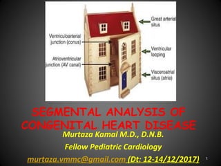
  • 1. SEGMENTAL ANALYSIS OF CONGENITAL HEART DISEASE Murtaza Kamal M.D., D.N.B. Fellow Pediatric Cardiology murtaza.vmmc@gmail.com (Dt: 12-14/12/2017) 1
  • 2. MAUDE ELIZABETH SEYMOUR ABBOTT 1936 McGill University, Canada 1st systematic study Based on study of 1000 heart specimen 2
  • 3. Richard Van praagh • 1964 • Pediatric Cardiologist • Boston Children Hospital, Massachusetts • Segmental approach to CHD 3
  • 4. History Cont… • Kirklin(1973): Modified the segmental approach using concordance/discordance to describe connections between 3 main cardiac segments • Shinebourne/Anderson/Tynan(1976): Popularized the sequential segmental approach to CHD 4
  • 5. 5
  • 6. 6
  • 7. THORACO-ABDOMINAL SITUS Situs: Topology/ spatial position of structureSitus: Topology/ spatial position of structure 1st segment evaluated is visceral situs Atrial and visceral situs considered together because they usually are concordant ( atrial and visceral situs are same) 7
  • 8. Pulmonary Situs (Sidedness) • Determined by positions of morphologic rt & lt lungs • Pulmonary morphology, in turn, defined by relationship of PAs to their adjacent bronchi, and not by the number of lobes • PA of a morphologic right lung travels anterior to its upper and intermediate bronchi, whereas that of a morphologic left lung travels over its main bronchus and posterior to the upper lobe bronchus 8
  • 9. Pulmonary Situs (Sidedness) Cont… • Rt bronchus branches, then RPA crosses it • LPA crosses lt bronchus, then it branches • Clinically, pulmonary situs may be inferred by comparing relative lengths of 2 main bronchi, as measured on a CXR that shows an air bronchogram • Distance from carina to origin of upper lobe bronchus is 1.5- 2.5 X greater for morphologic left lung than for right lung, and this ratio holds true regardless of the sidedness of the aortic arch 9
  • 10. 10
  • 11. The morphologically left bronchus is long, and it branches only after it has been crossed by its accompanying pulmonary artery, making the bronchus hyparterial. In contrast, the morphologically right bronchus is short, and is crossed by its pulmonary artery only after it has branched, giving an eparterial pattern of branching LR 11
  • 14. 14
  • 15. Cardiac malposition • Often an integral part of complex associated anomalies of visceral atrial situs • Prevalence 1 in 10, 000 live births Cardiac malpositions: Dextrocardia Mesocardia Levocardia (isolated) Ectopia cordis 15
  • 17. 17
  • 18. DEXTROCARDIA • Detro position • Dextro version • Mirror image dextrocardia 18
  • 19. Dextroposition: Heart pushed/ pulled into right chest Push: Lt sided tension pneumothoax Lt congenital lobar emphysema Lt diaphragmatic hernia Pull: Hypoplasia/ agenesis of rt. lung DEXTROCARDIA 19
  • 20. DEXTROCARDIA Cont… Dextroversion: Failure of pivoting of cardiac apex to left, frequently associated with A-V discordance Mirror-image dextrocardia with atrial situs inversus There may be major and complex pathology associated with this type of dextrocardia 20
  • 21. 21
  • 22. MESOCARDIA Location of heart with cardiac base apex axis directed to midline of thorax or with ventricular apices equally directed to both right and left sides This is often ignored as being infrequent and atypical 22
  • 23. Isolated levocardia Occurring in conjunction with situs inverses and situs ambiguous  An abnormal atrial and visceral situs, the heart is in its normal location in the left hemi thorax with the apex pointing to left 23
  • 24. Ectopia Cordis • Partially/ completely exteriorized • Pentalogy of Cantrell: – 1. Deficiency of ant. Diaphragm – 2. Midline supra umbilical abdominal wall defect – 3. Defect in diaphragmatic pericardium – 4. CHD – 5. Defect of lower sternum 24
  • 25. BASIC CARDIAC POSITIONS 95% CHD 100% CHD 3-5%CHD<1%CHD 25
  • 26. 26
  • 28. SEGMENT CONNECTION Great veins Atria Ventricles Great arteries Venoatrial Atrioventricular Ventriculoarterial 28
  • 29. 29
  • 30. 30
  • 31. VENOUS SEGMENT Systemic veins: IVC, hepatic veins, SVC, CS Pulmonary veins HEPATIC VEINS: SC view, drains into RA CS – RA IVC – RA 31
  • 32. Venous Segment Cont… ABNORMALITY OF IVC: Subcostal view Interrupted IVC (Polysplenia): Large azygous vein entering SVC seen along the spine in abdomen, can be either right or left sided Even then there is another vein draining into RA called: Suprahepatic IVC Suprahepatic IVC always drains into RA Suprahepatic IVC differentiated from IVC in being small and can’t be traced below liver 32
  • 33. SVC • SVC: Single right sided • Subcostal view, high parasternal or suprasternal view • LSVC: • Connected to CS posteriorly/ laterally • Border of LA (dilated CS) • Directly visualized in a long axis plane • Parasternal short axis images: Circular vessel anterior to LPA near bifurcation • SVC less reliable than IVC for situs 33
  • 35. Pulmonary veins • Subcostal, suprasternal, apical • Color Doppler: Distinguishes from atrial appendage • 4 in number • Left 2 veins may join: 3 entries into LA 35
  • 36. ATRIAL SITUS The identification of the morphologic right atrium is important for establishing atrial situs Anatomic hallmark is the limbus of the fossa ovalis 36
  • 37. Atrial Situs • Describes location of atria • Solitus: Morphological RA on rt • Inverses: Morphological RA on lt • Ambiguous: Undifferentiated atria 37
  • 38. Right atrium Left atrium SEPTUM: Limbus of fossa ovalis Flap valve of fossa ovalis APPENDAGES: Broad based, triangular, anterior Long and narrow, finger like, posterior MYOCARDIAL FEATURES: Crista terminalis, tinea sagittalis, extension of pectinate muscles towards AV valve Pectinate muscle confined to appendage VEINS: Receives IVC, suprahepatic IVC, SVC, coronary sinus Receives pulmonary veins 38
  • 40. 40
  • 41. Subcostal sagittal views demonstrating eustachian valve (arrow in A) and flap valve (arrow in B). 41
  • 43. 43
  • 44. Viscero atrial situs and its abnormality Isomerism of Lt atrial appendage: •B/l lt atrial appendage: Smooth pectinate muscles •Interrupted IVC: 85% Isomerism of Rt atrial appendage: • B/l rt atrial appendage: Coarse pectinate muscles •Abscent coronary sinus: 100% •Abnormal relation of aorta and IVC 44
  • 46. 46
  • 47. VENTRICULAR SITUS AND MORPHOLOGY Right ventricle Left ventricle Large apical trabeculations Small apical trabeculations Coarse septal surface Smooth upper surface Moderator band No moderator band Receives tricuspid valve Receives mitral valve Tricuspid-pulmonary discontinuity Mitral-aortic continuity Crescentic in cross section Circular in cross section Thin free wall (3-5 mm) Thick free wall (12-15 mm) 47
  • 48. Apical four-chamber view in a patient with ventricular inversion with L-TGA 48
  • 49. VENTRICULAR LOOPING After morphology Determine looping Bulboventricular loop: Describes location of ventricles d-Loop: Morphologic RV on right l-Loop: Morphologic RV on left 49
  • 50. VENTRICULAR LOOPING Cont…  Performed by imagining one is standing on the right ventricle side facing the right ventricular face of the interventricular septum  The palm of one hand is placed against the septum  The looping is determined by establishing which of the two hands allow the thumb to point into the atrioventricular valve and the fingers to point into the outflow tract  If rt. hand meets criteria: d-looped, if lt hand meets criteria: l-looped 50
  • 51. The palmar surface of the right hand can be placed on the septal surface of the normal morphologically right ventricle with the thumb in the inlet component and the fingers extending into the ventricular outlet. (d-ventricular loop.) 51
  • 52. The mirror-imaged normal heart. In this setting, it is the palmar surface of the left hand that can be placed on the septal surface of the morphologically right ventricle with the thumb in the inlet and the fingers in the outlet. (l-ventricular loop) 52
  • 53. VENTRICULAR LOOPING Cont…  In general, convexity of aorta points to the position of the right ventricle and thus helps indicate bulboventricular loop  Definitive indicator of bulboventricular loop, however, is relative positioning of ventricular inlets/ AV valves  Thus, in a d-loop the tricuspid valve is to the right of the mitral valve, and in an l-loop the tricuspid valve is to the left of the mitral valve  In a normal d-loop the apex pivots to the left hemithorax; in a "normal" l-loop (i.e., one in the setting of situs inversus) the apex pivots to the right hemithorax 53
  • 54. SINGLE VENTRICLE • Single ventricle: Determined by morphology of ventricle • Single vent of RV morphology: Hypo plastic LV remnant, posterior to main ventricular chamber • LV morphology: Rudimentary RV, anterior 54
  • 55. 55
  • 56. 56
  • 57. AV Connections and AV Valve Morphology First see: Bi/uni ventricular connection If double see: AV concordance/discordance If single ventricle see: Double inlet, single inlet, common inlet 57
  • 58. 50% rule  Anderson et al: Rule of 50% for determining whether a cardiac chamber is a ventricle  For the assessment of AV connections, an atrium is considered to join the ventricle into which >50% of the valve orifice empties  This rule states that a chamber is a ventricle if it receives 50% more of an inlet  The inlet consists of the fibrous ring of the AV valve and need not always include a patent AV valve with well formed valve leaflets 58
  • 59. 50% RULE Cont…  Eg: In HLHS with aortic and mitral atresia, the fibrous ring of the MV contains an imperforate membrane and is situated over the small LV  Thus, this small left sided chamber is a ventricle because it receives 100% of an inlet  A chamber need not have an outlet to be a ventricle  The rule of 50% has also been used to define VA connections  Thus, if 50% or more of a great artery arises above a chamber, the great artery is defined as being 59
  • 60. 50% RULE IN AV, VA • A great vessel should be related to a ventricle by 50% of its dimension to be considered committed to it • Av valve is committed to that ventricle to which >50% of that valve is connected 60
  • 62. ATRIOVENTRICULAR VALVE MORPHOLOGY Tricuspid valve Mitral valve Septal chordal attachments No septal chordal attachments Low septal annular attachment High septal annular attachment Triangular orifice (midleaflet) Elliptical orifice (midleaflet) Three leaflets Two leaflets Several papillary muscles Two large papillary muscles Empties into right ventricle Empties into left ventricle 62
  • 64. OVERRIDING AV VALVE Abnormality of AV valve alignment or connection in which one valve annulus opens into another chamber through a VSD 64
  • 65. STRADDLING A feature of the tensor apparatus (chordae tendineae and papillary muscles) of an AV valve and indicates anomalous insertion into the contralateral ventricle, either along its septum or its free wall 65
  • 66. 66
  • 67. 67
  • 68. VENTRICULOARTERIAL CONNECTION If Ao from LV and PA from RV: Concordant If Ao from RV and PA from LV: Discordant Origins of Ao and PA are evident on PLAX view, sweeping the transducer inferiorly from the basal short-axis view, A5CV and SC coronal and sagittal views 3rd type of VA connection: Double outlet, almost always from RV Final type of VA connection: Single outlet (truncus arteriosus) 68
  • 69. VA Connection Aorta: •Coronary artery •Carotids •Absence of proximal division PA: •Bifurcation 69
  • 70. Parasternal short-axis images demonstrating different relationships between the great vessels. An echocardiogram from an infant with tetralogy of Fallot and pulmonary atresia demonstrates the identifying features of a pulmonary artery (MPA) with its bifurcation into right and left pulmonary arteries (RPA, LPA). The great vessels are normally related 70
  • 71. 71
  • 72. 72
  • 73. CONAL MORPHOLOGY Conus/ infundibulum: Cavitary space formed by muscular segment of heart that connects ventricles with great arteries and separates the AV and semilunar valves Abnormalities in conal development consist of variations in the presence, length and diameters of subpulmonary and subaortic conus These variations can lead to (or be associated with) complex malformations, such as TOF, IAA,TGA and DORV 73
  • 74. Subpulmonary conus - Best identified in subcostal views In normal heart: Conus- Nearly vertical tubular outflow portion of RV Separated from nearly horizontal right ventricular inflow portion by distinct muscle bands These muscle bands form a near- circular rim formed by parietal band anteriorly, crista supraventricularis posteriorly, and septal band medially and prohibit PV to AV valve continuity 74
  • 75. Conus Cont…  Subaortic conus is evident on subcostal coronal and sagittal views  Persistence of subaortic conus and involution of subpulmonic conus is the usual conal relationship in d- (or l-) TGA  Persistence of subaortic conus prohibits continuity of the aortic valve to either AV valve, and involution of subpulmonary conus allows continuity between PV and both AV valves in TGA 75
  • 76. BILATERAL CONUS  B/l persistence of the subarterial conus usually results in DORV  Because the main goal of surgical correction of DORV is to connect the aorta with the morphologic left ventricle through VSD, it is important to determine the conal relationships with each other and with the great vessels 76
  • 77. BILATERAL CONUS When 2 coni are present, their relationship may be classified as either anterior/posterior or side-by-side With the anterior/posterior conal relationship, the ventricular septal defect is usually subaortic; with the side-by-side relationship, the defect is usually subpulmonic  The conal relationship can be determined by subcostal coronal and sagittal imaging with anterior/posterior and left/right sweeping, respectively 77
  • 78. ABSENT CONUS  A rare type of d-transposition can exist in the context of bilaterally deficient subarterial conus  This results in an unusual heart in which d-TGA exists with a doubly committed VSD and a posterior aorta  Associated with DOLV 78
  • 79. 79
  • 81. Great Artery Relations 8 basic types of great artery relationships are possible based on the aortic and pulmonary valve positions at the level of the semilunar valves • Right posterior aorta (normally related) Right lateral aorta (side by side) Right anterior aorta (d-malposed) Directly anterior aorta Left anterior aorta (l-malposed) Left lateral aorta (left side by side) Left posterior aorta (inverted normal) Directly posterior aorta. 81
  • 82. 82
  • 83. 83
  • 84. ARCH OF AORTA  Unexpected position of aortic arch: Well recognized associated anomaly of conditions such eg. TOF/ Truncus  Distinction should be made b/w position of arch and side of descending aorta, particularly in describing vascular rings  Side of aortic arch depends on whether it passes to right or left of the trachea  Position of descending aorta is defined relative to vertebral column 84
  • 85. ARCH OF AORTA Cont…  Aorta lies more posterior in center of  Visualized in PLAX,PSAX, A5CV,SC , and suprasternal notch views  Arch best seen in subcostal oblique view and suprasternal views  Side of arch diagnosed by sweeping transducer in suprasternal long-axis view and noting the relationship of the arch to the trachea, the rings of which resemble a stack of coins 85
  • 86. This transducer position allows visualization of the ascending aorta (Asc), aortic arch (Arch), origin of the brachiocephalic vessels (arrows), descending thoracic aorta (Dsc), and right pulmonary artery (*). ARCH OF AORTA LONG-AXIS 86
  • 87. The short-axis view of the aortic arch is obtained by rotating the transducer clockwise, which also allows visualization of the right pulmonary artery (RPA) in its long-axis format, located inferiorly to the aortic arch (Arch). Inferior to RPA is the left atrial (LA) cavity with connections of the four pulmonary veins (arrows SHORT-AXIS VIEW 87
  • 88. 88
  • 89. FINAL DIAGNOSIS • SS, LEVOCARDIA, NORMAL SYSTEMIC & PULMONARY VENOUS COMMUNICATIONS, NRGAs, AV & VA CONCORDANCE, INTACT IA & IV SEPTUM, NO PDA, GOOD SIZED CONFLUENT BPAs, NORMAL ARCH, NO COA, NORMAL CORONARIES, NORMAL SIZED CHAMBERS, GOOD BV FUNCTION, NO VEGETATION/ CLOT OR PE 89
  • 90. THANKS FOR UR PATIENCE 90