

Recomendados
Mais conteúdo relacionado
Mais procurados
Mais procurados (20)
Semelhante a Respiratory system
Semelhante a Respiratory system (20)
Mais de Dr Motawei
Último
Último (20)
Respiratory system
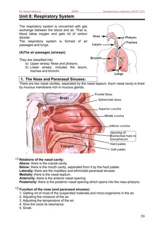
- 1. Dr. Kamal Motawei HIMT Introduction to Anatomy (ANAT 215) Unit 8: Respiratory System The respiratory system is concerned with gas exchange between the blood and air. That is, blood takes oxygen and gets rid of carbon dioxide. The respiratory system is formed of air passages and lungs. (A)The air passages (airways): They are classified into: a) Upper airway: Nose and pharynx. b) Lower airway: includes the larynx, trachea and bronchi. 1. The Nose and Paranasal Sinuses: There are two nasal cavities, separated by the nasal septum. Each nasal cavity is lined by mucous membrane rich in mucous glands. Relations of the nasal cavity: Above: there is the cranial cavity. Below: there is the mouth cavity, separated from it by the hard palate. Laterally: there are the maxillary and ethmoidal paranasal sinuses Medially: there is the nasal septum. Anteriorly: there is the anterior nasal opening. Posteriorly: there is the posterior nasal opening which opens into the naso-pharynx. Function of the nose (and paranasal sinuses): 1. Getting rid of most of the suspended materials and micro-organisms in the air. 2. Adjusting the moisture of the air. 3. Adjusting the temperature of the air. 4. Give the voice its resonance. 5. Smell. 59
- 2. Dr. Kamal Motawei HIMT Introduction to Anatomy (ANAT 215) 2. The pharynx: It is a muscular tube that lies behind the nose, mouth and larynx. It collects air from the nose and food from the mouth, then it transfers the air to the larynx and the food to the esophagus. It is divided into three parts: 1) Naso-pharynx (behind the nose), 2) oro-pharynx (behind the mouth), and 3) laryngo-pharynx behind the larynx. ☺In each lateral wall of the nasopharynx there is an opening for the Eustachian tube (which connects the middle ear with the nasopharynx). ☺ The lateral walls of the oropharynx show the palatine tonsils. Each tonsil lies between two longitudinal folds of mucous membrane. 3. The Larynx: The larynx produces voice and acts as a valve. It is formed of cartilages and muscles. It is lined with mucous membrane which shows a fold on each side called VOCAL CORD. The inlet of the larynx is directed backwards and is guarded by the EPIGLOTTIS. The epiglottis closes the inlet of the larynx during swallowing to prevent entrance of food and drink into the larynx. 60
- 3. Dr. Kamal Motawei HIMT Introduction to Anatomy (ANAT 215) 4. The Trachea It is a midline structure, but it inclines to the right while descending downward It is formed of rings of hyaline cartilages connected together by smooth muscles. Ring of cartilage are incomplete posteriorly (to allow for dilatation of the esophagus during swallowing). It passes through the neck and thorax where it ends by dividing into two bronchi. Relations: Anteriorly, it is related to: isthmus of thyroid gland, major vessels of the thorax, and base of the heart, Posteriorly, it is related to the esophagus. 5. The Bronchi The trachea ends by dividing into two main (primary) bronchi. The right bronchus is shorter and wider than the left bronchus. The right bronchus lies in line with the trachea, while the left bronchus is more oblique. The primary bronchi divide in the lungs into secondary bronchi, then tertiary and so on, until they get very small (bronchioles). The alveoli of the lung open into the respiratory bronchioles. 61
- 4. Dr. Kamal Motawei HIMT Introduction to Anatomy (ANAT 215) 6. The Lungs There are two lungs, right and left. Each lung is pyramidal in shape, having apex, base and side walls. The right lung is shorter and broader than the left lung. The left lung shows a cardiac notch. The right lung is divided into three lobes, while the left lung is divided into two lobes. Each lung has a hilum which contains the following structures: 1) a main bronchus 2) a pulmonary artery 3) two pulmonary veins 4) Lymphatics 5) Autonomic nerves. Each lung is covered with a serous membrane called PLEURA. The pleura is a serous sac which has: 1. a parietal layer lining the thoracic wall. 2. a visceral layer covering the lung surface. 3. a cavity which contain a little amount of serous fluid. 62
- 5. Dr. Kamal Motawei HIMT Introduction to Anatomy (ANAT 215) Surface marking of the Pleura and Lungs This is represented by the following points: The apex of the lung: one inch above the junction of the medial and intermediate one third of the clavicle. The anterior border: a line passing through the following points; A) Apex of lung. B) Sternoclavicular joint C) Sternal angle in the midline D) On the right side, midline at the level of 4th costal cartilage. On the left side, some distance to the left of the edge of the sternum at the level of 4 th costal cartilage. E) On the right side, midline at the level of 6th costal cartilage. On the left side, some distance to the left of the edge of the sternum at the level of 6 th costal cartilage. The inferior border of the lung: It is represented by a line passing through the following points: E) 6th costal cartilage in the midline (on the right side) or to the left of the edge of sternum (to the left side). F) 6th rib in the midclavicular line G) 8th rib in the midaxillary line H)10th rib close to the vertebral column. The inferior border of the pleura: the pleura extends to a level lower than the lung by two ribs E) 6th costal cartilage in the midline (on the right side) or to the left of the edge of sternum (to the left side). F) 8th rib in the midclavicular line G) 10th rib in the midaxillary line H) 12th rib close to the vertebral column. The posterior border of the lung and pleura: It is represented by a line connecting point (H) with point (A). 63