Ventilador Pulmonar
•Transferir como PPTX, PDF•
4 gostaram•2,540 visualizações

Descripción elemental y funcional de los Ventiladores Pulmonares.

Recomendados
Mais conteúdo relacionado
Mais procurados
Mais procurados (20)
Destaque
Destaque (19)
Semelhante a Ventilador Pulmonar
Semelhante a Ventilador Pulmonar (20)
Mais de Rigoberto José Meléndez Cuauro
Mais de Rigoberto José Meléndez Cuauro (20)
Último
Último (20)
Ventilador Pulmonar
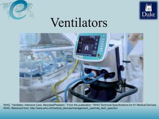
- 1. Ventilators WHO. “Ventilator, Intensive Care, Neonatal/Pediatric.” From the publication: “WHO Technical Specifications for 61 Medical Devices. WHO. Retrieved from: http://www.who.int/medical_devices/management_use/mde_tech_spec/en/
- 2. Summary • Respiration System Anatomy • Respiratory Control • Clinical Use • Specifications • History • Principles of Operation • Variables • Operation Modes • Block Diagram • Commercial Examples • Patient’s Safety • Preventive Maintenance • Common Problems • Test Procedures • Artificial Lung
- 3. Respiration System Anatomy Sunshineconnelly at English Wikibooks [CC BY 3.0 (http://creativecommons.org/licenses/by/3.0)], via Wikimedia Commons
- 4. Respiration System Anatomy National Heart Lung and Blood Institute (National Heart Lung and Blood Institute) [Public domain], via Wikimedia Commons
- 5. Respiration System Anatomy 3D Yoga (2008), View of diaphragm during respiration [video]. Retrieved from https://www.youtube.com/watch?v=hp-gCvW8PRY
- 6. • The amount of air flowing into and out of the lungs with each breath is called the tidal volume (TV). • Typical adult = 500 mL (quiet breathing) • Possible to inhale a volume 7 x TV Respiration System Anatomy
- 7. Respiration Volumes Kapwatt (2014), Output of a spirometer [image]. Retrieved from https://en.wikipedia.org/wiki/File:Lungvolumes_Updated.png
- 8. O2 saturation vs PO2 Ratznium at English Wikipedia Later versions were uploaded by Aaronsharpe at en.wikipedia. (Transferred from en.wikipedia to Commons.) [Public domain], via Wikimedia Commons
- 9. Respiratory Failure • Lungs, or the heart and lungs, are not able to sufficiently oxygenate the blood and body tissue. • Often, the ability to excrete CO2 is also impaired. • Examples – Apnea – Tuberculoses – Pneumonia – Edema PaO2 (partial pressure ) < 50 mm Hg PaCO2 > 50 - 60 mm Hg
- 10. Respiratory Control Brain Nerves Bellows AirwaysAlveoli It only requires one disrupted “link” to cause respiratory failure ! Patrick J Lynch (2006), Lungs [drawing]. Retrieved from https://en.wikipedia.org/wiki/Lung#/media/File:Lungs_diagram_detailed.svg
- 11. Respiratory Control CHEMORECEPTORS BARORECEPTORS INFLATION RECEPTORS PROPIROCEPTORS IRRITATION ARTERIAL BLOOD RESPIRATORY MUSCLES MEDULLARY AND PONS RESPIRATORY CENTERS CEREBRAL CORTEX, LIMBIC SYSTEM, HYPOTHALMUS Virginia Reid (2016), Respiratory Control [drawing]. Adapted from previous image (unknown)
- 12. Respiratory Control During exercise: – Respiration rate increases – Inhalation time is faster than exhalation time – Respiratory flow wave shapes become more trapezoidal – Expiratory reserve volume decreases.
- 13. Ventilator Clinical Use • When should a patient be ventilated: – Lung injury and respiratory failures – All thoracic surgery cases
- 14. Effects of Major Surgery & Anesthesia • Respiratory Center in Brain – Narcotic drugs • Neuromuscular Connections – Paralyzing agents • Thoracic Bellows (rib cage and diaphragm) Chest or abdominal incision WHO. “Ventilator, Intensive Care.” From the publication: “WHO Technical Specifications for 61 Medical Devices. WHO. Retrieved from: http://www.who.int/medical_devices/management_use/mde_tech_spec/en/ Ventilator Clinical Use
- 15. Effects of Major Surgery & Anesthesia • Airways (upper & lower) – Dry, irritating gases • Lungs (bronchioles, alveoli and capillaries) – Pain, and ineffective cough Ventilator Clinical Use
- 16. Respirator vs Ventilator • Respirator is a device that supplies or filters air in a harsh environment By John Dimos and Paul Satti [Public domain], via Wikimedia Commons, retrieved from https://commons.wikimedia.org/wiki/File:Air- Purifying_Respirator.jpg
- 17. • Input/ Output : – Atmosphere air or/and – Oxygen or/and – Nitrogen Specifications
- 18. Mechanical Ventilators History • Negative-pressure ventilators (“iron lungs”) • Non-invasive ventilation first used in Boston Children’s Hospital in 1928 • Used extensively during polio outbreaks in 1940s – 1950s
- 19. History Problems • Venous blood pool in the abdomen (reducing cardiac output) • Less accessible for patient care and monitoring • Difficult synchronization with patient’s effort Negative-pressure ventilators Photo Credit: Content Providers(s): CDC [Public domain], via Wikimedia Commons, retrieved from https://commons.wikimedia.org/wiki/File:Poumon_artificiel.jpg
- 20. History Positive-pressure ventilators • Invasive ventilation first used at Massachusetts General Hospital in 1955 • Now the modern standard of mechanical ventilation Allows treatment of patients with high lung resistance and low compliance Average of 833 patient days of mechanical ventilation per hospitals per year in USA.
- 21. Principles of Operation National Heart Lung and Blood Institute (NIH) (National Heart Lung and Blood Institute (NIH)) [Public domain], via Wikimedia Commons. Retrieved from https://commons.wikimedia.org/wiki/File:Ventilators.jpg
- 22. Principles of Operation Malkin, Robert. “2.2 Ventilators.” Medical Instrumentation in the Developing World. Engineering World Health, 2006.
- 23. Wiley - Encyclopedia of Biomedical Engineering - 6 Vol. Set - RESPIRATION MEASUREMENTS Pressure Principles of Operation
- 24. Input Variables • Modes of Ventilation • Output – Exponential – Ramp – Rectangular – Sinusoidal • Alarm Systems • Frequency • Volume • Flow • Pressures Malkin, Robert. “2.2 Ventilators.” Medical Instrumentation in the Developing World. Engineering World Health, 2006.
- 25. Operation Modes • Mandatory Ventilation – Volume controlled (limited) ventilation – Pressure controlled ventilation – Timed cycle (combination of volume and pressure) • Spontaneous Ventilation (Assisted Mode) (still controlling the breath rate, flow rate, and the tidal volume) – Continuous Positive Airway Pressure (CPAP) – Pressure Support
- 26. Operation Modes Pressure regulated volume control By Eduardo Mireles-Cabodevila, MD (The Cleveland Clinic Foundation) [CC BY-SA 3.0 (http://creativecommons.org/licenses/by-sa/3.0)], via Wikimedia Commons. Retrieved form https://commons.wikimedia.org/wiki/File:Pressure_regulated_volume_control_graphic.jpg
- 27. Operation Modes Spontaneous Ventilation – Continuous Positive Airway Pressure (CPAP) L. Droll (2005), Cpap cycle [image]. Retrieved from https://commons.wikimedia.org/wiki/File:CPAP.jp g
- 28. Operation Modes Spontaneous Ventilation – Pressure Support This mode is similar to the CPAP mode with the exception that during the inspiration the ventilator attempts to maintain the patient airway pressure at a level above PEEP. In fact, CPAP may be considered a special case of pressure support ventilation in which the support level is fixed at the atmospheric level. In this mode, when the patient’s airway pressure drops below the therapist-set sensitivity line, the ventilator inspiratory breath delivery system raises the airway pressure to the pressure support level (>PEEP), selected by the therapist. The ventilator stops the flow of breathable gases when the patient starts to exhale and controls the exhalation valve to achieve the set PEEP level.
- 29. Ventilators Parts • Power supply • Compressed air and oxygen • A drive mechanism to push oxygen • A control mechanism • Humidifier
- 30. Block Diagram Malkin, Robert. “2.2 Ventilators.” Medical Instrumentation in the Developing World. Engineering World Health, 2006.
- 31. Diagram • Boyle’s Ether Vaporizer WHO. “Anesthetic and Resuscitation Equipment.” Maintenance and Repair of Laboratory, Diagnostic Imaging, and Hospital Equipment (WHO: 1996), p. 121-134.
- 32. Air Moisture • Bottled gases delivered from cylinders are too dry for the human body to moisturize comfortably • Sterile water should be used for humidification (but often isn’t in the developing world)
- 33. Air Moisture • Humidifier Pass air over liquid to pick up natural vapor • Vaporizer Heat liquid to increase amount of vapor • Nebulize Turn liquid to fine spray by passing gas through liquid or by ultrasonic agitation
- 34. Air Moisture • Some ventilators heat the tubing or air to prevent “rain-out” of the vapor delivered to the patient. • Older systems may have water traps
- 35. Review • Forced breathing or assisted breathing for patient • Inputs: Gases (usually air & oxygen) plus water for humidification • Outputs: Humidified (and warmed) gas to patient • Modes of operation: Pressure limited, volume limited or timed cycle • Modes of initiation: controlled and assisted
- 36. Commercial Examples WHO. “Ventilator, Intensive Care, Neonatal/Pediatric.” From the publication: “WHO Technical Specifications for 61 Medical Devices. WHO. Retrieved from: http://www.who.int/medical_devices/management_use/mde_tech_spec/en/
- 37. Patient’s Safety • Barotrauma – PIP(peak inspiratory pressure) > 45 cmH2O • Volutrauma – VT > 6-8 ml/Kg • Oxygen Toxicity – Prolonged FIO2 (fraction of inspired oxygen) of 1.0
- 38. Patient’s Safety • Pneumonia (most common) • Hypotension
- 39. Preventive Maintenance • Filters cleaning • Leak troubleshooting • Calibration • Change O2 sensor • Use of gloves LadyInGrey (2005), Disposable Nitrile Gloves [photograph]. Retrieved from https://commons.wikimedia.org/wiki/File:Disposable_nitrile_ glove.jpg
- 40. Common Problems • In the US it is illegal for uncertified engineers/technicians to work on ventilators • Never work on a ventilator when connected to a patient
- 41. Common Problems • User error • Power supply • Filtration • Tubing
- 42. Common Problems • User error – Controls are not standardized between manufacturers – Manuals were either not supplied with the donation or were supplied in a language that the hospital staff does not speak
- 43. Common Problems • Tubing – Disposable tubing is being reused...The non- rebreathing valve may break or the tubing may leak.
- 44. Common Problems • If the problem is not one of these problems, it is probably better NOT to attempt to fix the ventilator without specialized training • However, your decision should be made in careful consultation with the physicians
- 45. Test Procedures • Measuring the breathing rate and measuring the I:E ratio (approximately 20% of the set ratio) • The pressure limit: Partially occluding the connection to the patient with your hand (light should flash)
- 46. Test Procedures • Assisted mode: – Place the patient tube in your mouth (The device will deliver gas only when you inhale) – Remove the tube from your mouth (The device should take over in a controlled mode) – Place the tube back in your mouth and breathe normally and the device should automatically return to assisted mode
- 47. Test Procedures • Breathing circuit: – Leak test - occlude one end and blow hard into the other end with the tube submerged in water…should be no bubbles – Soapy water Erich Schulz, Brisbane (en:Image:Syringe.jpg) [Public domain], via Wikimedia Commons
- 48. Test Procedures • Breathing circuit: – Leak test - occlude one end and blow hard into the other end with the tube submerged in water…should be no bubbles – Non-rebreathing • Blow into the patient connection end and making sure that the air goes down the expiratory tube. • Then suck from the patient end and make sure the air is coming in from the inspiratory tubing
- 49. Test Procedures • Pressure Limit: – Connect the patient tubing to a u-shaped bend of tubing filled with water – The ventilator should push the far end of the column of water the height of the pressure setting, and then indicate a pressure limit alarm