SlideShare uma empresa Scribd logo
1 de 14
Baixar para ler offline
THE HEART
Formation of the heart in the embryo
The finished heart has two separate major pumping systems - the left and right
ventricles (ventricle = small cavity), two minor pumping systems (the left and right
atria as seen in Fig. 10). The right atrium (=entrance) delivers blood from the body
into the right ventricle which then pumps blood to the lungs. Blood from the lungs
then returns to the left atrium which delivers blood to the left ventricle which then
pumps blood to the body. Each ventricle will require two valves, one at the entrance
and one at the exit.
The heart begins embryonic life as a tube and thus major remodelling is required but
during the change from the primitive tube to a four-chambered heart blood must
continue to flow at all times. Knowledge of the embryological changes are necessary
to understand the malformations which may occur.
An impulse conducting system also has to develop to co-ordinate atrial and
ventricular contractions (so that the atrial muscles contract when the ventricular
muscle is relaxing) and vice versa.
In the embryo a contracting heart tube forms, with veins entering and arteries exiting
(Fig. 11). However a contracting tube with no valves is no use as a pump.
Accordingly this primitive tube receives blood from the great veins and empties into
an atrium, then via atrioventricular valves into a ventricle then into the arteries via
semilunar valves.
The heart tube is attached at its ends and is free to grow and fold (Fig. 12). The
caudally situated Y junction (the sino-atrial chamber where venous blood enters the
primitive heart tube) develops two swellings which will later form the right and left
atria. The atria move cranially, passing dorsal to the ventricles (Fig. 13). ). Veins
(which become the superior and inferior vena cavae which return deoxygenated blood
from the body) later enter directly into the right atrial cavity wherea four pulmonary
veins carrying oxygenated blood enter the left atrium.
The truncus, the artery that will form the outflow tracts of the ventricles, indents the
groove separating the atria. The truncus splits longitudinally into two tubes which
provide separate outlets for the left and right ventricles (these outlets will become the
aortic and pulmonary arteries respectively), and each of the two tubes develops one-
way valves. Three valves (the tricuspid, aortic, and pulmonary) have three cusps and
one (the mitral) has two cusps (Fig. 14). The ventricular walls grow thicker, become
contractile, and the communication between the two ventricles becomes smaller as an
interventricular septum develops.
The right and left atria are separated by a thin sheet of tissue (the septum primum)
but, prior to birth, blood has to continue to flow from the right to the left atrium
through an opening, the ostium primum in order to bypass the “useless” lungs. The
ostium primum subsequently closes but, because blood must still bypass the lungs,a
second opening develops (the ostium secundum), which is oval in shape (the foramen
ovale).
The ostium secundum is surrounded by tissue which provides a flap-like valve (Fig.
15) which remains open only if the pressure in the right atrium exceeds that in the left
atrium (at birth the pressure in the left atrium will rise as blood returns to it from the
lungs and this communicating hole will close).
Initially the outer muscle wall which surrounded the primitive heart tube contracted as
a whole, but with subsequent convolutions of the tube and the need for reciprocal
contractions of the atria and ventricles, modified heart muscle fibres that transmit
depolarisation waves (faster than would occur between normal heart muscle cells)
were developed. An electrical initiating point, the sinoatrial node, forms in the right
atrium and sends out a depolarisation wave that causes contraction to spread radially
over the atria. A fibrous ring develops to separate the atria from the ventricles and a
“focal penetrating area,” the atria-ventricular node, develops and connects with a
short stem of conducting tissue (the Bundle of His) which then divides into right and
left branches to transmit depolarisation waves to the right and left ventricles (Fig. 16).
All the above begs the question “Why do we need a two-sided heart?” There are two
answers. Firstly, a high pressure system is required to perfuse the head in an upright
posture (otherwise we would faint when we stood up). Secondly, a lower pressure
system is required to supply the lungs - if blood pressure in the pulmonary arteries
were at aortic pressure then the pulmonary capillaries would have to have walls that
were too thick to allow gas exchange (fish which, by and large, do not stand up thus
only need one ventricle to pump blood through their gills to the body).
Anatomy
Figure 17 shows the situation and anatomy of the heart.
The heart weighs 300 grams and is much smaller than is commonly supposed (the size
is exaggerated by chest X-rays). Until the advent of cardiac transplantation the heart
was only guaranteed to last for a lifetime “three score years and ten” and thus a heart
had to contract about 2,500 million times to pump about 170 million litres of blood.
Heart function
The function of the heart is to pump blood to the lungs for oxygenation and then to
the tissues for utilisation. To achieve this, a two-sided pump (the heart), a distribution
system (the systemic and pulmonary arteries) and return system (the systemic and
pulmonary veins) are required. Cardiac contraction (systole) or relaxation (diastole)
refers to ventricular events. Both atria contract together (during ventricular diastole)
and, later, both ventricles contract together during ventricular systole. When both
ventricles are contracting, both atria are relaxing and vice versa (Fig. 18). Although
there are no true valves, there are muscle rings around the entry sites of the superior
and inferior vena cavae into the right atrium and around the entrysites of pulmonary
veins into the left atrium (both to minimise retrograde blood flow during atrial
contraction).
Cardiac muscle is a specialised form of striated muscle. As muscles can only contract
in a linear fashion the heart muscle has to be wound so they can compress the blood
within the heart chambers (Fig. 19).
Venous blood returns to the right atrium via the superior and inferior vena cavae.
This blood then flows (with the assistance of right atrial contraction) through the
tricuspid valve into the right ventricle during ventricular relaxation. At the start of
ventricular contraction, the tricuspid valve shuts and the venous blood derived from
the tissues is ejected into the lungs, passing through the pulmonary valve into the
pulmonary arteries (Fig.20).
After passing through the lungs the blood, now oxygenated, returns to the left atrium
via the pulmonary veins. During ventricular relaxation blood flows (with the
assistance of atrial contraction) from the left atrium through the mitral valve into the
left ventricle. During ventricular contraction the mitral valve shuts and blood is
ejected through the aortic valve into the aorta and, thence, to the body tissues.
All four heart valves lie at much the same level in the area which separates the atria
from the ventricles.
The pericardium, the sac that contains the heart, consists of two layers, an outer
fibrous and inner serous layer (Fig. 21). The fibrous layer act as a limiting enclosure
for the heart, preventing overstretching of the heart muscle. A small amount of fluid
between the two layers serves to lubricate movement.
With increasing heart rates the ventricular relaxation (filling) time becomes shorter,
rather than the contraction time becoming shorter. Fast heart rates, above about 120
per minute, do not allow sufficient time for ventricular filling and thus predispose to
heart failure. With atrial fibrillation, in which the atria contract haphazardly “like a
bag of worms,” the lack of atrial assistance for ventricular filling means that heart
failure can occur at lower heart rates than this.
The power of heart contraction -inotropy- is related to metabolic factors and
workload. Preload is heart work required to cope with the venous blood returning to
the heart and is related to the length of cardiac muscle fibres when contraction
commences. Afterload is the heart work required when the heart muscle contracts to
push blood around the systemic circulation and this is in effect the resistance to blood
flow in the arteries.
In general the heart tries to function as a constant flow pump rather than a constant
pressure pump (variation of arteriolar constriction provide most of the pressure
regulation). If peripheral resistance increases then arterial blood pressure hase to rise
to maintain perfusion, which requires more work by the heart. As an approximation,
the more the stretching of heart muscle in ventricular relaxation the more strong the
ventricular muscle contraction and the greater the resulting stroke volume (Starling’s
Law of the Heart).
Blood is supplied to the heart by two coronary arteries which arise from near the
origin of the aorta (Fig. 22). These coronary arteries are functionally end arteries, and
partial or complete blockage is likely to result in heart muscle death distal to the
blockage. The left coronary artery supplies most of the left ventricle and the right
coronary artery supplies most of the right ventricle but also supplies the conducting
tissue comprising the sino-atrial node and the interventricular node. Thus
dysrythmias (abnormal heart rhythms caused by abnormal conduction) are
particularly likely to occur if there is right coronary artery narrowing or obstruction.
The Heart

Mais conteúdo relacionado

Mais procurados (20)

Anatomy of the heart
Anatomy of the heartAnatomy of the heart
Anatomy of the heart
 
Heart anatomy
Heart anatomyHeart anatomy
Heart anatomy
 
Anatomy of the heart
Anatomy of the heartAnatomy of the heart
Anatomy of the heart
 
Cardio vascular system Anatomy and physiology
Cardio vascular system Anatomy and physiology Cardio vascular system Anatomy and physiology
Cardio vascular system Anatomy and physiology
 
Anatomy and physiology of the heart by Adeboye Oluwajuyitan
Anatomy and physiology of the heart by Adeboye OluwajuyitanAnatomy and physiology of the heart by Adeboye Oluwajuyitan
Anatomy and physiology of the heart by Adeboye Oluwajuyitan
 
Anatomy of Heart..!!
Anatomy of Heart..!!Anatomy of Heart..!!
Anatomy of Heart..!!
 
The heart walls
The heart wallsThe heart walls
The heart walls
 
Heart (Its Structure & Function).pptx
Heart (Its Structure & Function).pptxHeart (Its Structure & Function).pptx
Heart (Its Structure & Function).pptx
 
The Anatomy Of Heart
The Anatomy Of HeartThe Anatomy Of Heart
The Anatomy Of Heart
 
Function of arterial & venous system
Function of arterial & venous systemFunction of arterial & venous system
Function of arterial & venous system
 
Heart Anatomy
Heart AnatomyHeart Anatomy
Heart Anatomy
 
Heart
HeartHeart
Heart
 
Anatomy of the heart
Anatomy of the heartAnatomy of the heart
Anatomy of the heart
 
Cvs anatomy
Cvs anatomyCvs anatomy
Cvs anatomy
 
Human circulatory system
Human circulatory systemHuman circulatory system
Human circulatory system
 
Anatomy and function of the coronary arteries
Anatomy and function of the coronary arteriesAnatomy and function of the coronary arteries
Anatomy and function of the coronary arteries
 
Heart
HeartHeart
Heart
 
Anatomy of the heart
Anatomy of the heartAnatomy of the heart
Anatomy of the heart
 
Cardiovascular system
Cardiovascular systemCardiovascular system
Cardiovascular system
 
The aorta
The aortaThe aorta
The aorta
 

Destaque (7)

Cardiology in a Heartbeat
Cardiology in a HeartbeatCardiology in a Heartbeat
Cardiology in a Heartbeat
 
Cardiology Presentation
Cardiology PresentationCardiology Presentation
Cardiology Presentation
 
Heart development i
Heart development iHeart development i
Heart development i
 
Hepatic Encephalopathy
Hepatic EncephalopathyHepatic Encephalopathy
Hepatic Encephalopathy
 
Respiratory examination - OSCE guide
Respiratory examination - OSCE guideRespiratory examination - OSCE guide
Respiratory examination - OSCE guide
 
Neurology book
Neurology bookNeurology book
Neurology book
 
Textbook of human neuroanatomy-
Textbook of human neuroanatomy-Textbook of human neuroanatomy-
Textbook of human neuroanatomy-
 

Semelhante a The Heart

The structure and Function of the Heart
The structure and Function of the Heart The structure and Function of the Heart
The structure and Function of the Heart TessMiller519
 
Cape biology unit 2 -_circulatory_system_in_humans_and_exercise
Cape biology unit 2 -_circulatory_system_in_humans_and_exerciseCape biology unit 2 -_circulatory_system_in_humans_and_exercise
Cape biology unit 2 -_circulatory_system_in_humans_and_exerciseHilton Ritch
 
L8 Cardiac Anatomy
L8 Cardiac AnatomyL8 Cardiac Anatomy
L8 Cardiac AnatomyMarc Potter
 
Chapter 22, sp 10
Chapter 22, sp 10Chapter 22, sp 10
Chapter 22, sp 10j2jpop
 
CARDIOVASCULAR SYSTEM.pptx
CARDIOVASCULAR SYSTEM.pptxCARDIOVASCULAR SYSTEM.pptx
CARDIOVASCULAR SYSTEM.pptxStephenAmoah10
 
The cardiovascular system.pptx
The cardiovascular system.pptxThe cardiovascular system.pptx
The cardiovascular system.pptxJagruti Marathe
 
Organ system for internal transport (circulatory system)
Organ system for internal transport (circulatory system)Organ system for internal transport (circulatory system)
Organ system for internal transport (circulatory system)Cesar Rentino
 
Anatomy and physiology of heart, lung ,
Anatomy and physiology of heart, lung ,Anatomy and physiology of heart, lung ,
Anatomy and physiology of heart, lung ,ligi xavier
 
The circulatoy sistem
The circulatoy sistemThe circulatoy sistem
The circulatoy sistemPlácido Cobo
 
The circulatoy sistem
The circulatoy sistemThe circulatoy sistem
The circulatoy sistemPlácido Cobo
 
Physiologyofheart 140406090507-phpapp01
Physiologyofheart 140406090507-phpapp01Physiologyofheart 140406090507-phpapp01
Physiologyofheart 140406090507-phpapp01Brijesh Pratap Singh
 
Physiologyofheart 140406090507-phpapp01
Physiologyofheart 140406090507-phpapp01Physiologyofheart 140406090507-phpapp01
Physiologyofheart 140406090507-phpapp01Brijesh Pratap Singh
 
the cardiovascular system and Physiology of heart
the cardiovascular system and Physiology of heartthe cardiovascular system and Physiology of heart
the cardiovascular system and Physiology of heartbhupendra kumar
 
Physiologyofheart 140406090507-phpapp01
Physiologyofheart 140406090507-phpapp01Physiologyofheart 140406090507-phpapp01
Physiologyofheart 140406090507-phpapp01Brijesh Pratap Singh
 
Physiologyofheart 140406090507-phpapp01
Physiologyofheart 140406090507-phpapp01Physiologyofheart 140406090507-phpapp01
Physiologyofheart 140406090507-phpapp01Brijesh Pratap Singh
 

Semelhante a The Heart (20)

The structure and Function of the Heart
The structure and Function of the Heart The structure and Function of the Heart
The structure and Function of the Heart
 
Cape biology unit 2 -_circulatory_system_in_humans_and_exercise
Cape biology unit 2 -_circulatory_system_in_humans_and_exerciseCape biology unit 2 -_circulatory_system_in_humans_and_exercise
Cape biology unit 2 -_circulatory_system_in_humans_and_exercise
 
Cardiac cycle (Human Heart)
Cardiac cycle (Human Heart)Cardiac cycle (Human Heart)
Cardiac cycle (Human Heart)
 
L8 Cardiac Anatomy
L8 Cardiac AnatomyL8 Cardiac Anatomy
L8 Cardiac Anatomy
 
Chapter 22, sp 10
Chapter 22, sp 10Chapter 22, sp 10
Chapter 22, sp 10
 
The Heart Lab Report
The Heart Lab ReportThe Heart Lab Report
The Heart Lab Report
 
Human heart
Human heartHuman heart
Human heart
 
CARDIOVASCULAR SYSTEM.pptx
CARDIOVASCULAR SYSTEM.pptxCARDIOVASCULAR SYSTEM.pptx
CARDIOVASCULAR SYSTEM.pptx
 
The cardiovascular system.pptx
The cardiovascular system.pptxThe cardiovascular system.pptx
The cardiovascular system.pptx
 
Organ system for internal transport (circulatory system)
Organ system for internal transport (circulatory system)Organ system for internal transport (circulatory system)
Organ system for internal transport (circulatory system)
 
Anatomy and physiology of heart, lung ,
Anatomy and physiology of heart, lung ,Anatomy and physiology of heart, lung ,
Anatomy and physiology of heart, lung ,
 
CARDIOVASCULAR SYSTEM
CARDIOVASCULAR SYSTEMCARDIOVASCULAR SYSTEM
CARDIOVASCULAR SYSTEM
 
Heart.pptx
Heart.pptxHeart.pptx
Heart.pptx
 
The circulatoy sistem
The circulatoy sistemThe circulatoy sistem
The circulatoy sistem
 
The circulatoy sistem
The circulatoy sistemThe circulatoy sistem
The circulatoy sistem
 
Physiologyofheart 140406090507-phpapp01
Physiologyofheart 140406090507-phpapp01Physiologyofheart 140406090507-phpapp01
Physiologyofheart 140406090507-phpapp01
 
Physiologyofheart 140406090507-phpapp01
Physiologyofheart 140406090507-phpapp01Physiologyofheart 140406090507-phpapp01
Physiologyofheart 140406090507-phpapp01
 
the cardiovascular system and Physiology of heart
the cardiovascular system and Physiology of heartthe cardiovascular system and Physiology of heart
the cardiovascular system and Physiology of heart
 
Physiologyofheart 140406090507-phpapp01
Physiologyofheart 140406090507-phpapp01Physiologyofheart 140406090507-phpapp01
Physiologyofheart 140406090507-phpapp01
 
Physiologyofheart 140406090507-phpapp01
Physiologyofheart 140406090507-phpapp01Physiologyofheart 140406090507-phpapp01
Physiologyofheart 140406090507-phpapp01
 

Mais de meducationdotnet

Mais de meducationdotnet (20)

No Title
No TitleNo Title
No Title
 
Spondylarthropathy
SpondylarthropathySpondylarthropathy
Spondylarthropathy
 
Diagnosing Lung cancer
Diagnosing Lung cancerDiagnosing Lung cancer
Diagnosing Lung cancer
 
Eczema Herpeticum
Eczema HerpeticumEczema Herpeticum
Eczema Herpeticum
 
The Vagus Nerve
The Vagus NerveThe Vagus Nerve
The Vagus Nerve
 
Water and sanitation and their impact on health
Water and sanitation and their impact on healthWater and sanitation and their impact on health
Water and sanitation and their impact on health
 
The ethics of electives
The ethics of electivesThe ethics of electives
The ethics of electives
 
Intro to Global Health
Intro to Global HealthIntro to Global Health
Intro to Global Health
 
WTO and Health
WTO and HealthWTO and Health
WTO and Health
 
Globalisation and Health
Globalisation and HealthGlobalisation and Health
Globalisation and Health
 
Health Care Worker Migration
Health Care Worker MigrationHealth Care Worker Migration
Health Care Worker Migration
 
International Institutions
International InstitutionsInternational Institutions
International Institutions
 
Haemochromotosis brief overview
Haemochromotosis brief overviewHaemochromotosis brief overview
Haemochromotosis brief overview
 
Ascities overview
Ascities overviewAscities overview
Ascities overview
 
Overview of the Liver
Overview of the LiverOverview of the Liver
Overview of the Liver
 
Overview of Antidepressants
Overview of AntidepressantsOverview of Antidepressants
Overview of Antidepressants
 
Gout Presentation
Gout PresentationGout Presentation
Gout Presentation
 
Review of orthopaedic services: Prepared for the Auditor General for Scotland...
Review of orthopaedic services: Prepared for the Auditor General for Scotland...Review of orthopaedic services: Prepared for the Auditor General for Scotland...
Review of orthopaedic services: Prepared for the Auditor General for Scotland...
 
Sugammadex - a revolution in anaesthesia?
Sugammadex - a revolution in anaesthesia?Sugammadex - a revolution in anaesthesia?
Sugammadex - a revolution in anaesthesia?
 
Ophthamology Revision
Ophthamology RevisionOphthamology Revision
Ophthamology Revision
 

The Heart

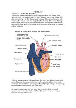
  • 1. THE HEART Formation of the heart in the embryo The finished heart has two separate major pumping systems - the left and right ventricles (ventricle = small cavity), two minor pumping systems (the left and right atria as seen in Fig. 10). The right atrium (=entrance) delivers blood from the body into the right ventricle which then pumps blood to the lungs. Blood from the lungs then returns to the left atrium which delivers blood to the left ventricle which then pumps blood to the body. Each ventricle will require two valves, one at the entrance and one at the exit. The heart begins embryonic life as a tube and thus major remodelling is required but during the change from the primitive tube to a four-chambered heart blood must continue to flow at all times. Knowledge of the embryological changes are necessary to understand the malformations which may occur. An impulse conducting system also has to develop to co-ordinate atrial and ventricular contractions (so that the atrial muscles contract when the ventricular muscle is relaxing) and vice versa.
  • 2. In the embryo a contracting heart tube forms, with veins entering and arteries exiting (Fig. 11). However a contracting tube with no valves is no use as a pump. Accordingly this primitive tube receives blood from the great veins and empties into an atrium, then via atrioventricular valves into a ventricle then into the arteries via semilunar valves. The heart tube is attached at its ends and is free to grow and fold (Fig. 12). The caudally situated Y junction (the sino-atrial chamber where venous blood enters the primitive heart tube) develops two swellings which will later form the right and left atria. The atria move cranially, passing dorsal to the ventricles (Fig. 13). ). Veins (which become the superior and inferior vena cavae which return deoxygenated blood from the body) later enter directly into the right atrial cavity wherea four pulmonary veins carrying oxygenated blood enter the left atrium.
  • 3. The truncus, the artery that will form the outflow tracts of the ventricles, indents the groove separating the atria. The truncus splits longitudinally into two tubes which provide separate outlets for the left and right ventricles (these outlets will become the aortic and pulmonary arteries respectively), and each of the two tubes develops one- way valves. Three valves (the tricuspid, aortic, and pulmonary) have three cusps and one (the mitral) has two cusps (Fig. 14). The ventricular walls grow thicker, become
  • 4. contractile, and the communication between the two ventricles becomes smaller as an interventricular septum develops. The right and left atria are separated by a thin sheet of tissue (the septum primum) but, prior to birth, blood has to continue to flow from the right to the left atrium through an opening, the ostium primum in order to bypass the “useless” lungs. The ostium primum subsequently closes but, because blood must still bypass the lungs,a second opening develops (the ostium secundum), which is oval in shape (the foramen ovale). The ostium secundum is surrounded by tissue which provides a flap-like valve (Fig. 15) which remains open only if the pressure in the right atrium exceeds that in the left atrium (at birth the pressure in the left atrium will rise as blood returns to it from the lungs and this communicating hole will close).
  • 5. Initially the outer muscle wall which surrounded the primitive heart tube contracted as a whole, but with subsequent convolutions of the tube and the need for reciprocal contractions of the atria and ventricles, modified heart muscle fibres that transmit depolarisation waves (faster than would occur between normal heart muscle cells) were developed. An electrical initiating point, the sinoatrial node, forms in the right atrium and sends out a depolarisation wave that causes contraction to spread radially over the atria. A fibrous ring develops to separate the atria from the ventricles and a “focal penetrating area,” the atria-ventricular node, develops and connects with a short stem of conducting tissue (the Bundle of His) which then divides into right and left branches to transmit depolarisation waves to the right and left ventricles (Fig. 16).
  • 6. All the above begs the question “Why do we need a two-sided heart?” There are two answers. Firstly, a high pressure system is required to perfuse the head in an upright posture (otherwise we would faint when we stood up). Secondly, a lower pressure system is required to supply the lungs - if blood pressure in the pulmonary arteries were at aortic pressure then the pulmonary capillaries would have to have walls that were too thick to allow gas exchange (fish which, by and large, do not stand up thus only need one ventricle to pump blood through their gills to the body).
  • 7. Anatomy Figure 17 shows the situation and anatomy of the heart. The heart weighs 300 grams and is much smaller than is commonly supposed (the size is exaggerated by chest X-rays). Until the advent of cardiac transplantation the heart
  • 8. was only guaranteed to last for a lifetime “three score years and ten” and thus a heart had to contract about 2,500 million times to pump about 170 million litres of blood.
  • 9. Heart function The function of the heart is to pump blood to the lungs for oxygenation and then to the tissues for utilisation. To achieve this, a two-sided pump (the heart), a distribution system (the systemic and pulmonary arteries) and return system (the systemic and pulmonary veins) are required. Cardiac contraction (systole) or relaxation (diastole) refers to ventricular events. Both atria contract together (during ventricular diastole) and, later, both ventricles contract together during ventricular systole. When both ventricles are contracting, both atria are relaxing and vice versa (Fig. 18). Although there are no true valves, there are muscle rings around the entry sites of the superior and inferior vena cavae into the right atrium and around the entrysites of pulmonary veins into the left atrium (both to minimise retrograde blood flow during atrial contraction). Cardiac muscle is a specialised form of striated muscle. As muscles can only contract in a linear fashion the heart muscle has to be wound so they can compress the blood within the heart chambers (Fig. 19).
  • 10. Venous blood returns to the right atrium via the superior and inferior vena cavae. This blood then flows (with the assistance of right atrial contraction) through the tricuspid valve into the right ventricle during ventricular relaxation. At the start of ventricular contraction, the tricuspid valve shuts and the venous blood derived from the tissues is ejected into the lungs, passing through the pulmonary valve into the pulmonary arteries (Fig.20).
  • 11. After passing through the lungs the blood, now oxygenated, returns to the left atrium via the pulmonary veins. During ventricular relaxation blood flows (with the assistance of atrial contraction) from the left atrium through the mitral valve into the left ventricle. During ventricular contraction the mitral valve shuts and blood is ejected through the aortic valve into the aorta and, thence, to the body tissues. All four heart valves lie at much the same level in the area which separates the atria from the ventricles.
  • 12. The pericardium, the sac that contains the heart, consists of two layers, an outer fibrous and inner serous layer (Fig. 21). The fibrous layer act as a limiting enclosure for the heart, preventing overstretching of the heart muscle. A small amount of fluid between the two layers serves to lubricate movement. With increasing heart rates the ventricular relaxation (filling) time becomes shorter, rather than the contraction time becoming shorter. Fast heart rates, above about 120 per minute, do not allow sufficient time for ventricular filling and thus predispose to heart failure. With atrial fibrillation, in which the atria contract haphazardly “like a
  • 13. bag of worms,” the lack of atrial assistance for ventricular filling means that heart failure can occur at lower heart rates than this. The power of heart contraction -inotropy- is related to metabolic factors and workload. Preload is heart work required to cope with the venous blood returning to the heart and is related to the length of cardiac muscle fibres when contraction commences. Afterload is the heart work required when the heart muscle contracts to push blood around the systemic circulation and this is in effect the resistance to blood flow in the arteries. In general the heart tries to function as a constant flow pump rather than a constant pressure pump (variation of arteriolar constriction provide most of the pressure regulation). If peripheral resistance increases then arterial blood pressure hase to rise to maintain perfusion, which requires more work by the heart. As an approximation, the more the stretching of heart muscle in ventricular relaxation the more strong the ventricular muscle contraction and the greater the resulting stroke volume (Starling’s Law of the Heart). Blood is supplied to the heart by two coronary arteries which arise from near the origin of the aorta (Fig. 22). These coronary arteries are functionally end arteries, and partial or complete blockage is likely to result in heart muscle death distal to the blockage. The left coronary artery supplies most of the left ventricle and the right coronary artery supplies most of the right ventricle but also supplies the conducting tissue comprising the sino-atrial node and the interventricular node. Thus dysrythmias (abnormal heart rhythms caused by abnormal conduction) are particularly likely to occur if there is right coronary artery narrowing or obstruction.