
Recomendados
Mais conteúdo relacionado
Semelhante a TB - an introduction to Infectious Diseases
Semelhante a TB - an introduction to Infectious Diseases (20)
Mais de meducationdotnet
Mais de meducationdotnet (20)
TB - an introduction to Infectious Diseases
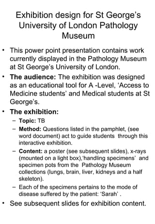
- 1. Exhibition design for St George’s University of London Pathology Museum • This power point presentation contains work currently displayed in the Pathology Museum at St George’s University of London. • The audience: The exhibition was designed as an educational tool for A -Level, ‘Access to Medicine students’ and Medical students at St George’s. • The exhibition: – Topic: TB – Method: Questions listed in the pamphlet, (see word document) act to guide students through this interactive exhibition. – Content: a poster (see subsequent slides), x-rays (mounted on a light box),‘handling specimens’ and specimen pots from the Pathology Museum collections (lungs, brain, liver, kidneys and a half skeleton). – Each of the specimens pertains to the mode of disease suffered by the patient: ‘Sarah’ . • See subsequent slides for exhibition content.
- 2. The pamphlet (back and front)
- 5. Why is TB an important disease to study? TB currently infecting ‘one third of the World’s population (2)’. The World Health Organisation (WHO) has ‘declared TB a public health emergency’ (4). TB is highly contagious - if left untreated, each infected person will pass TB on to ‘between 10 and 15 people every year’ (7). TB is not limited to the Developing World. Today, in the UK, over ‘6,500 cases are reported each year.’ (5) For this reason TB is a ‘notifiable disease’ – by law an incident of TB must be reported to Governmental and Medical authorities to ensure effective treatment and control. Most importantly - today, TB is both curable and controllable. But, less than 50 years ago, before the discovery of the antibiotic Streptomycin, this was not the case. Follow the story of Sarah Finnegan to learn more about the transmission, Infection, control and cure of TB.
- 6. A Case Study: Sarah Finnegan She had lost a considerable amount of weight, was suffering from diarrhea and painful, frequent urination. Upon examination Sarah was pale, restless, sweating, and complained of pain in her chest, back and head. There was a large swelling over her right hip. Tests • A chest x-ray • A cerebro-spinal fluid (CSF) sample, taken by lumbar puncture. • Blood sample was taken. • Sputum sample taken. Upon admission, Sarah was given a course of streptomycin. Initially her condition improved, but after a number of weeks Sarah’s headaches became more frequent and severe. Unable to combat the TB that had infected her brain, sadly, Sarah slipped into a coma and died, four weeks after admission. Sarah Finnegan is a fictitious character inspired by the specimens on display in the Pathology Museum. Examine the display specimens to see how Sarah's body may have been affected by TB On December the 4th 1941 at 21:34, Sarah Finnegan*, aged 19 years, was admitted to St George’s Hospital with a fever (37.8ºC), severe chest pain and a violent cough.
- 7. Specimen B TB with 'cold abscess' formation in dorsal spine A ‘cold abscess’ destroyed several of Sarah’s vertebrae, causing them to collapse, bend and fuse to one another, resulting in severe curvature of her spine. Specimen C Cerebral tuberculoma - TB meningitis. White granulomatous lesions inside the brain cause it to become inflamed and swollen. The resultant increase in intracranial pressure accounts for Sarah’s severe headaches, nausea and vomiting. Specimen E TB of the lungs This is Miliary tuberculosis - tiny Granulomas, resembling millet seeds, throughout the lung, causing the porous tissue to harden. Inflammation would have caused the surrounding capillaries to rupture, accounting for Sarah’s violent, blood stained cough.
- 8. Poster depicting mode of infection in TB