SlideShare uma empresa Scribd logo
1 de 5
Baixar para ler offline
SOME SPECIFIC HEART CONDITIONS
Congential heart disease
In cyanotic congenital (= existing from birth) heart disease there is excess
deoxygenated blood in the systemic circulation (because some blood bypasses the
lungs), whereas in non-cyanotic congenital heart disease blood does go through the
lungs (sometimes twice) but at the expense of the systemic circulation.
Atrial septal defect (Fig. 49)
In atrial septal defects oxygenated blood returns from the lungs into the left atrium,
passes through the defect into the (lower pressure) right atrium and thence (again) to
the lungs. Cyanosis thus does not occur in uncomplicated atrial septal defect.
In theory atrial septal defect murmurs should be heard during ventricular diastole
when the atria will be in systole (as if the murmur were exclusively caused by passage
of blood through the defect between the right and left atrium). In practice a murmur
generated at the defect itself is rarely audible because of the low pressures involved
and the largeness of most defects. The murmur of atrial septal defect occurs during
ventricular systole and is caused by the augmented blood volume passing through the
pulmonary valve (thus simulating a pulmonary stenosis murmur). The augmented
blood flow through the right heart may also cause a tricuspid flow murmur during
right ventricular filling.
Normally the second heart sound is split in inspiration with the aortic valve closing
before the pulmonary valve because of the higher aortic pressures. During inspiration
more blood can be accommodated in the lungs, this extra blood passes into the right
ventricle during ventricular relaxation which then caused prolonged right ventricular
contraction (it takes longer to eject the increased amount of blood). The prolonged
contraction time delays pulmonary valve closure. Thus a normal second heart sound
may be split and the split is normally accentuated during inspiration.
With an atrial septal defect the two atria can be regarded as one chamber and blood
flow into both ventricles is similar. Both ventricular contraction times are therefore
similar and both valve closures are delayed equally. Thus there is a split second
1
sound which does not vary with respiration (fixed splitting). The right ventricle may
also enlarge.
Cardiac tamponade (Fig. 50)
Fluid accumulates between the two layers of pericardium. This fluid preferentially
compresses the right atrium and ventricle (which are at a lower internal pressure and
thus more easily compressed than their left sided counterparts). Impaired filling of
the heart, predominantly the right side during ventricular relaxation, is impaired. The
neck veins become distended particularly with inspiration as the expanded lungs
compress the heart further. Also less blood returns from the lungs to the left heart and
the pulse volume therefore falls in inspiration (pulsus paradoxus). The blood pressure
will be low and the heart rate fast because the heart attempts to maintain output by
increasing the number of fixed volume contractions. Treatment is usually by drainage
of the fluid.
Cardiomyopathy
With hypertrophic cardiomyopathy there is hypertrophy of the ventricular septum or
ventricular muscle. Ventricular contraction is distorted and ventricular filling during
ventricular diastole may be impaired. There may be an abrupt carotid pulse because
there is an abrupt obstruction to left ventricular emptying. With restrictive
cardiomyopathy ventricular filling is restricted because of ventricular muscle
abnormality. This produces symptoms and signs similar to those of constrictive
pericarditis.
2
Constrictive pericarditis (Fig. 51)
The heart is imprisoned by the tight outer layer of pericardium.
Fallot’s tetrad (Fig.52)
This illustrates one congenital heart problem which is explicable on an embryological
basis. The basic feature is that the truncus does not divide appropriately into the aorta
and pulmonary arteries with:
• The pulmonary artery being poorly developed
• The aorta straddling the interventricular septum
• Failure of the interventricular septum to develop fully resulting in a ventricular
septal defect
• Pressure in the left and right ventricles tends to be equal (a combined effect of the
ventricular septal defect and the pulmonary stenosis) with the right ventricle thus
becoming hypertrophied
3
Infective endocarditis
Micro-organisms, almost invariably bacteria, infect heart valves, causing them to be
incompetent. Infective endocarditis may have a rapid onset when caused by virulent
micro-organisms which may affect normal heart valves. Infective endocarditis caused
by less virulent micro-organisms may develop slowly and usually only affects
previously damaged valves. Right sided endocarditis is a particular risk for
intravenous drug abusers who usually inject into veins.
Ischaemic heart disease
Ischaemia (= a local and temporary deficiency of blood caused by interruption of the
blood supply) is almost always associated with coronary artery atheroma. Intrinsic
heart muscle disease or overdevelopment of ventricular muscle caused by overwork
can outstrip the available blood supply to cause ischaemic symptoms.
Heart muscle has a limited ability to metabolise without oxygen and increased oxygen
demands caused by increased work have to be met by increased blood flow brought
about by coronary artery vasodilatation (which may be able to compensate for
narrowing caused by atheroma).
When heart muscle is starved of oxygen angina (a pressing, constricting discomfort
or pain which is brought on by exertion and relieved by rest) may result. Angina may
also be felt in the left arm, which has a similar somatic nerve supply as the autonomic
supply to the heart. Treatments of an anginal attack includes stopping the causative
activity, sitting down and using a nitrate preparation which dilates peripheral arteries
and thereby reduces the afterload.
4
Myocardial infarction
Myocardial infarction (infarction = muscle death) occurs when ischaemic heart
muscle dies. Myocardial infarction causes about 200,000 deaths a year in the United
Kingdom. Symptoms may be similar to angina but may begin at rest, last for longer
(more than 20 minutes) and be more severe.
Treatment of myocardial infarction may include:
• Pain relief
• Aspirin to reduce clotting
• Early attempts to dissolve the thrombus (thrombolysis)
• Interventions include drugs to dilate blood vessels to reduce heart workload, and
drugs that reduce heart work by direct action on heart muscle
Patent ductus arteriosus (Fig. 53)
If the ductus arteriosus fails to close at birth then blood will flow continuously from
the aorta into the pulmonary veins because the aortic pressure after birth is higher
than pulmonary vein pressure at all stages of the cardiac cycle. Blood in the aorta is
always fully oxygenated and therefore patients do not develop cyanosis. There is a
wide pulse pressure because the aortic diastolic pressure is lower than normal due to
the continued drainage of blood from the aorta into the pulmonary artery.
Pregnancy and the heart
Pregnancy may exacerbate heart failure because in pregnancy there is:
• An increased cardiac output
• An increased blood and extracellular volume
• A slightly increased heart rate
• A 30 percent increase in heart oxygen requirement
5

Mais conteúdo relacionado

Mais procurados

Mais procurados (20)

COLLATERAL ARTERIES
COLLATERAL ARTERIESCOLLATERAL ARTERIES
COLLATERAL ARTERIES
 
Aortic stenosis
Aortic stenosisAortic stenosis
Aortic stenosis
 
Arrhythmia
ArrhythmiaArrhythmia
Arrhythmia
 
06 circulatory disorders
06   circulatory disorders06   circulatory disorders
06 circulatory disorders
 
Cardiac output
Cardiac outputCardiac output
Cardiac output
 
Traumatic heart injuries a brief
Traumatic heart injuries a briefTraumatic heart injuries a brief
Traumatic heart injuries a brief
 
Aneurysm
AneurysmAneurysm
Aneurysm
 
Pulmonary circulation, pulmonary edema, pleural fluid
Pulmonary circulation, pulmonary edema, pleural   fluidPulmonary circulation, pulmonary edema, pleural   fluid
Pulmonary circulation, pulmonary edema, pleural fluid
 
Circulatory diseases and disorders
Circulatory diseases and disordersCirculatory diseases and disorders
Circulatory diseases and disorders
 
Mitral stenosis (propeduetics)
Mitral stenosis (propeduetics)Mitral stenosis (propeduetics)
Mitral stenosis (propeduetics)
 
Pulmonary circulation
Pulmonary circulationPulmonary circulation
Pulmonary circulation
 
Cardiovascular ststem
Cardiovascular ststemCardiovascular ststem
Cardiovascular ststem
 
8. Congintal anomalies of the cardiovascular system
8. Congintal anomalies of the cardiovascular system8. Congintal anomalies of the cardiovascular system
8. Congintal anomalies of the cardiovascular system
 
Pulmonary circulation
Pulmonary circulationPulmonary circulation
Pulmonary circulation
 
Aortic stenosis- Dr Shaz Pamangadan
Aortic stenosis- Dr Shaz PamangadanAortic stenosis- Dr Shaz Pamangadan
Aortic stenosis- Dr Shaz Pamangadan
 
coronary circulation
coronary circulationcoronary circulation
coronary circulation
 
Aortic stenosis
Aortic stenosisAortic stenosis
Aortic stenosis
 
Aortic Stenosis
Aortic StenosisAortic Stenosis
Aortic Stenosis
 
Aortic valve stenosis
Aortic valve stenosisAortic valve stenosis
Aortic valve stenosis
 
cardiovascular diseases
cardiovascular diseasescardiovascular diseases
cardiovascular diseases
 

Destaque (17)

Body Systems: Homeostasis, blood, cardio and respiratory
Body Systems: Homeostasis, blood, cardio and respiratoryBody Systems: Homeostasis, blood, cardio and respiratory
Body Systems: Homeostasis, blood, cardio and respiratory
 
Shock pathophysiology
Shock pathophysiologyShock pathophysiology
Shock pathophysiology
 
Diabetic Feet
Diabetic FeetDiabetic Feet
Diabetic Feet
 
Bus Bites- Cardiac Arrhythmias
Bus Bites- Cardiac ArrhythmiasBus Bites- Cardiac Arrhythmias
Bus Bites- Cardiac Arrhythmias
 
Cardiomyopathies
CardiomyopathiesCardiomyopathies
Cardiomyopathies
 
Acute Coronary Syndrome and the ECG
Acute Coronary Syndrome and the ECGAcute Coronary Syndrome and the ECG
Acute Coronary Syndrome and the ECG
 
Blood Physiology
Blood PhysiologyBlood Physiology
Blood Physiology
 
Overview of the Liver
Overview of the LiverOverview of the Liver
Overview of the Liver
 
Hypertension in Pregnancy
Hypertension in PregnancyHypertension in Pregnancy
Hypertension in Pregnancy
 
Ischemic Heart Disease
Ischemic Heart DiseaseIschemic Heart Disease
Ischemic Heart Disease
 
M.Tuberculosis
M.TuberculosisM.Tuberculosis
M.Tuberculosis
 
Gastroenterology Tutorial
Gastroenterology TutorialGastroenterology Tutorial
Gastroenterology Tutorial
 
Headaches at a Glance - Usman Ahmed
Headaches at a Glance - Usman AhmedHeadaches at a Glance - Usman Ahmed
Headaches at a Glance - Usman Ahmed
 
Abdominal Exam
Abdominal ExamAbdominal Exam
Abdominal Exam
 
The Basics of ECG Interpretation
The Basics of ECG InterpretationThe Basics of ECG Interpretation
The Basics of ECG Interpretation
 
Endocrinology Notes
Endocrinology NotesEndocrinology Notes
Endocrinology Notes
 
Respiratory Notes
Respiratory NotesRespiratory Notes
Respiratory Notes
 

Semelhante a SOME SPECIFIC HEART CONDITIONS

Cynotic heart disease plane radiography
Cynotic heart disease plane radiography Cynotic heart disease plane radiography
Cynotic heart disease plane radiography Dr-Girish Gunari
 
Guides on Heart Disease
Guides on Heart DiseaseGuides on Heart Disease
Guides on Heart DiseaseDr. Rubz
 
Anatomy of cardiovascular system
Anatomy of cardiovascular systemAnatomy of cardiovascular system
Anatomy of cardiovascular systembutterflyfairy
 
Valvular heart disease (1).pdf
Valvular heart disease (1).pdfValvular heart disease (1).pdf
Valvular heart disease (1).pdfMisbah660782
 
cardiovascular system
cardiovascular systemcardiovascular system
cardiovascular systemJADHAVDEEPALI
 
The_Heart__anatomy and physiology AHscience
The_Heart__anatomy and physiology AHscienceThe_Heart__anatomy and physiology AHscience
The_Heart__anatomy and physiology AHsciencesudheendrapv
 
Heart and its disease
Heart and its diseaseHeart and its disease
Heart and its diseasebhagya shree
 
Systemic pathology cardiovascular system
Systemic  pathology  cardiovascular systemSystemic  pathology  cardiovascular system
Systemic pathology cardiovascular systemTofiqueAhmed5
 
Rheumatic Heart Disease ( RHD )
Rheumatic Heart Disease ( RHD )Rheumatic Heart Disease ( RHD )
Rheumatic Heart Disease ( RHD )RxSlides PPT
 
CARDIAC OUTPUTdhdhdhsdfhhfhjfjefefgefgef
CARDIAC OUTPUTdhdhdhsdfhhfhjfjefefgefgefCARDIAC OUTPUTdhdhdhsdfhhfhjfjefefgefgef
CARDIAC OUTPUTdhdhdhsdfhhfhjfjefefgefgefwaytoway91
 
CARDIOMYOPATHY.pptx
CARDIOMYOPATHY.pptxCARDIOMYOPATHY.pptx
CARDIOMYOPATHY.pptxZellanienhd
 
1 introduction to cardiovascular diseases
1 introduction to cardiovascular diseases1 introduction to cardiovascular diseases
1 introduction to cardiovascular diseaseskarima Akool AlSalihi
 
Heart Anatomy and Physiology:Review
Heart Anatomy and Physiology:ReviewHeart Anatomy and Physiology:Review
Heart Anatomy and Physiology:ReviewTosca Torres
 

Semelhante a SOME SPECIFIC HEART CONDITIONS (20)

Cynotic heart disease plane radiography
Cynotic heart disease plane radiography Cynotic heart disease plane radiography
Cynotic heart disease plane radiography
 
Guides on Heart Disease
Guides on Heart DiseaseGuides on Heart Disease
Guides on Heart Disease
 
Anatomy of cardiovascular system
Anatomy of cardiovascular systemAnatomy of cardiovascular system
Anatomy of cardiovascular system
 
Cardiovascular System Pathology 2014 edited by @jennings argwing
Cardiovascular System Pathology 2014 edited by @jennings argwingCardiovascular System Pathology 2014 edited by @jennings argwing
Cardiovascular System Pathology 2014 edited by @jennings argwing
 
Valvular heart disease (1).pdf
Valvular heart disease (1).pdfValvular heart disease (1).pdf
Valvular heart disease (1).pdf
 
cardiovascular system
cardiovascular systemcardiovascular system
cardiovascular system
 
The_Heart__anatomy and physiology AHscience
The_Heart__anatomy and physiology AHscienceThe_Heart__anatomy and physiology AHscience
The_Heart__anatomy and physiology AHscience
 
Heart and its disease
Heart and its diseaseHeart and its disease
Heart and its disease
 
Systemic pathology cardiovascular system
Systemic  pathology  cardiovascular systemSystemic  pathology  cardiovascular system
Systemic pathology cardiovascular system
 
Congestive heart failure
Congestive heart failureCongestive heart failure
Congestive heart failure
 
The heart
The heartThe heart
The heart
 
Cardiac cycle
Cardiac cycleCardiac cycle
Cardiac cycle
 
Rheumatic Heart Disease ( RHD )
Rheumatic Heart Disease ( RHD )Rheumatic Heart Disease ( RHD )
Rheumatic Heart Disease ( RHD )
 
Samir rafla principles of cardiology pages 62 86
Samir rafla principles of cardiology pages 62 86Samir rafla principles of cardiology pages 62 86
Samir rafla principles of cardiology pages 62 86
 
CARDIAC OUTPUTdhdhdhsdfhhfhjfjefefgefgef
CARDIAC OUTPUTdhdhdhsdfhhfhjfjefefgefgefCARDIAC OUTPUTdhdhdhsdfhhfhjfjefefgefgef
CARDIAC OUTPUTdhdhdhsdfhhfhjfjefefgefgef
 
Cardiac disorders
Cardiac disordersCardiac disorders
Cardiac disorders
 
CARDIOMYOPATHY.pptx
CARDIOMYOPATHY.pptxCARDIOMYOPATHY.pptx
CARDIOMYOPATHY.pptx
 
Cardiac cycle
Cardiac cycleCardiac cycle
Cardiac cycle
 
1 introduction to cardiovascular diseases
1 introduction to cardiovascular diseases1 introduction to cardiovascular diseases
1 introduction to cardiovascular diseases
 
Heart Anatomy and Physiology:Review
Heart Anatomy and Physiology:ReviewHeart Anatomy and Physiology:Review
Heart Anatomy and Physiology:Review
 

Mais de meducationdotnet

Mais de meducationdotnet (20)

No Title
No TitleNo Title
No Title
 
Spondylarthropathy
SpondylarthropathySpondylarthropathy
Spondylarthropathy
 
Diagnosing Lung cancer
Diagnosing Lung cancerDiagnosing Lung cancer
Diagnosing Lung cancer
 
Eczema Herpeticum
Eczema HerpeticumEczema Herpeticum
Eczema Herpeticum
 
The Vagus Nerve
The Vagus NerveThe Vagus Nerve
The Vagus Nerve
 
Water and sanitation and their impact on health
Water and sanitation and their impact on healthWater and sanitation and their impact on health
Water and sanitation and their impact on health
 
The ethics of electives
The ethics of electivesThe ethics of electives
The ethics of electives
 
Intro to Global Health
Intro to Global HealthIntro to Global Health
Intro to Global Health
 
WTO and Health
WTO and HealthWTO and Health
WTO and Health
 
Globalisation and Health
Globalisation and HealthGlobalisation and Health
Globalisation and Health
 
Health Care Worker Migration
Health Care Worker MigrationHealth Care Worker Migration
Health Care Worker Migration
 
International Institutions
International InstitutionsInternational Institutions
International Institutions
 
Haemochromotosis brief overview
Haemochromotosis brief overviewHaemochromotosis brief overview
Haemochromotosis brief overview
 
Ascities overview
Ascities overviewAscities overview
Ascities overview
 
Overview of Antidepressants
Overview of AntidepressantsOverview of Antidepressants
Overview of Antidepressants
 
Gout Presentation
Gout PresentationGout Presentation
Gout Presentation
 
Review of orthopaedic services: Prepared for the Auditor General for Scotland...
Review of orthopaedic services: Prepared for the Auditor General for Scotland...Review of orthopaedic services: Prepared for the Auditor General for Scotland...
Review of orthopaedic services: Prepared for the Auditor General for Scotland...
 
Sugammadex - a revolution in anaesthesia?
Sugammadex - a revolution in anaesthesia?Sugammadex - a revolution in anaesthesia?
Sugammadex - a revolution in anaesthesia?
 
Ophthamology Revision
Ophthamology RevisionOphthamology Revision
Ophthamology Revision
 
Dermatology Atlas
Dermatology AtlasDermatology Atlas
Dermatology Atlas
 

SOME SPECIFIC HEART CONDITIONS

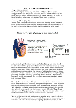
  • 1. SOME SPECIFIC HEART CONDITIONS Congential heart disease In cyanotic congenital (= existing from birth) heart disease there is excess deoxygenated blood in the systemic circulation (because some blood bypasses the lungs), whereas in non-cyanotic congenital heart disease blood does go through the lungs (sometimes twice) but at the expense of the systemic circulation. Atrial septal defect (Fig. 49) In atrial septal defects oxygenated blood returns from the lungs into the left atrium, passes through the defect into the (lower pressure) right atrium and thence (again) to the lungs. Cyanosis thus does not occur in uncomplicated atrial septal defect. In theory atrial septal defect murmurs should be heard during ventricular diastole when the atria will be in systole (as if the murmur were exclusively caused by passage of blood through the defect between the right and left atrium). In practice a murmur generated at the defect itself is rarely audible because of the low pressures involved and the largeness of most defects. The murmur of atrial septal defect occurs during ventricular systole and is caused by the augmented blood volume passing through the pulmonary valve (thus simulating a pulmonary stenosis murmur). The augmented blood flow through the right heart may also cause a tricuspid flow murmur during right ventricular filling. Normally the second heart sound is split in inspiration with the aortic valve closing before the pulmonary valve because of the higher aortic pressures. During inspiration more blood can be accommodated in the lungs, this extra blood passes into the right ventricle during ventricular relaxation which then caused prolonged right ventricular contraction (it takes longer to eject the increased amount of blood). The prolonged contraction time delays pulmonary valve closure. Thus a normal second heart sound may be split and the split is normally accentuated during inspiration. With an atrial septal defect the two atria can be regarded as one chamber and blood flow into both ventricles is similar. Both ventricular contraction times are therefore similar and both valve closures are delayed equally. Thus there is a split second 1
  • 2. sound which does not vary with respiration (fixed splitting). The right ventricle may also enlarge. Cardiac tamponade (Fig. 50) Fluid accumulates between the two layers of pericardium. This fluid preferentially compresses the right atrium and ventricle (which are at a lower internal pressure and thus more easily compressed than their left sided counterparts). Impaired filling of the heart, predominantly the right side during ventricular relaxation, is impaired. The neck veins become distended particularly with inspiration as the expanded lungs compress the heart further. Also less blood returns from the lungs to the left heart and the pulse volume therefore falls in inspiration (pulsus paradoxus). The blood pressure will be low and the heart rate fast because the heart attempts to maintain output by increasing the number of fixed volume contractions. Treatment is usually by drainage of the fluid. Cardiomyopathy With hypertrophic cardiomyopathy there is hypertrophy of the ventricular septum or ventricular muscle. Ventricular contraction is distorted and ventricular filling during ventricular diastole may be impaired. There may be an abrupt carotid pulse because there is an abrupt obstruction to left ventricular emptying. With restrictive cardiomyopathy ventricular filling is restricted because of ventricular muscle abnormality. This produces symptoms and signs similar to those of constrictive pericarditis. 2
  • 3. Constrictive pericarditis (Fig. 51) The heart is imprisoned by the tight outer layer of pericardium. Fallot’s tetrad (Fig.52) This illustrates one congenital heart problem which is explicable on an embryological basis. The basic feature is that the truncus does not divide appropriately into the aorta and pulmonary arteries with: • The pulmonary artery being poorly developed • The aorta straddling the interventricular septum • Failure of the interventricular septum to develop fully resulting in a ventricular septal defect • Pressure in the left and right ventricles tends to be equal (a combined effect of the ventricular septal defect and the pulmonary stenosis) with the right ventricle thus becoming hypertrophied 3
  • 4. Infective endocarditis Micro-organisms, almost invariably bacteria, infect heart valves, causing them to be incompetent. Infective endocarditis may have a rapid onset when caused by virulent micro-organisms which may affect normal heart valves. Infective endocarditis caused by less virulent micro-organisms may develop slowly and usually only affects previously damaged valves. Right sided endocarditis is a particular risk for intravenous drug abusers who usually inject into veins. Ischaemic heart disease Ischaemia (= a local and temporary deficiency of blood caused by interruption of the blood supply) is almost always associated with coronary artery atheroma. Intrinsic heart muscle disease or overdevelopment of ventricular muscle caused by overwork can outstrip the available blood supply to cause ischaemic symptoms. Heart muscle has a limited ability to metabolise without oxygen and increased oxygen demands caused by increased work have to be met by increased blood flow brought about by coronary artery vasodilatation (which may be able to compensate for narrowing caused by atheroma). When heart muscle is starved of oxygen angina (a pressing, constricting discomfort or pain which is brought on by exertion and relieved by rest) may result. Angina may also be felt in the left arm, which has a similar somatic nerve supply as the autonomic supply to the heart. Treatments of an anginal attack includes stopping the causative activity, sitting down and using a nitrate preparation which dilates peripheral arteries and thereby reduces the afterload. 4
  • 5. Myocardial infarction Myocardial infarction (infarction = muscle death) occurs when ischaemic heart muscle dies. Myocardial infarction causes about 200,000 deaths a year in the United Kingdom. Symptoms may be similar to angina but may begin at rest, last for longer (more than 20 minutes) and be more severe. Treatment of myocardial infarction may include: • Pain relief • Aspirin to reduce clotting • Early attempts to dissolve the thrombus (thrombolysis) • Interventions include drugs to dilate blood vessels to reduce heart workload, and drugs that reduce heart work by direct action on heart muscle Patent ductus arteriosus (Fig. 53) If the ductus arteriosus fails to close at birth then blood will flow continuously from the aorta into the pulmonary veins because the aortic pressure after birth is higher than pulmonary vein pressure at all stages of the cardiac cycle. Blood in the aorta is always fully oxygenated and therefore patients do not develop cyanosis. There is a wide pulse pressure because the aortic diastolic pressure is lower than normal due to the continued drainage of blood from the aorta into the pulmonary artery. Pregnancy and the heart Pregnancy may exacerbate heart failure because in pregnancy there is: • An increased cardiac output • An increased blood and extracellular volume • A slightly increased heart rate • A 30 percent increase in heart oxygen requirement 5