

Recomendados
Mais conteúdo relacionado
Mais procurados
Mais procurados (20)
Semelhante a Renal pathology iv
Semelhante a Renal pathology iv (20)
Mais de med_students0
Mais de med_students0 (20)
Último
Último (20)
Renal pathology iv
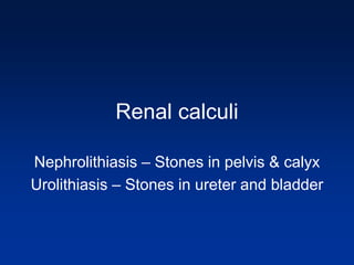
- 1. Renal calculi Nephrolithiasis – Stones in pelvis & calyx Urolithiasis – Stones in ureter and bladder
- 2. Renal calculi Pathogenesis: 1)Supersaturation 2)Absence of normal stone inhibitors 3)Presence of infection
- 3. Renal calculi Types of stones: 1) Calcium stones (Most common) – Oxalate, Phosphate 2) Struvite stones (Triple phosphate, Staghorn calculi) – Mg NH3 Phosphate, proteus infection, Alkaline pH 3) Uric acid stones – Acidic pH 4) Cystine stones - rare All stones are Radio-opaque except “Uric acid stones”
- 4. Renal Calculi • Urinary tract calculi are usually unilateral and about 1 to 3 mm in size. Their passage is marked by intense abdominal or back or flank pain. This pain can be paroxysmal, known as renal or ureteral "colic". • Hematuria may also be present. • Larger stones that cannot pass may produce hydronephrosis or hydroureter.
- 5. Nephrolithiasis • Plain X-ray – Showing a staghorn calculi • Gross – Appearance of triple stone resembling the horns of an antler
- 6. Management of renal stones MAJORITY : 85 to 90% of all stones can be treated by - EXTRA - CORPOREAL SHOCK WAVE LITHOTRIPSY (ESWL) MINORITY : 10 to15 % SHOULD NEED MINIMALLY INVASIVE SURGERY (PCNL / URETEROSCOPY) (LESS THAN 1 % NEED OPEN SURGERY)
- 8. Fetal lobulation in adult kidney • Persistance of fetal lobulation can be seen in routine autopsies in certain adults • Renal function is usually preserved and no symptoms occur • At the lower right is a smooth- surfaced, small, clear fluid-filled simple renal cyst. Such cysts occur either singly or scattered around the renal parenchyma and are not uncommon in adults.
- 9. Horseshoe kidney • Its incidence is 1/500. • Seen in Turner’s syndrome • 90% of case there is fusion in the lower poles • The possible problem here is that the ureters take an abnormal course across the "bridge" of renal tissue and this can lead to partial obstruction with hydronephrosis.
- 10. Horseshoe kidney • Fusion in the lower poles
- 12. Multicystic Renal Dysplasia • a sporadic disorder without familial clustering. • results from abnormal differentiation of the metanephric parenchyma • most cases - unilateral • bilateral lesions – Potter’s Sequence, renal failure
- 13. Multicystic Renal Dysplasia • Gross -The kidney is irregular and multicystic, the cysts are variably sized, from 1 mm to 1 cm in size, and filled with clear fluid. • Micro - There are few recognizable glomeruli and tubules • The hallmark of renal dysplasia is the presence of "primitive ducts" lined by cuboidal to columnar epithelium and surrounded by a collagenous stroma. This increased stroma may contain small islands of cartilage.
- 14. Multicystic Renal Dysplasia • Gross – Multiple cyst with distortion of the architecture • Micro –Note cartilage & cyst spaces
- 15. AD (Adult) polycystic Kidney disease • Incidence 1:400-1000 live births • Presentation – Cysts not present at birth but presents at 4th or 5th decade with Bilateral involvement • Genetics: • PKD 1 gene (polycystin I)–Ch 16 • PKD 2 gene(Polycystin II) – Ch 4 • PKD 3 gene
- 16. ADPKD - Gross • Kidneys are enlarged (Massive sizes) with diffuse variably sized cysts. Cysts may contain clear fluid or turbid
- 17. ADPKD • Clinical - usually presents in 4th decade with hypertension and hematuria-renal failure within a decade • Other associations : • Berry aneurysms - may present with subarachnoid hemorrhage • Polycystic liver disease • Mitral valve prolapse • Colonic Diverticulosis
- 18. AR(Childhood) PKD • Incidence – 1 in 30,000 • The kidneys are affected bilaterally • May occur in utero, infancy & childhood • Present with Potter’s sequence • Genetics: • Fibrocystin gene
- 19. ARPKD • Grossly, the kidneys are markedly enlarged • The cysts are quite small and uniform, perhaps 1 to 2 mm on average, arranged at right angles to the cortex. • Extra-renal manifestation – Congenital hepatic fibrosis
- 20. recessive polycystic kidney disease (RPKD).
- 21. recessive polycystic kidney disease (RPKD).
- 22. Cystic diseases of Renal medulla • Medullary sponge kidney: • Occurs in adults • Cysts are small representing cystic dilatation of collecting ducts in the medulla • Pathogenesis unknown and usually renal function is good
- 23. Familial Juvenile Nephronophthisis- Uremic Medullary Cystic Disease • Kidneys are shrunken with cysts in the corticomedullary junction • Associated with cortical tubular atrophy and interstitial fibrosis • Renal failure, salt wasting, polyuria, tubular acidosis, growth retardation, anemia • Juvenile form is autosomal recessive and presents around age 11 • Adult form is autosomal dominant and presents around age 20 • Both together account for 10-25% of cases of end-stage renal failure in the first 3 decades
- 24. Uremic Medullary Cystic Disease • Note cysts in the cortico-medullary areas
- 25. Acquired Cysts (Dialysis- associated) Disease • Seen in Pts with endstage renal disease relying on dialysis • Gross – exhibit numerous cortical and medullary cysts 0.5 to 2 cms size often contain calcium oxalate crystals • Associated with renal cell carcinoma (10 yrs duration)
- 26. Simple cysts • Common Post mortem finding • Hemorrhage in the cyst occurs produce pain • Has to be differentiated from renal tumours because of their massive sizes(1-5 cms) – USG will give clue to the cystic nature with fluid levels
- 27. Simple renal cyst • Note the enormous size of the cyst with shiny glistening surface
- 28. Tumours of kidney Benign – Papillary adenoma Angio-myolipoma Oncocytoma Malignant – Renal cell carcinoma Transitional cell carcinoma
- 29. Malignant – Renal cell carcinoma • Also called Hypernephroma, Grawitz tumour, renal adenocarcinoma • Older individuals affected M:F=2-3:1 Predisposing factors: smoking, Dialysis-associated cystic disease, tuberous sclerosis, obesity, exposure to industrial chemicals, and autosomal dominant polycystic renal disease Most tumours are sporadic but some are familial • Familial types: • Von Hippel Lindau Syndrome – CNS tumours – Hemangioblastoma of cerebellum + Renal carcinoma • Hereditary clear cell ca – VHL gene mutation with renal tumour but no CNS tumours • Hereditary Papillary carcinoma – Mutations in MET protooncogene
- 30. Gross features of RCC • Arise in any portion – Common Upper pole • Size -3-15 cms • Color – solid, yellowish white • Some are cystic with areas of necrosis • Tumour may involve renal vein rarely emboli to IVC and Rt heart
- 31. Gross - RCC • 2 cases of renal cell ca
- 32. RCC • Histological types: • Clear cell type – VHL mutations, rounded or polygonal cells with clear cytoplasm • Papillary type – MET oncogene mutation, papillary configuration lined by cuboidal columnar cells with psammoma bodies • Chromophobe renal ca – Cells with pale eosinophilic cytoplasm around blood vessels
- 33. RCC – Clear cell type • Cell arranged in nest with clear cytoplasm
- 34. Papillary RCC • Numerous branching papillae and small dark structures (Psammoma bodies)
- 35. Chromophobe renal carcinoma • Notice the cells showing pale eosinophilic cytoplasm – Compare with earlier slide with Clear cell type
- 36. RCC • Clinical: – Cost-vertebral pain, Palpable mass, Hematuria – Occasionally fever, weakness, Weight loss – Symptoms relating to paraneoplastic syndrome PTH producing hypercalcemia Erythropoietin producing polycythemia (3%) Renin producing hypertension Gonadotropins producing feminization or masculinization Cortisol producing Cushing’s syndrome
- 37. RCC • Metastasis - spread to the lungs(60% have “cannon-ball” lesions), bone, brain, liver, and paraaortic lymph nodes • Some cases – Involvement of the renal vein and spermatic veins by thrombo- embolism can cause varicocele in the scrotum of male pts
- 38. Transitional Cell Carcinoma, Renal Pelvis • This type of carcinoma constitutes 5 to 10% of all renal carcinomas. • They are usually detected because they produce prominent hematuria. • They invade early through the relatively thin wall of the pelvic mucosa.
- 39. TCC The cut surfaces of the kidney removed surgically here demonstrate normal cortex and medulla, but the calyces show focal papillary tumor masses of transitional cell carcinoma. Note the multiplicity of tumors.
- 40. TCC • At high power, the transitional cell carcinoma does resemble urothelium, but the thickness is much greater than normal and the cells show more pleomorphism
- 41. Benign - Renal Papillary Adenoma • Renal adenomas are small,usually0.5 to 1cm ,renal cortical tumors. • They are usually papillary in histological appearance but may be clear cell in type. • The problem is that they are histologically similar to renal cell carcinomas and differ from them only in size. • Because of increased use of imaging studies these are now being picked up. • The present formula is to consider tumors from 1 to 3 cm as potentially malignant.
- 42. Benign - Angio-myolipoma • Benign tumour - occurs in patients with Tuberous Sclerosis (CNS cortical tubers with epilepsy and MR & skin lesions) in which cases it is often bilateral and sometimes multifocal. • It is made up of a mixture of fat cells, smooth muscle and thick walled vessels. • These often have a pleomorphic appearance (mistaken for malignancy)
- 43. Angio-myolipoma • Blood vessels • Smooth muscle • Adipose tissue
- 44. Benign - Oncocytoma • This is a benign tumor which can be mistaken for a carcinoma. • Gross - large Mahogany brown tumor with a central stellate hyaline scar • Micro – Uniform tumour cells with granular eosinophilic cytoplasm with benign nucleus • With the electron microscope these cells show markedly increased mitochondria
- 45. Oncocytoma • Notice the dark brown color and Central scar
- 46. Oncocytoma • Note the cells with uniform ,eosinophilic granular cytoplasm and the hyaline fibrous tissue of the central scar.
- 47. Oncocytoma, Electron Microscopy • Note the markedly increased number of mitochondria
- 48. • A 4 yr old child presents with painless mass in the abdomen, hematuria and hypertension. Diagnosis? A)MCD B)PSGN C)Neuroblastoma D)Wilms tmour E)Renal cell carcinoma