vascular access for dialysis access: seminar
•Transferir como PPT, PDF•
19 gostaram•4,545 visualizações

vascular access including fistula, graft and catheterization procedure

Recomendados
Mais conteúdo relacionado
Mais procurados
Mais procurados (20)
Semelhante a vascular access for dialysis access: seminar
Semelhante a vascular access for dialysis access: seminar (20)
Último
Último (20)
vascular access for dialysis access: seminar
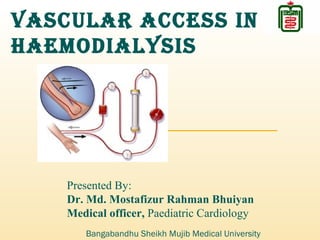
- 1. Bangabandhu Sheikh Mujib Medical University Vascular access in Haemodialysis Presented By: Dr. Md. Mostafizur Rahman Bhuiyan Medical officer, Paediatric Cardiology
- 2. What is Dialysis: The word Dialysis was taken from Greek "διάλυσις " which means “Dissolution through splitting” It is the process of removing excess water, solutes and toxins from the blood in people whose kidneys can no longer perform these functions naturally. This is referred to as renal replacement therapy. When kidneys fail, dialysis is often only option to save life. The first successful dialysis was performed in 1943.
- 3. Dialysis Options There are two form of dialysis. – Peritoneal dialysis – Hemodialysis
- 4. Pie Chart shows among 635,645 patients with end-stage renal disease (ESRD) 64.2% undergo hemodialysis https://www.ajronline.org/doi/full/10.2 214/AJR.15.14650
- 5. Hemodialysis Hemodialysis circulates blood through a machine outside the body to remove toxins and excess fluid & then pumps the cleansed blood back into the body.
- 6. Hemodialysis Access Dialysis access refers to the creation of an entranceway into the bloodstream so that the blood can be cleansed by the dialysis procedure.
- 7. Types of Hemodialysis Access Fistula (arteriovenous fistula) Graft (arteriovenous graft) Venous catheter 1. Cuffed 2. Uncuffed
- 8. Types of Hemodialysis Access: Among 4,07,811 U.S. end-stage renal disease patients undergoing hemodialysis : 64.2% are dialyzed through AVFs 18.5% through AVG 19.5% through hemodialysis catheters https://www.ajronline.org/doi/full/1 0.2214/AJR.15.14650
- 9. 2017 Annual Data Report Change in type of vascular access during the first year of dialysis among ESRD patients starting via hemodialysis in 2013 quarterly: (a) type of vascular access in use (cross-sectional) (b) longitudinal changes in vascular access use and other outcomes, Data Source: Special analyses, USRDS ESRD Database. Data from January 1, 2013 to May 30, 2016 CROWNWeb, Consolidated Renal Operations in a Web-enabled Network; ESRD, end-stage renal disease; HD, hemodialysis; PD, peritoneal dialysis.
- 10. Preparation for Haemodialysis Access: Ideally before dialysis an venous access should be made – Fistula should be placed 6 months prior to start dialysis – Graft should be placed 3-6 weeks prior to start dialysis – Venous catheter can be used instantly
- 11. Preparation for Access Before an access is made, patient is evaluated by a surgeon – – vein mapping with an doppler ultrasound to see – – Vessel with a 2-2.5 mm and above diametre are acceptable for fistula. – US also help to determine course of the veins – Blood lab tests for anaesthetic and surgical fitness
- 12. FISTULA/ AVF
- 13. An arteriovenous fistula is a connection between an artery and a vein surgically created for hemodialysis by the vascular surgeon It is the preferred access of all the types of hemodialysis access and is often referred to as the “gold standard.” This access results in an extra pressure and extra blood to flow into the vein, which helps to enlarge and strengthen the vein. What is a fistula or AVF?
- 14. An AV fistula allows a higher rate of blood to flow back and forth from the vein to a dialysis machine. Untreated veins cannot withstand repeated needle insertions, because they would collapse under strong suction. What is an arteriovenous fistula?
- 15. Hemodialysis Access There are only about ten sites in the body where an AV fistula or graft can be made. They are commonly located in the – Arm (non-dominate forearm or upper arm) – Leg – Neck
- 16. Different fistula: Radiocephalic End to side anastomosis of radial artery and forearm cephalic vein, Brescia- Cimino fistula (proximal forearm) Original fistula created by Dr. James Cimino in 1966. Technically simple Distal patency rates at one year are approximately 50% to 80%. The use of this distal access site preserves more proximal vessels for subsequent attempts at creating a fistula. Ref: Types of Arteriovenous Fistulas Michael Segal; Erion Qaja. Last Update: March 16, 2019.
- 17. Proximal forearm AVF: Anastomosis between proximal radial artery and median antecubital vein
- 18. Proximal forearm AVF: Anastomosis between perforating vein and proximal radial artery
- 19. Brachiocephalic AVF: Anastomosis between Cephalic vein and Brachial artery
- 20. Transposed brachiobasilic fistula Anastomosis between Basilic vein & brachial artery
- 21. Maturation of the fistula: Rule of 6s includes: – The flow should be greater than 600 mL per minute – Greater than 6 mm diameter – Less than 6 mm below the skin – At least 6 cm of the vein for cannulation – A thorough examination and expected maturation at six weeks Ref: Types of Arteriovenous Fistulas Michael Segal; Erion Qaja. Last Update: March 16, 2019.
- 22. Postoperative management of all arteriovenous fistulas (AVFs) Outcomes for forearm and upper arm arteriovenous fistula creation with the transposition technique. Journal of Vascular Surgery Volume 63, Issue 3, March 2016, Pages 764-771
- 23. Outcomes for forearm and upper arm arteriovenous fistula creation with the transposition technique. Journal of Vascular Surgery Volume 63, Issue 3 , March 2016, Pages 764-771
- 24. Advantages of AVFs The gold standard for vascular access because – – it provides adequate blood flow, – lasts a long time, usually 20 plus years – has a lower complication rate than other types of access. It is done as minor outpatient surgery Usually take 6 to 12 weeks to develop Fewer infections & thrombus than grafts and catheters Pt can take Bath
- 25. Disadvantages of AVFs May require another temporary type of access during the healing and maturation phase Maturation may be delayed, or it may fail to mature Visible as a bulge under the skin Not always possible for all patients Needles are required to access the AV fistula for hemodialysis
- 26. Caring for Your AV Fistula Daily care of AV fistula is essential for it’s proper functioning Look – Check for – signs of infection, such as – swelling, – redness, – warmth and – drainage, as well as – bleeding, peeling of the skin over the access or bulging areas.
- 27. Caring for Your AV Fistula Listen – Check for Bruit Feel – Check for “thrill.” Ask the patient – – not to squeeze an access arm with elastic, a watch, or by carrying something across it. – To visit whenever there is chills or a fever.
- 28. Graft
- 29. Graft AV graft is the second most common vascular access of choice in hemodialysis patients Arteriovenous graft is a surgically created anastomosis between an artery and vein via prosthetic conduit. The conduit can be straight or looped and placed superficially under skin for easy cannulation The graft becomes an artificial vein that can be used repeatedly for needle placement and blood access during hemodialysis
- 30. AV Graft Location: Grafts can be placed in your arm or leg but most are placed in the forearm Grafts can be used after 3-6 weeks of placement Indications – – Small, weak or hypoplastic peripheral vein – obesity – severe arterial occlusive disease .
- 31. Biological Synthetic – polytetrafluorethylene (PTFE) , Dacron, silicon, and polyurethane. Polytetrafluoroethylene (PTFE) grafts are preferred over biological and other synthetic grafts due to low thrombosis risk, longer patency, ease of implantation, and low risk of disintegration with infection Ref: Comparative study of use of Diastat versus standard wall PTFE grafts in upper arm hemodialysis access.Almonacid PJ, Pallares EC, Rodriguez AQ, Valdes JS, Rueda Orgaz JA, Polo JR Ann Vasc Surg. 2000 Nov; 14(6):659-62. AV Graft material:
- 32. AV Graft material, newer options: The HeRO Graft (Hemodialysis Reliable Outflow) HeRO Graft is the only fully subcutaneous AV access clinically proven to maintain long-term access for catheter-dependent patients with central venous stenosis Ref: Merit Medical dialysis devices
- 33. AV Graft material, newer options: TEVG (Tissue Engineered Vascular Graft) : Built to tolerate hemodynamic loads, heal and remodel in response to needle sticks, resist infection, no post operative maturation period. Currently the major draw back is cost effectiveness The Tissue-Engineered Vascular Graft—Past, Present, and Future; Tissue Eng Part B Rev. 2016 Feb 1; 22(1): 68–100. doi: 10.1089/ten.teb.2015.0100
- 34. Types of AVGs depending on location: Straight forearm (radial artery to cephalic vein)
- 35. Looped forearm Graft (brachial artery to cephalic vein)
- 36. Straight upper arm (brachial artery to axillary vein)
- 37. Looped upper arm (axillary artery to axillary vein)
- 38. looped lower extremity graft
- 39. Advantages of Graft Implanted during minor outpatient surgery Can be used within 3-4 weeks Initial high blood flow rates Less primary failure than AVFs
- 40. Disadvantages of Graft Usually only lasts 3-5 years More likely to get infected than AVF More likely to have infection & blood clots than an AVF Longer bleeding time than an AVF after dialysis needles are removed
- 41. Catheter
- 42. Catheter Dialysis catheters are artificial indwelling transcutaneous conduits that are used to access the venous space for renal replacement therapy (RRT). Ref: http://meditechdevices.com/dura- flow-acute-hemodialysis-catheter/
- 43. Catheter Once a catheter is placed, needle insertion is not necessary. Though Catheters are not ideal for permanent access, but they are useful to start hemodialysis immediately & will work for several weeks or months while fistula / graft matures. Catheterization should be carried out in operating theatre or high-dependency care areas, always using a fully aseptic technique.
- 44. Sites – Right Subclavian Vein – Internal Jugular vein – Femoral Vein
- 45. Advantage of Catheter Dialysis can be performed immediately after placement Easy to remove and replace
- 46. Disadvantage of Catheter Highest infection rate Direct line to the heart contributes to more serious life threatening infections Clots more frequently Often difficult to obtain sufficient blood flow to allow for effective removal of waste materials through dialysis Bathing and swimming are not recommended due to infection risks
- 50. Different size of catheters:
- 51. Different size of catheters:
- 52. Complication during jugular / subclavian catheterization: Common : Minor hematoma formation at insertion site Local infection Arterial (carotid, subclavian, vertebral) puncture Arrhythmias,
- 53. Complication during jugular / subclavian catheterization: Rare Complications Major hematoma compressing airway Major trauma to large vessels with hemorrhage Cardiac perforation with tamponade Pneumothorax or hemothorax (diagnosis via chest radiograph) Thoracic duct injury, usually associated with left subclavian or internal jugular approach (diagnosis established by the presence of chyle in pleural fluid) Sepsis Venous air embolism Nerve injury Venous thrombosis and pulmonary emboli
- 54. PROCEDURE: HOW TO OBTAIN A CENTRAL VENOUS ACCESS
- 55. EQUIPMENTS : Haemodialysis kit containing: Seldinger needle 5/10 cc syringe without lure lock Guidewire Dilator central venous catheter / Haemodialysis catheter anchoring clips.
- 56. EQUIPMENTS : Other instruments: Sterile mask, gloves, and gown Sterile drapes Monitors (ECG, pulse oximeter & BP) Peripheral IV with infusion Suture material Scalpel / BP blade – 15 no Sterile gauze Syringes Disinfectant (2% chlorhexidine, iodine solution) Gallipot 0.9% normal saline Heparin Needle holder Sponge holding forceps
- 57. EQUIPMENTS : Seldinger needle : designed for single wall puncture – small in diameter, – thin walled, – short beveled – very sharp. – Hub clear
- 58. Wire:
- 60. Procedure: Obtain informed written consent Choose the site for insertion Position the patient Put on your gloves and gown. Clean and drape the site: The iodine solution should be applied vigorously to an area of skin approximately 30cm in diameter, in a circular motion from centre to periphery for at least 30 seconds. Do not use a forward and backward movement. repeat this step three times using a new swab for each application allow the antiseptic to air dry, do not wipe or blot
- 61. Procedure: Draw 5 ml of lidocaine; raise a bleb on the skin with a 27-gauge needle. Infiltrate local anesthetic all around the site, working down toward the vein. Pull back on the plunger before injecting each time to ensure that you don’t inject into the vein. open the dialysis catheter Kit, Flush each port of the catheter with saline or heparinized saline (1:10), and close off each line
- 62. Procedure: The length of the catheter planned to be inserted should be noted prior to insertion and documented Attach a syringe to the 18/19 G needle, keeping the beveled surface along Numeric marking on syringe. Catheterization with tip at desired position Dressing
- 63. Procedure: How to puncture:
- 65. Anatomical consideration Rt Femoral vein catheterization: Find the arterial pulse and enter the skin 1 cm medial to this, at a 45° angle to the vertical and heading parallel to the artery. Advance slowly, aspirating all the time, until you enter the vein
- 66. Anatomical consideration Rt subclavian vein Catheterization: Pt positioning Selection of puncture site Puncture Wire advancement with angled tip toward the heart
- 68. Anatomical consideration (IJV & CA) Relation of internal carotid artery with internal jugular vein
- 69. US view of the Internal Juvular vein & Carotid artery
- 70. Procedure: Catheter tip position in RA
- 71. Procedure: Catheter tip position in RA
- 72. Follow up X- ray: Catheter tip is at rt upper atrium.
- 76. Journal inference Infusion Nurses Society. Standards of Practice. J Intrav Nurs 2000; 23(suppl):6S “central catheters should have the distal tip dwelling in the vena cava” NationalKidney Foundation. K/ DOQI Clinical Practice Guidelines for Vascular Access. Am J Kidney Dis 2001; 37(suppl1):S137–S181. • for tunneled (cuffed) catheters - states that the tip should be positioned at the SVC/right atrialjunction or into the right atrium to ensure optimal blood flow. • For nontunneled hemodialysis catheters, position the catheter tip at the SVC/atrial junction or in the SVC. Central Venous Catheter Tip Position: A Continuing Controversy J Vasc Interv Radiol 2003; 14:527–534 The majority of centralvenous catheters used for routine applications should be positioned with the distal tip in the SVC. However, to achieve optimal performance of a hemodialysis or pheresis catheter, it may be necessary to position the tip within the upper right atrium
- 78. Catheter exit site review: CVCs should be reviewed for signs of infection at each haemodialysis treatment or whenever accessed The insertion site should be examined by the clinician for erythema, exudate, tenderness, pain, redness, swelling, suture integrity and catheter position
- 79. THANK YOU