
Recomendados
Mais conteúdo relacionado
Mais procurados
Mais procurados (19)
Destaque
Semelhante a Marina bozilovic heart medical terminology
Semelhante a Marina bozilovic heart medical terminology (20)
Último
Último (20)
Marina bozilovic heart medical terminology
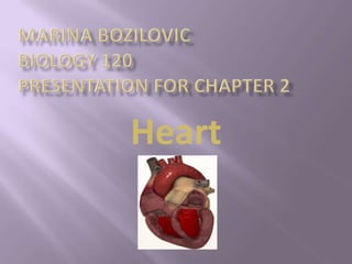
- 1. Heart
- 2. 1. Generating blood pressure o Required for blood flow through the blood vessels 2. Routing blood o Two pumps, moving blood through the pulmonary and systematic circulations 3. Regulating blood supply o Adjusts blood flow by changing the rate and force of heart contractions as needed
- 3. Pulmonary circulation o The flow of blood from the heart through the lungs back to the heart o Picks up oxygen and releases carbon dioxide in the lungs System circulation o The flow of blood from the heart through the body back to the heart o Delivers oxygen and picks up carbon dioxide in the body’s tissues
- 4. Heart is located just behind and slightly left of the breastbone, in ventral thoracic cavity which is separated from abdominal cavity by the diaphragm. Approximately the size of the fist, weighs about 10.5oz The shape of the heart is like a cone, or an upside-down pear.
- 6. Heart Chambers o Heart is divided into four chambers: two atria and two ventricles Heart Layers o The wall of the heart is composed of three layers: endocardium, myocardium and epicardium Heart Valves o Valves act as restraining gates to control direction of blood flow : tricuspid, pulmonary, mitral and aortic valve
- 7. There are 2 atria or upper chambers, and 2 ventricles or lower chambers Chambers are divided into right and left sides by walls called interatrial septum and interventricular septum
- 8. Endocardium is the inner layer of the heart lining the heart chambers. Endocardium lines the inner cavities of the heart, covers heart valves and is continuous with the inner lining of the blood vessels. Myocardium is muscular middle layer of the wall of the heart. Myocardium stimulates heart contractions to pump blood from the ventricles and relaxes the heart to allow the atria to receive blood. Epicardium is outer layer of the wall of the heart. Provides an outer protective layer for the heart.
- 9. Tricuspid valve controls the opening between the right atrium and the right ventricle. Once the blood enters the right ventricle it cannot go back up into the atrium again. Pulmonary valve prevents blood that has been ejected into the pulmonary artery from returning to the right ventricle as it relaxes. Mitral valve has two cusps. The blood flows through this atrioventricular valve to the left ventricle and cannot go back up into the left atrium. Aortic valve is located between the left ventricle and the aorta. Blood leaves the left ventricle through this valve and cannot return to the left ventricle.
- 11. Blood from the body flows through the right atrium into the right ventricle and than to the lungs. Blood returns from the lungs to the left atrium, enters the left ventricle, and is pumped back to the body. This is the video where we can see the blood flow through the heart: route of blood flow through the heart
- 12. Heart is regulated by autonomic nervous system. 1. Sinoatrical node(pacemaker) – contracts and generates nerve impulses that travel throughout the heart wall causing both atria to contract. 2. Atrioventricular bundle – carries impulses down the septum to the ventricles via the Purkinje fibers. 3. Atrioventricular node – delays cardiac impulses from the sinoatrial node to allow the atria to contract and empty their contents first. 4. Purkinje fibers – relays cardiac impulses to the ventricular cells causing the ventricles to contract.
- 14. 1. Arteries are the large, thick-walled vessels that carry the blood away from the heart. 2. Capillaries are a network of tiny blood vessels referred to as a capillary bed. Arterial blood flows into a capillary bed, and venous blood flows back out. 3. Veins carry blood back to the heart. Veins have valves that allow the blood to move only toward the heart. THERE ARE THREE TYPES OF BLOOD VESSELS:
- 15. Myocardial infraction – also known as a heart attack. Myocarditis – inflammation of the muscle layer of the heart wall. Hypertension – blood pressure above the normal range. Coronary artery disease (CAD) – insufficient blood supply to the heart muscle due to an obstruction of one or more coronary arteries. Thrombus – blood clot forming within a blood vessel.
- 16. arteriole – a small artery cardiac – pertaining to the heart interventricular – pertaining to between the ventricles interatrial – pertaining to between the atria myocardial – pertaining to heart muscle valvular – pertaining to a valve ventricular – pertaining to a ventricle venule – a small vein