VETERINARIA Oftalmologia exophtalmos
•
2 gostaram•2,224 visualizações

El documento describe la exoftalmia, o protrusión del ojo, causada por enfermedades retrobulbares. Las causas más comunes son el absceso/celulitis retrobulbar y la neoplasia retrobulbar. Estos comparten signos clínicos como la protrusión del tercer párpado, hinchazón periorbital y resistencia a la retropulsión del ojo. El absceso retrobulbar se caracteriza además por un inicio agudo y dolor severo, y a menudo requiere sedación para examinar al paciente. El tratamiento del

Recomendados
Mais conteúdo relacionado
Mais procurados
Mais procurados (20)
Semelhante a VETERINARIA Oftalmologia exophtalmos
Semelhante a VETERINARIA Oftalmologia exophtalmos (20)
Mais de David Mendez Rascon
Mais de David Mendez Rascon (18)
VETERINARIA Oftalmologia exophtalmos
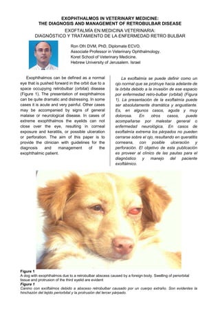
- 1. EXOPHTHALMOS IN VETERINARY MEDICINE: THE DIAGNOSIS AND MANAGEMENT OF RETROBULBAR DISEASE EXOFTALMÍA EN MEDICINA VETERINARIA: DIAGNÓSTICO Y TRATAMIENTO DE LA ENFERMEDAD RETRO BULBAR Ron Ofri DVM, PhD, Diplomate ECVO. Associate Professor in Veterinary Ophthalmology. Koret School of Veterinary Medicine. Hebrew University of Jerusalem. Israel Exophthalmos can be defined as a normal eye that is pushed forward in the orbit due to a space occupying retrobulbar (orbital) disease (Figure 1). The presentation of exophthalmos can be quite dramatic and distressing. In some cases it is acute and very painful. Other cases may be accompanied by signs of general malaise or neurological disease. In cases of extreme exophthalmos the eyelids can not close over the eye, resulting in corneal exposure and keratitis, or possible ulceration or perforation. The aim of this paper is to provide the clinician with guidelines for the diagnosis and management of the exophthalmic patient. La exoftalmía se puede definir como un ojo normal que se protruye hacia adelante de la órbita debido a la invasión de ese espacio por enfermedad retro-bulbar (orbital) (Figura 1). La presentación de la exoftalmía puede ser absolutamente dramática y angustiante. Es, en algunos casos, aguda y muy dolorosa. En otros casos, puede acompañarse por malestar general o enfermedad neurológica. En casos de exoftalmía extrema los párpados no pueden cerrarse sobre el ojo, resultando en queratitis corneana, con posible ulceración y perforación. El objetivo de esta publicación es proveer al clínico de las pautas para el diagnóstico y manejo del paciente exoftálmico. Figure 1 A dog with exophthalmos due to a retrobulbar abscess caused by a foreign body. Swelling of periorbital tissue and protrusion of the third eyelid are evident Figura 1 Canino con exoftalmos debido a absceso retrobulbar causado por un cuerpo extraño. Son evidentes la hinchazón del tejido periorbital y la protrusión del tercer párpado.
- 2. DIFFERENTIAL DIAGNOSIS The most common causes of exophthalmos are retrobulbar abscess/cellulitis and retrobulbar neoplasia. Together with retrobulbar traumatic hematoma, they account for more than 90 percent of all exophthalmos cases. There are a number of other causes for the presentation, including myositis of the extraocular muscles, and salivary cysts/mucoceles. However, as these are less common, their workup and management will not be discussed in this paper. It is worthwhile noting that some clinicians may be confused between exophthalmos and buphthalmos. The latter is a globe that is enlarged due to elevation in intraocular pressure (i.e., glaucoma), but one that is situated normally in the orbit. However, despite differences in globe size, globe position and causes, clinicians may find it difficult to differentiate between the two syndromes, as both present with an asymmetric appearance of the globe. A number of tests can be used to distinguish exophthalmos from buphthalmos. The easiest is probably to evaluate the patient’s orbit from the side or above to detect the forward displacement of the globe in exophthalmos. Estimating the corneal curvature, which is increased in buphthalmos, is also an easily- performed test and a reliable indicator. However, ultimate differentiation between exophthalmos and buphthalmos relies on measurement of intraocular pressure, or ultrasound/CT imaging of a retrobulbar disease process. DIAGNÓSTICO DIFERENCIAL Las causas más comunes de la exoftalmía son absceso/inflamación y neoplasia retro bulbar. Junto con el hematoma traumático retro bulbar, explican el 90 por ciento de todos los casos de exoftalmía. Hay un número de otras causas para la exoftalmia, incluyendo la miositis de los músculos extra oculares y quistes/mucoceles salivales. Sin embargo, como éstos son menos comunes, no los trataremos en este informe. Vale la pena observar que algunos clínicos pueden estar confundidos entre exoftalmía y buftalmos. Este último representa un globo ocular agrandado debido a la elevación en la presión intraocular (es decir glaucoma), pero que normalmente se sitúa dentro de la órbita. Sin embargo, a pesar de las diferencias de tamaño del globo, la posición y las causas, algunos clínicos pueden encontrar difícil distinguir entre los dos síndromes, siendo que ambos se presentan en un aspecto asimétrico del globo. Se pueden utilizar un número de pruebas para distinguir exoftalmía de buftalmia. La más fácil es, probablemente, evaluar la órbita del paciente del lado lateral o superior para detectar la dislocación delantera del globo en la exoftalmía. El cálculo de la curvatura corneana, que aumenta en la buftalmia, es también una prueba fácil de realizar y un indicador confiable. Sin embargo, la diferenciación definitiva entre la exoftalmía y el buftalmos es la medición de la presión intraocular, o la proyección de imagen de ultrasonido/TAC de un proceso de enfermedad retro bulbar. CLINICAL SIGNS ASSOCIATED WITH EXOPHTHALMOS As noted, the two most common causes of exophthalmos are retrobulbar abscess/cellulitis and retrobulbar neoplasia. Each of these disease processes has its unique clinical signs that aid in reaching a final diagnosis (see below). However, they both share a number of clinical signs, in addition to exophthalmos, that are common to both diseases: 1. Protrusion of 3rd eyelid: Due to the presence of a space occupying lesion in the orbit, there is forward displacement of the eye (exophthalmos). At the same time, the third eyelid is also displaced from its normal position. It becomes elevated, and protrudes to cover part of the eye (Figures 1 and 2). 2. There is swelling of orbital and SIGNOS CLÍNICOS ASOCIADOS A EXOFTALMÍA Según lo observado, las dos causas más comunes de la exoftalmía son absceso/inflamación y neoplasia retro bulbar. Cada uno de estos procesos tiene sus signos clínicos característicos que ayudan a alcanzar una diagnostico definitivo (ver abajo). Sin embargo, ambas comparten un número de signos clínicos que les son comunes a ellas: 1. Protrusión del 3º párpado: Debido a la presencia de una lesión que ocupa espacio en la órbita, hay dislocación delantera del ojo (exoftalmía). Al mismo tiempo, el tercer párpado también se desplaza de su posición normal. Se eleva, y protruye para cubrir parte del ojo (Figuras 1 y 2). 2. Se presenta hinchazón del tejido orbital y
- 3. periorbital tissue, as the disease process disrupts venous and lymphatic circulation in the orbit and surrounding tissue. Marked conjunctival edema (chemosis) and hyperemia are also common signs (Figures 1 and 2). 3. Resistance to retropulsion: A retropulsion test is conducted by placing your fingers on the eyelids, and applying gentle pressure to try and push the eye back into the orbit (Figure 3). In the case of a space occupying retrobulbar disease, you will feel resistance to the retropulsion. This test can also help you differentiate exophthalmos from buphthalmos as you should be able to push a buphthalmic eye into the orbit without resistance. In this context it is worthwhile noting that the two aforementioned signs also help differentiating buphthalmos from exophthalmos, as glaucoma does not cause protrusion of the third eyelid or periorbital tissue swelling. 4. Unilateral presentation: There are reports of bilateral, retrobulbar abscess or neoplasia. However, these are rare presentations, and the diseases are nearly always unilateral. 5. Signs of systemic disease. A retrobulbar abscess is very painful, and characterized by loss of appetite. Depending on the duration and extent of the abscess, pyrexia, leukocytosis, neutrophilia and other signs of sepsis may be present. A retrobulbar tumor may likewise present with signs of systemic illness if it is a metastasis from elsewhere in the body, or with signs of neurological disease if it is a primary tumor that extended into the brain. 6. Vision in the eye may be affected if the disease process involves the optic nerve directly (e.g., tumor) or indirectly (e.g., compression). peri orbitario, pues el proceso interrumpe la circulación venosa y linfática en la órbita y el tejido circundante. El edema conjuntival marcado (quemosis) y la hiperemia son también signos comunes (Figuras 1 y 2). 3. Resistencia a la retropulsión: Se efectúa una prueba de retropulsión colocando los dedos en los párpados y aplicando presión suave para intentar empujar el ojo nuevamente dentro de la órbita (Figura 3). En el caso de presencia de alguna patología que ocupe el espacio retrobulbar, usted sentirá resistencia a la retropulsión. Esta prueba puede también ayudarle a distinguir exoftalmía de buftalmia, debido a que debería poder empujar un ojo buftálmico dentro de la órbita sin resistencia. En este contexto, vale la pena observar que los dos signos mencionados anteriormente también ayudan a distinguir buftalmos de la exoftalmía, pues el glaucoma no causa protrusión del tercer párpado o la hinchazón del tejido periorbitario. 4. Presentación unilateral: Existen informes sobre presencia de abscesos o neoplasias retrobulbares bilaterales. Sin embargo, éstas son presentaciones raras, y las enfermedades son casi siempre unilaterales. 5. Signos de enfermedad sistémica: Un absceso retro-bulbar es muy doloroso, y caracterizado por la pérdida de apetito. Dependiendo de la duración y del grado del absceso, la pirexia, la leucocitosis, la neutrofilia y otros signos de sepsis pueden estar presentes. Un tumor retro-bulbar puede presentarse además con signos de enfermedad sistémica si es una metástasis de otra parte del cuerpo, o con signos de enfermedad neurológica si es un tumor primario que se extendió al cerebro. 6. La visión en el ojo puede ser afectada si el proceso de la enfermedad implica al nervio óptico directamente (ej. tumor) o indirectamente (ej. compresión).
- 4. Figure 2 Marked chemosis and third eyelid protrusion are 2 clinical signs of retrobulbar disease. Photo courtesy of DJ Maggs, UC Davis Ophthalmology Service Figura 2 La marcada quemosis y la protrusión del tercer párpado son dos signos clínicos de enfermedad retrobulbar. Foto cortesía de DJ Maggs, UC Davis Ophthalmology Service Figure 3 Retropulsion test in a Gordon setter. The examiner is using her fingers to gently push the eyes into the orbit. In cases of retrobulbar disease, resistance to retropulsion will be encountered. Figura 3 Prueba de Retropulsión en un Gordon setter. El examinador usa sus dedos para empujar suavemente los ojos dentro de la órbita. En casos de enfermedad retrobulbar, se encontrará resistencia a la retropulsión.
- 5. RETROBULBAR CELLULITIS/ABSCESS PATHOGENESIS It is believed that most common cause of retrobulbar abscess is foreign body penetration through the oral cavity, as the animal chews sticks or other sharp objects. Hematogenous spread of micro-organisms from other infected organs, or from adjacent tooth roots or nasal sinuses, is also believed to be a common cause. However, in many cases the cause remains unknown. Pasteurella and Aspegillus spp are commonly isolated from dogs and cats, respectively. But once again, in many cases no organism is isolated. The disease process will usually begin as orbital cellulitis, which is characterized by clinical signs that are less noticeable. After localization occurs and an abscess forms, clinical signs, including pain, become much more significant. CLINICAL SIGNS & DIAGNOSIS OF RETROBULBAR ABSCESS In addition to the aforementioned clinical signs that generally characterize exophthalmos, retrobulbar abscess has a few clinical signs that are almost pathognomonic. The disease is characterized by acute onset and by severe pain. The pain is caused when the condyle of the mandible presses on the abscess whenever the animal opens its mouth. This leads to refusal to eat and great resistance to opening the mouth for examination. It is often necessary to sedate the animal in order to open its mouth. Once the mouth has been opened, it is often possible to see a red swelling, or even an open draining tract, in the oral mucosa, behind the last upper molar tooth (Figure 4). If no gross lesion is visible in the oral cavity, it is possible to use imaging techniques, such as ultrasound or CT (Figure 5), to image the retrobulbar space. This may also demonstrate foreign bodies, or allow to perform guided fine needle aspirations for cytological diagnosis. TREATMENT & MANAGEMENT OF RETROBULBAR ABSCESS Treatment of a retrobulbar abscess requires general anesthesia. This is because the patient must be intubated to avoid aspiration of exudate when the abscess is drained. Make an incision in the mucosa behind the last upper molar tooth (unless an open draining tract is present), and slowly inset a pair of INFLAMACIÓN/ABSCESO RETRO- BULBAR PATOGÉNESIS Se cree que la causa más común de absceso retro-bulbar es la penetración de un cuerpo extraño a través de la cavidad bucal, mientras el animal mastica palillos u otros objetos punzantes. La expansión hematógena de microorganismos desde otros órganos infectados, o de raíces de dientes adyacentes o de senos nasales, también pueden ser una causa común. Sin embargo, en muchos casos la causa sigue siendo desconocida. Pasteurella y Aspegillus spp se aíslan comúnmente de perros y gatos, respectivamente. Pero, nuevamente, en muchos casos no se aísla ningún organismo. La enfermedad comenzará generalmente como inflamación orbital, caracterizada por signos clínicos menos evidentes. Luego de ocurrida la localización y la formación del absceso, los signos clínicos, incluyendo dolor, llegan a ser mucho más significativos. SIGNOS CLÍNICOS Y DIAGNÓSTICO DEL ABSCESO RETRO-BULBAR Además de los signos clínicos mencionados anteriormente que caracterizan generalmente a la exoftalmía, el absceso retro bulbar tiene algunos signos que son casi patognomónicos. La enfermedad se caracteriza por inicio agudo y por dolor severo. El dolor se produce cuando el cóndilo de la mandíbula presiona el absceso siempre que el animal abre su boca. Esto lleva a la negación para comer y a la gran resistencia a abrir la boca para la examinación. Es a menudo necesario sedar al animal para abrir su boca. Una vez que se ha abierto la boca, es posible ver una hinchazón roja, y hasta una fístula de drenaje abierta, en la mucosa oral detrás del último molar superior (Figura 4). Si no hay lesión gruesa visible en la cavidad bucal, es posible utilizar técnicas de proyección de imagen, tales como ultrasonido o TAC (Figura 5), para ver el espacio retro bulbar. Esto puede también demostrar cuerpos extraños, o permitir realizar una aspiración con aguja fina guiada para diagnóstico citológico. TRATAMIENTO Y MANEJO DEL ABSCESO RETRO-BULBAR El tratamiento de un absceso retro bulbar
- 6. curved hemostats. These are used to blindly explore the orbit and open pockets of exudate (Figure 6). One must never close the hemostats while they are in the orbit, as this could lead to crushing of the orbital vessels or optic nerve and blindness. Instead, the hemostats are inserted closed into the tract; they are opened in the orbit, withdrawn while open, closed in the oral cavity and re-inserted into the orbit in the closed position. If a pocket of exudate is encountered, copious amounts of exudate will flow out. This can be collected for cytology, and culture & sensitivity. In cases of retrobulbar cellulitis, no massive drainage of exudate will be seen. However, the very act of establishing a draining tract is usually sufficient to achieve a cure. After creating a draining tract, the clinician should gently flush the orbit with saline and antibiotics. The wound is not sutured. Systemic antibiotics are administered for 10- 14 days, and the animal fed soft food. Hot packs, and lubrication of the cornea to prevent desiccation, should be considered. However, dramatic, and most rewarding, improvement is usually observed within 1-2 days. If the animal does not respond to therapy, or in case of recurrence, surgical exploration for a foreign body may be required. requiere anestesia general. Esto es porque el paciente debe intubarse para evitar la aspiración del exudado cuando se drena el absceso. Haga una incisión en la mucosa detrás del último molar superior (a menos que ya exista un drenaje abierto) y lentamente, inserte una pinza hemostática curva. Ésta se utiliza para explorar a ciegas la órbita y para romper el absceso y liberar el exudado (Figura 6). Uno nunca debe cerrar la pinza hemostática mientras está en la órbita, pues podría llevar a la ruptura de los vasos orbitales o del nervio óptico y producir ceguera. En su lugar, la pinza hemostática se inserta cerrada en la zona; se abre en la órbita, se retira abierta, se cierra en la cavidad bucal y se reinserta cerrada en la órbita. Si se encuentra el absceso el exudado fluirá en cantidades copiosas. Esto se puede recolectar para el examen citológico, cultivo y antibiograma. En casos de inflamación retro bulbar, no se verá ningún drenaje masivo del exudado. Sin embargo, el mismo acto de establecer una zona de drenaje es generalmente suficiente para alcanzar una curación. Después de crear una zona de drenaje, el clínico debe limpiar la órbita con un chorro suave de solución fisiológica y aplicar antibióticos. La herida no se sutura. Los antibióticos sistémicos se administran por 10-14 días, y el alimento debe ser de consistencia blanda. Aplicar fomentos tibios, y lubricar la córnea para prevenir la desecación. A pesar de ser un cuadro dramático la mejoría se observa generalmente en el plazo de 1-2 días. Si el animal no responde a la terapia, o en caso de recurrencia, puede ser requerida la exploración quirúrgica en busca de un cuerpo extraño.
- 7. Figure 4 A draining tract is evident behind the last upper molar tooth of the dog shown in Figure 1. This finding is pathognopmonic to a retrobulbar abscess. The dog had to be anesthetized, as it it was in great pain and would not allow its mouth to be opened for examination Figura 4 Se evidencia un drenaje detrás del último molar del paciente mostrado en la Figura 1. Este hallazgo es patognomónico de absceso retrobulbar. El paciente debió ser anestesiado debido al intenso dolor y a que no permitía abrir su boca para la examinación. Figure 5 CT imaging is used to diagnose retrobulbar disease in this dog. In this case, a retrobulbar abscess was caused by a stick foreign body. Photo courtesy of DJ Maggs, UC Davis Ophthalmology Service. Figura 5. Imágenes de TAC se utilizan para diagnosticar enfermedad retrobulbar en el canino. En este caso, un absceso retrobulbar causado por un objeto extraño. Foto cortesía de DJ Maggs, UC Davis Ophthalmology Service.
- 8. Figure 6 Draining the retrobulbar abscess of the dog in Figures 1 & 5. The patient is intubated to prevent aspiration of draining exudate. Hemostats are used to explore the retrobulbar space and drain pockets of exudate. Closed hemostats may be opened in the retrobulbar space to increase drainage. However, open hemostats should not be closed in the retrobulbar space, as this may lead to crushing of the optic nerve. Instead, the hemostats are withdrawn open from the retrobulbar space , and then closed in the oral cavity Figura 6 Drenando el absceso retrobulbar del paciente de las figuras 1 y 5. El paciente es intubado para prevenir la aspiración del exudado drenado. Se utilizan hemostáticas para explorar el espacio retrobulbar y drenar el absceso. Se debe abrir la hemostática dentro del espacio retrobulbar para incrementar el drenaje. Sin embargo, la hemostática abierta no debe cerrarse en el espacio retrobulbar, debido a que podría causar el colapso del nervio óptico. En su lugar, la hemostática debe retraerse abierta del espacio retrobulbar y luego cerrarse en la cavidad oral. RETROBULBAR TUMOR As with any other organ, tumors in the orbit can be primary, or metastasis from nearby or distant regions. In dogs, most tumors are primary, with meningioma being the most commonly diagnosed tumor. In cats, a majority of retrobulbar tumors are secondary. In both species, however, the majority of tumors are malignant. CLINICAL SIGNS & DIAGNOSIS As noted, retrobulbar tumors share a number of signs with retrobulbar abscesses. However, in contrast to retrobulbar abscesses, retrobulbar tumors are usually very slowly progressive, and non painful (at least in the initial stages). Furthermore, patients with retrobulbar tumors are 10-11 years old, on average, significantly older than patients with retrobulbar abscesses. A retrobulbar tumor can cause deformation of the posterior part of the globe. This deformation can be visualized TUMOR RETROBULBAR Como con cualquier otro órgano, los tumores en la órbita pueden ser primarios, o ser metástasis de regiones próximas o distantes. En perros, la mayoría de los tumores son primarios, como el meningioma siendo el tumor más comúnmente diagnosticado. En gatos, la mayoría de los tumores retro-bulbares son secundarios. En ambas especies, sin embargo, la mayoría de los tumores son malignos. SIGNOS CLÍNICOS Y DIAGNÓSTICO Como se ha mencionado anteriormente, muchos tumores retro-bulbares comparten signología con los abscesos retro-bulbares. Sin embargo, en contraste con los abscesos, los tumores progresan muy lentamente y no son dolorosos (por lo menos en las etapas iniciales). Además, los pacientes con tumores retro-bulbares tienen 10-11 años de edad, en promedio,
- 9. ophthalmoscopically, or using an ultrasound. In addition to exophthalmos, the tumor can also cause deviation of the globe, with the direction of the deviation hinting at the location of the mass. However, ultimate localization relies on ultrasonography, CT or MRI imaging. The final diagnosis is usually made by guided fine needle aspiration and cytology. TREATMENT AND PROGNOSIS Solitary tumors discovered in early stages may be removed surgically. In such cases, the best surgical approach is usually oribitotmy, and it may be possible to preserve the globe and vision. Advanced cases may require radical orbitectomy, combined with radiation therapy and/or chemotherapy. However, most tumors are discovered in advanced stages, and due to their malignant nature they carry a very poor prognosis. One retrospective study reported a mean survival time of 1 month in cats and 10 months in dogs, with 35% of patients euthanized at the time of diagnosis. perceptiblemente más viejos que los pacientes con abscesos retro-bulbares. Un tumor retro-bulbar puede causar la deformación de la parte posterior del globo. Esta deformación se puede visualizar oftalmoscópicamente, o con ultrasonido. Además de exoftalmía, el tumor puede también causar la desviación del globo, con la dirección de la desviación haciendo alusión a la localización de la masa. Sin embargo, la localización final, más confiable, se obtiene por ultrasonografía, TAC o RM. El diagnóstico definitivo se realiza, generalmente, por aspiración con aguja fina y citología. TRATAMIENTO Y PRONÓSTICO: Los tumores solitarios descubiertos en estadios tempranos se pueden extirpar quirúrgicamente. En tales casos, el mejor abordaje quirúrgico es, generalmente, la orbitotomía, con posibilidad de preservar el globo y la visión. Los casos avanzados pueden requerir orbitectomía radical, combinado con radioterapia y/o quimioterapia. Sin embargo, la mayoría de los tumores se descubren en etapas avanzadas, y debido a su naturaleza maligna, tienen pronóstico muy malo. Un estudio retrospectivo reportó un tiempo de supervivencia promedio de 1 mes en gatos y de 10 meses en perros, con el 35% de pacientes eutanasiados al momento de realizado el diagnóstico. SUGGESTED READING / BIBLIOGRAFÍA SUGERIDA 1. Miller PE. Orbit. In: Maggs DJ, Miller PE, Ofri R. eds. Slatter's Fundamentals of Veterinary Ophthalmology, 4th edition. St Louis: Saunders Elsevier, 2007; 352-73. 2. Spiess BM. Diseases and surgery of the canine orbit. In: Gelatt KN. ed. Veterinary Ophthalmology, 4th ed. Ames, Iowa: Blackwell Publishing, 2007;539-62. 3. Boroffka SA et al. Assessment of ultrasonography and computed tomography for the evaluation of unilateral orbital disease in dogs. J Am Vet Med Assoc 2007; 230:671-80 4. Attali-Soussay K et al. Retrobulbar tumors in dogs and cats: 25 cases. Vet Ophthalmol. 2001;4:19-27 5. Hendrix DV et al. Diagnosis, treatment and outcome of orbital neoplasia in dogs: a retrospective study of 44 cases. J Small Anim Pract. 2000;41:105-8. 6. Mason DR et al. Ultrasonographic findings in 50 dogs with retrobulbar disease. J Am Anim Hosp Assoc. 2001;37:557-62. 7. O’Brien MG et al. Total and partial orbitectomy for thte treatment of periorbital tumors in 24 dogs and 6 cats: a retrospective study. Vet Surg 1996;25:471-9 Este envío es una atención de Laboratorio LOVE Sudamericana www.laboratoriolove.com.ar