SlideShare uma empresa Scribd logo
1 de 102
Gait disorders
Introduction
 The maintenance of an upright posture and the act
of walking are among the first, and ultimately most
complex, motor skills humans acquire.
 individual’s pattern of walking may be so distinctive
they can be recognized by the characteristics of
their gait or even the sound of their steps.
 Many diseases of the nervous system are identified
by the disturbances of gait and posture.
Anatomical aspects of gait
 2 basic systems responsible for equilibrium and
locomotion
1. Brainstem locomotor centers
 pedunculopontine &
 adjacent nuclei of midbrain
2. Spinal locomotor centers
 Additional level of supraspinal control is needed to
maintain bipedal stance and gait
1. Frontal cortex
2. Parietal cortex
3. cerebellum
Brainstem locomotor centers:
 Projects from the pontomedullary reticular formation, through
reticulospinal pathways into ventromedial spinal cord.
 Stimulation results in increase in axial and limb muscle tone
to assume an upright posture before stepping begins
Spinal locomotor centers:
 Assemblies of spinal interneurons
 Activate motor neurons of limb and trunk muscles in a
patterned and repetitive manner to drive stepping
movements
 propriospinal networks that link the trunk and limbs to
facilitate synergic coordinated limb and trunk movements of
Frontal cortex
prefrontal cortex
◦ via corticobulbar and corticostriatal tracts
◦ modulates midbrain and cerebellar locomotor
regions.
Frontal motor projections
 to the pontomedullary reticular formation
◦ innervate axial muscles modulate postural
responses associated with stepping
 to the spinal motor neurons,
◦ enables precision of foot movements.
Parietal cortex
 It integrates sensory inputs
 indicates position and orientation in space, the
relationship to gravitational forces,
Cerebellum
 Modulates the rate,rhythm,amplitude and force of
stepping
 contributes to medial brainstem efferent system
through projections from flocculonodular and
anterior lobes
◦ Controlls the equilibrium and truncal posture
PHYSIOLOGICAL ASPECTS OF GAIT
 Stability when standing is based on
◦ musculoskeletal linkages between the trunk and
legs
◦ neurological control detecting and correcting
body sway i.e, postural responses.
 Synergitic action axial and proximal limb muscle
and postural responses maintain static postural
control.
Postural reflexes:
2 types
◦ anticipatory postural reflexes:
 feed-forward
 occurring before limb movement.
◦ Reactive postural reflexes:
 feedback
 postural adjustments counteracting body
perturbations during movement.
◦ modified by voluntary control.
◦ generated by the integration of visual,
vestibular, and proprioceptive inputs in the
context of voluntary intent.
 Once the trunk is upright and stable, locomotion can
begin.
 The initiation of gait is heralded by a series of shifts in
the center of pressure beneath the feet.
◦ first posteriorly,
◦ then laterally toward the stepping foot
◦ finally toward the stance foot to allow the stepping
foot to swing forward
 This sequence is then followed by the stereotyped
stance, swing, and step phases of the gait cycle.
GAIT CYCLE
Period between two successive points at which the
heel of the same foot strikes the ground
Cadence:“the rhythm and number of steps per minute”
Gait
cycle
2 phases
8 events
Phases
stance
swing
Events
4 stance phase
events
4 swing phase
events
Stance phase Swing phase
Accounts for 60% gait
cycle.
Consists of 4 phases
 initial contact
 loading
 mid-stance
 terminal stance.
Accounts for 40% gait
cycle.
Consists of 4 phases
 Preswing
 Initial swing
 Mid swing
 Terminal swing
Events during stance phase
1. Heel strike:
 initiates gait cycle
 body’s center of gravity is at its lowest position
2. Foot flat :
 plantar surface of the foot touches the ground
3. Mid stance
 Swinging contra lateral foot passes the stance foot
 Center of gravity at its highest position
4. Heel off
 Heel looses contact with ground.
5. Toe off
 terminates the stance phase as foot leaves the ground
Events during swing phase
1. Acceleration:
 begins as soon as foot leaves the ground
 activation of hip flexors to accelerate leg forward
2. Mid swing:
 foot passes directly beneath the body
 coincident with mid stance of other foot
3. Deceleration:
 describes the action of muscles as they slow
 the leg and stabilize the foot in preparation for next
heel strike
Distance measures:
Stride length
◦ distance between heels from one heel strike to the next heel strike on
same side
◦ distance travelled by a person during a gait cycle
Step length
◦ length between heel strike of one foot to other
◦ Rt + Lt step length = stride length
Step width
◦ medio lateral distance between feet
 A typical adult walking parameters comfortably on a
level surface includes:
◦ velocity of about 80 m per minute,
◦ about 113 steps per minute,
◦ stride length of 1.41 m.
◦ About 60% of the gait cycle is spent in stance phase
◦ 40% in swing phase
◦ 10% in double limb support.
 The body's center of mass is located just anterior to
the S2 vertebral body.
Evaluation of gait
HISTORY
 A detailed history provides the first clues to diagnosis.
 note the circumstances in which walking difficulty
occurs.
 Ask for any associated symptoms, especially falls.
 Walking over uneven ground exacerbates most
walking difficulties.
 Often an individual is unaware of their gait
abnormality, and family or friends note altered
cadence, shuffling or slowness.
Weakness:
 weakness of certain muscle groups produces
characteristic difficulties during particular movements
of the gait cycle.
◦ weakness of ankle dorsiflexion - foot drop
◦ Weakness of knee extension - buckling attacks
◦ Weakness of ankle plantar flexion - shallow stepped
gait.
◦
◦ proximal muscle weakness
- Difficulty in climbing stairs or rising from a
seated position
 Catching or scraping a toe on the ground and a
Slowness and Stiffness:
 Slowness of gait is normal reaction when walking
on slippery surfaces.
 Similarly, when the balance was threatened any
musculoskeletal or neurological disorder patient
will have slowness of gait.
 In Parkinson disease (PD) and other basal ganglia
diseases, slowness of walking is due to shuffling
with short, shallow steps.
 Difficulty rising from a chair or turning in bed may
be clues to loss of truncal mobility in diffuse
cerebro vascular disease, hydrocephalus, and
basal ganglia diseases.
 Complaints of stiffness, heaviness in the limbs are
the symptoms of a spasticity.
 It is important to remember that leg muscle tone in
some upper motor neuron syndromes and dystonia
may be normal when the patient is examined in the
supine position but is increased during walking.
 In childhood, an action dystonia of the foot is a
common initial symptom, walking on the toes only
becoming evident after walking or running.
Imbalance:
 poor balance and unsteadiness are cardinal features
of cerebellar ataxia and sensory ataxia.
 cerebellar gait ataxia complains of
◦ Unsteadiness and inability to walk in a straight line
and near falls.
◦ Turning and suddenly changing direction results in
veering to one side.
◦ Symptoms are exacerbated by an uneven support
surface.
Sensory ataxia:
◦ presents with unsteadiness when walking in the
dark.
◦ Patients complain of being uncertain of the exact
position of their feet when walking.
◦ impression of walking on a spongy surface or cotton
wool.
Acute vestibulopathy:
◦ Is associated with vertigo and severe imbalance.
Chronic vestibular dysfunction
◦ often causes veering and problems in an
environment with many moving objects.
 A wide-based unsteady gait also is a feature of frontal
lobe diseases
◦ normal pressure hydrocephalus,
◦ diffuse small vessel ischemia of frontal sub cortical
white matter.
 Imbalance in sub cortical cerebro vascular disease
commonly manifests when
◦ turning while walking,
◦ stepping backwards,
◦ bending over to pick up something.
Falls:
 Toppling Falls
◦ muscle tone is retained
◦ falling like a tree trunk
◦ due to impaired static and dynamic postural
responses that control body equilibrium
 collapsing falls
◦ muscle tone is lost
◦ imply a loss of consciousness characteristic of
syncope or seizures
 Tripping falls:
◦ many people attribute falls to tripping when in
fact tripping did not occur.
◦ Tripping may be due to foot drop or shallow
steps
◦ also be a consequence of secondary to
inattention, dementia, or poor vision.
 Proximal muscle weakness may result in the legs
giving way and falls.
 Unsteadiness and poor balance in an ataxic
syndrome may lead to falls.
 falls associated with postural adjustments, or falls
occurring when performing multiple tasks suggest
an impairment of postural responses.
 Backward falls in early stages
◦ Feature of an akinetic-rigid syndromes
◦ Favors a diagnosis of MSA or PSP rather than
PD
◦ Falls do occur in PD but are late feature.
• In PD falls are usually forward and may also occur
while turning and changing the posture.
 Collapsing falls related to orthostatic hypotension, a
Sensory Symptoms and Pain:
 distribution of sensory complaints provides a clue
to the site of the lesion.
◦ Ex: Distal symmetrical paresthesias of limbs
S/O peripheral neuropathy.
 Skeletal pain due to degenerative joint disease is
aggravated by movement of the affected joints.
 The normal pattern of walking is frequently
modified by joint disease.
 strategies to minimize pain by avoiding full weight
bearing on the affected limb will produce limping.
Urinary Incontinence:
 Urinary urgency and urge incontinence seen in
◦ spinal cord lesion
◦ parkinsonism
◦ subcortical white-matter ischemia.
 frontal lobe tumors, frontal lobe infarction, and
hydrocephalus also produce urinary
incontinence.
Cognitive Changes:
 Slowing of gait may be a marker of impending
cognitive impairment and dementia.
 executive dysfunction including inattention may
produce falls.
 disinhibition and impulsivity are associated with
reckless gait problems and falls.
EXAMINATION OF POSTURE AND GAIT
 First observe the overall pattern of limb and body
movement during walking.
 Normal walking progresses in a smooth and
effortless manner.
 The truncal posture is upright, and the legs swing in a
fluid motion with a regular stride length.
 Synergistic head, trunk, and upper-limb movement
flow with each step.
 After the overall walking pattern is observed, the
specific aspects of posture and gait should be
examined.
specific aspects of posture and gait includes:
1. Arising to stand from seated position
2. Stance and posture
3. Walking
4. Turning while walking
5. Other maneuvers
◦ Tandem walking
◦ Walking backwards
◦ Running
◦ Walking on toes, heels
Arising from Sitting:
 informs about
◦ pelvic girdle strength,
◦ control of truncal movement,
◦ coordination, and balance.
 proximal muscle weakness:
Inability to rise when the feet are appropriately
placed under the body.
 incoordination or imbalance:
An abnormally wide stance base when
standing.
 Frontal lobe diseases:
Feet are not positioned directly under the
body
Stance:
 Stance base = the distance between the feet
 gives an indication of balance.
 Wide stance base indicates poor balance.
 Wide-based gaits are
◦ typical of cerebellar or sensory ataxia
◦ diffuse cerebro vascular disease
◦ frontal lobe lesions.
 Eversion of the feet to increase stability seen in
diffuse cerebrovascular disease.
 In mild ataxia, a widened base may only be evident
with turning.
 Tandem (heel to toe) walking is a good objective
measure of walking stability.
Trunk Posture:
 Flexion of the trunk and a stooped posture - PD.
 Upright posture with neck and trunk extension- PSP.
 Slight flexion at the hips to lower the trunk
- feature of cautious gait.
 Neck flexion – MND
Myasthenia gravis
MSA
Parkinsonism
 Tilt of the trunk to one side - dystonia.
 exaggerated lumbar lordosis
◦ stiff person syndrome[d/t Paraspinal muscle spasm]
◦ Proximal myopaties.
Truncal tilt away from the side of the lesion
◦ acute vascular lesions of the thalamus( postero
lateral) and basal ganglia.
Truncal tilt away from the side of the lesion
◦ Acute vestibular imbalance in the lateral medullary
syndrome
Truncal flexion (camptocormia)
◦ paraspinal myopathies that produce weakness of
trunk extension.
 restricted range of spinal movement and persistence
of the abnormal spinal posture when supine or
during sleep are useful pointers toward a bony spinal
deformity.
Postural Responses:
1.Reactive postural responses
2. anticipatory postural responses.
3.protective postural responses.
.
pull test
Reactive postural responses
are examined by “the pull
test”.
 Examiner sharply pulls the
upper trunk backward or
forward while the patient is
standing.
 The pull should be sufficient
to require the patient to
step to regain their balance.
 The examiner must be
prepared to prevent the
patient from falling.
 A few short, shuffling steps
backward or an impending
fall backward or forward
suggests impairment of
righting reactions
 Falls after postural changes such as arising from a
chair or turning while walking suggest impaired
anticipatory postural responses.
 Falls without rescue arm movements or stepping
movements to break the fall indicate loss of
protective postural responses.
 Injuries sustained during falls provide a clue to the
loss of postural responses
 A tendency to fall backward spontaneously is a
sign of impaired postural reflexes in PSP and gait
disorders associated with frontal lobes
Romberg test
 Patients with proprioceptive loss depends on visual
cues.
 the removal of vision greatly exaggerates the ataxia.
 This is the basis of the Romberg test.
 In which eye closure leads to a dramatic increase in
unsteadiness and even falls in the patient with sensory
ataxia.
 When performing the Romberg test, it is important the
patient is standing comfortably before eye closure and
to remember that a modest increase in body sway is a
normal response to eye closure.
Walking:
Initiation of Gait:
start hesitation – difficulty in gait initiation.
slipping clutch phenomenon
◦ few short, shuffling steps on the spot without
forward progress.
magnetic foot phenomenon.
◦ complete immobility with the feet seemingly glued
to the floor
 Patients may make exaggerated upper-body
movements or alter the step pattern.
 Start hesitation seen in
◦ frontal lobe disease,
◦ diffuse cerebrovascular disease,
◦ hydrocephalus.
◦ gait ignition failure.
◦ PD
 Isolated start hesitation is seen in the syndrome of
gait ignition failure.
Stepping:
 length and trajectory of each step and the rhythm of
stepping should be noted.
 Short, regular, shallow steps or shuffling and a slow gait
-
akinetic-rigid
syndromes.
 Freezing typically occurs when turning, when walking
has been interrupted by an obstacle.
 Festination (increasingly rapid, small steps) is common
in PD but rare in other akinetic-rigid syndromes.
 Unilateral loss of synergistic arm swing while walking
1. valuable sign of early PD
2. Unilateral UMN lesion
3. acute cerebellar syndrome
Turning:
 Requires more balance more than walking in a straight
line.
 Slowing on turns may be the first abnormality in walking in
PD
 Multiple steps on turning
◦ PD
More Challenging Tests of Walking:
Tandem walking:
 Subtle degrees of cerebellar ataxia unmasked by
tandem walking.
Walking on toes and heels:
 may bring out abnormal movements as well as
deficits in the strength of dorsiflexion and plantar
flexion of the ankle.
Walking backward:
 will sometimes reduce or abolish the dystonic foot
posturing observed walking forward in action dystonia
of the foot.
Running:
 examine the patient running to identify an action
dystonia of the legs in early stages.
Associated and Synergistic Limb Movements While
Walking:
Dystonic posturing of an arm or leg
◦ dystonia,
◦ parkinsonism,
◦ old hemiparesis.
Choreiform movements are more prominent during
walking than at rest in
◦ most chorea syndromes,
◦ levodopa-induced dyskinesia,
◦ tardive dyskinesia.
Parkinsonian tremor of the dependent upper limb is
often observed while walking.
MOTOR AND SENSORY EXAMINATION:
 motor and sensory function in the limbs is examined
with the patient sitting or supine.
 The size and length of the limbs should be measured
in any child presenting with a limp.
 The spinal column should be inspected for scoliosis,
and the lumbar region for skin defects or hairy
patches indicative of spinal dysraphism.
DISCREPANCIES ON EXAMINATION OF GAIT:
 minimal abnormal signs on physical examination of
the recumbent patient, in contrast to the observed
difficulty when walking.
Cerebellar gait ataxia caused by a vermis lesion:
◦ perform the heel-to-shin test normally when
supine but is ataxic when walking.
Action dystonia:
◦ Abnormal posturing of the feet only when
walking.
◦ A dystonic gait may be evident only when
running or walking forward but not when walking
backward.
Frontal lobe lesion, hydrocephalus,or diffuse
cerebrovascular disease :
◦ Patient totally unable to walk when standing.
◦ Gegenhalten (paratonia), with or without brisk
tendon reflexes, may be the only abnormal sign in
the recumbent patient
◦ Such patients perform the heel-shin test and make
bicycling movements of their legs normally when
lying on a bed.
Leg tremor of orthostatic tremor
◦ only appears during weight bearing, especially
when standing still.
GAIT DISORDERS
CLASSIFICATION OF GAIT PATTERNS:
 A scheme based on Hughlings Jackson’s three orders
of neurological function.
 Lower Level Gait Disorders (simplest)
 Middle Level Gait Disorders
 Higher Level Gait Disorders
(complex, integrative )
 Each level contributes sensory and motor function,
but that of higher centers is more complex and
dispersed within the nervous system.
Lower Level Gait Disorders
 manifest physical signs such as weakness or
sensory loss.
 are due to diseases of the muscle and peripheral
nerves that produce muscle weakness.
 Lower level sensory gait disorders follow loss of
one of the three basic senses important for gait and
balance:
◦ proprioception
◦ vision
◦ vestibular sensation.
Low level (sensory and motor) gait disorders:
1. Waddling gait - proximal weakness (myopathy)
2. high stepping gait - distal weakness (neuropathy)
3. Sensory ataxia – proprioception loss.
4. Vestibular ataxic gait -acute peripheral vestibulopathy
5. Gait difficulty due to acute visual impairment
Myopathic Weakness and Gait:
 Weakness of proximal leg and hip-girdle muscles
 interferes with stabilizing the pelvis and legs on the trunk.
 produces exaggerated rotation of the pelvis with each step
and a
waddling gait.
 The hips are slightly flexed (d/t hip extensor weakness).
 exaggerated lumbar lordosis.
 Gower’s sign positive
 myopathy is the most common cause of proximal muscle
weakness,
 neurogenic weakness of proximal muscles can also
Neurogenic Weakness and Gait:
 Muscle weakness of peripheral nerve origin.
 typically affects distal leg muscles
 results in a steppage gait.
 The patient lifts the leg and foot high above
the ground with each step because of
weakness of ankle dorsiflexion and foot drop.
 unilateral foot drop:
◦ common peroneal or sciatic nerve palsy or
an L5 radiculopathy
 Less common is foot drop caused by
myopathic weakness, as in the
scapuloperoneal syndromes.
 Weakness of ankle plantar flexion - shallow
stepped gait.
 A femoral neuropathy as in diabetes mellitus:
◦ produces weakness of knee extension and
buckling of the knee when walking or standing.
 This may first be evident when walking down stairs.
 similar weakness:
◦ Progressive muscular atrophy in MND
◦ quadriceps myopathy
◦ inclusion body myositis
Sensory Ataxia:
 wide stance base
 advance cautiously
 taking slow steps under visual guidance.
 the feet are thrust forward with variable direction and
height.
 The sole of the foot strikes the floor forcibly with a
slapping sound - slapping gait.
 Romberg test positive.
 Walking on uneven surfaces is particularly difficult.
 Results from
◦ peripheral neuropathies,
◦ posterior root or dorsal root ganglionopathies,
◦ dorsal column lesions.
Vestibular Imbalance, Vertigo, and Gait:
 Acute peripheral vestibulopathy
◦ produce veering to the side of the lesion.
◦ Veering less prominent while running than walking.
◦ Patients prefer to lie stand still.
 In chronic vestibular failure,
◦ gait may be normal,
◦ unsteadiness can be unmasked during eye closure
and rotation of the head from side to side while
walking.
Middle-Level Gait Disorders
 Related to impaired modulation of force generated by
the lower level motor system
include:
(1) spastic gait from corticospinal tract lesions
(2) ataxic gait arising from disturbances of the
cerebellum and its connections.
(3) hypokinetic gaits associated with parkinsonism.
(4) hyperkinetic gaits associated with chorea,dystonia,
and
Spastic Gait:
Spasticity of the arm and leg on one side:
 The leg is slightly flexed at the hip and
extended at the knee, with plantar flexion
and inversion of the foot.
 During the swing phase
◦ lateral flexion of the trunk toward the
unaffected side.
◦ circumduction of the extended paretic
leg.
◦ dragging the foot or catching the toe on
the ground.
◦ Minimal associated arm swing occurs on
the affected side.
 Muscle tone was increased on effected
side along with brisk DTR and extensor
plantar response.
Spasticity of both legs:
 legs are stiffly extended at the knees,
plantar flexed at the ankles, and
slightly flexed at the hips.
 Both legs circumduct, and the toes of
the plantar flexed feet catch on the
floor with each step.
 There is a tendency to adduct the
legs, particularly when the disorder
begins in childhood. [scissoring gait]
 Occasionally, bilateral leg dystonia
(dystonic paraparesis) mimics a
spastic paraparesis.
 This typically occurs in doparesponsive dystonia in
childhood.
 may be misdiagnosed as HSP or cerebral diplegia.
 Clinical differentiation between these conditions can
be difficult.
 Brisk tendon reflexes occur in both, and spontaneous
extension of a great toe in patients with striatal
disorders may be interpreted as a Babinski response.
 Fanning of the toes and knee flexion suggest spastic
paraplegia.
 Other distinguishing features include changes in
muscle tone, such as spasticity in HSP and rigidity in
dystonic paraparesis.
Cerebellar Ataxic gait:
 Lesions of spino cerebellum produces gait
ataxia.
 wide-based stance.
 Steps are taken slowly and carefully.
 tendency to lurch and stagger.
 Accentuated by tandem walking
 Gait with lurching and staggering quality
- midline cerebellar
pathology.
 Swaying to one side
A pure truncal ataxia
- midline (vermis) cerebellar syndrome.
- normal examination in supine position
Patients with anterior lobe atrophy:
◦ develop a 3-Hz antero posterior sway of the
trunk and a rhythmic truncal and head tremor
(titubation) that is superimposed on the gait
ataxia.
Lesions of vestibulo cerebellum:
 exhibit multidirectional body sway, & dysequilibrium.
Lesions of neo cerebellum:
 produce limb ataxia
 characterized by a decomposition of normal leg
movement.
 Steps are irregular and variable in timing
(dyssynergia), length, and direction (dysmetria).
 With lesions confined to one cerebellar hemisphere,
ataxia is limited to the ipsilateral limbs
 there is little postural instability or truncal imbalance
if the vermis is not involved.
 Cerebellar gait ataxia is exacerbated by the rapid
postural adjustments like turning.
Spastic Ataxic gait:
 A combination of spasticity and ataxia produces a
distinctive “bouncing” gait.
 Gait is wide based
 clonus is precipitated by standing or walking,
creating a bouncing motion.
 Compensatory movements, made in an effort to
regain balance, set up a vicious cycle of ataxic
movements, clonus, and increasing unsteadiness,
rendering the patient unable to stand or walk.
 Bouncing gaits must be distinguished from action
myoclonus of the legs and cerebellar truncal tremors.
Hypokinetic (Parkinsonian) Gait:
Early PD:
◦ Asymmetrical reduction of arm swing and
◦ Mild slowing in gait, particularly when turning.
Advanced PD:
◦ stooped posture with flexion of the shoulders,
neck, trunk.
◦ little associated limb movements while walking.
◦ the arms are held immobile at the sides or
slightly forward.
◦ Rest tremor of the upper limbs is often apparent
when walking but leg tremor is rare during
walking.
◦ Begins with start hesitation with a few rapid,
short, shuffling steps.
◦ Followed by shuffling gait with small, shallow
steps on a narrow base with freezing episodes
◦ In the long term, levodopa therapy may induce
 The posture of generalized flexion of the patient with
PD exaggerates the normal tendency to lean forward
when walking.
 To maintain balance when walking and avoid falling
forward, the patient may advance with a series of
rapid, small steps (festination).
 The shuffling gait of PD that is responsive to
levodopa characterizes the mid-level gait pattern.
 As the disease progresses, dysequilibrium and falls
emerge as features of a higher level gait disorder.
Choreic Gait:
 random movements of chorea - increased during
walking.
 The superimposition of chorea on the trunk and leg
movements gives the gait a dancing quality.
 Chorea can also interrupt the walking pattern,
leading to a hesitant gait.
 Huntington disease
◦ causes a stumbling and stuttering gait with
steps forward, backward, or to one side.
◦ Variable stance but generally wide-based
◦ Excessive swaying of trunk.
◦ Balance and equilibrium are maintained until the
terminal stages
Dystonic Gait:
 Produce bizzare gait pattetns.
 classic presentation of childhood-onset primary
torsion dystonia:
◦ action dystonia of a leg, with sustained abnormal
posturing of the foot (typically plantar flexion and
inversion) on attempting to run.
 In contrast, walking backward or even running
backward may be normal at an early stage.
Striatal toe:
 tonic extension of the great toe.
 Important early sign.
 A characteristic feature common to dystonic gaits is
excessive flexion of the hip when walking.
 Patients may hop or walk sideways in a crab-like
fashion.
 Hyperflexion of the hips and knee produce an
attitude of general body flexion in a simian
posture.
 excessive flexion of the hip and knee and plantar
flexion of the foot produce a birdlike (peacock)
gait during the swing phase of each step.
 Many patients have been thought to be hysterical
Dopa-responsive dystonia
◦ characteristically presents in childhood with
walking difficulties
◦ child walks normally in the early morning
◦ dystonic posturing of the legs increases as the
day progresses or after exercise.
◦ Symptoms may be relieved by a nap (“sleep
benefit”).
◦ Examination reveals dystonic plantar flexion and
inversion of the foot, with brisk tendon reflexes.
◦ Some of these patients respond dramatically to
levodopa.
◦ Indeed, all children presenting with a dystonic
foot or leg should have a therapeutic trial of
levodopa before other therapies such as
anticholinergic drugs are commenced.
Paroxysmal kinesogenic choreoathetosis:
◦ present with difficulty walking as the result of
dystonic postures and involuntary movements of
the legs.
◦ often appearing after standing from a seated
position.
◦ These attacks are typically brief, lasting a matter of
seconds.
Mixed Movement Disorders and Gait:
Athetoid cerebral palsy:
 Produce motor signs reflecting abnormalities at
many levels of the nervous system.
 These include spasticity of the legs, truncal and gait
ataxia, dystonia, and dystonic trunk and limb
spasms.
 DD - primary torsion dystonia.
 patient with cerebral palsy usually has a
◦ history of perinatal injury or birth asphyxia.
◦ history of hypotonia.
◦ delayed developmental motor milestones.
◦ presence of spasticity and ataxia.
Tremor of the trunk and Legs:
 Leg tremor in benign essential tremor occasionally
symptomatic.
 Trunk and leg tremor may contribute to unsteadiness in
cerebellar disease.
 Orthostatic tremor:
◦ has a unique frequency (16 Hz) and distribution.
◦ rapid tremor produces an intense sensation of
unsteadiness often with little obvious shaking of the
legs.
◦ relieved by walking or sitting down.
◦ Patients avoid standing still (e.g., in a queue) and
may shuffle on the spot in an effort to relieve the
unsteadiness.
◦ Examination reveals a rippling of the quadriceps
muscles during standing.
Action Myoclonus:
 Postanoxic action myoclonus of the legs is often
accompanied by negative myoclonus (asterixis)
that disrupts standing and walking.
 Repetitive action myoclonus produces jerky
movements of the legs.
 Lapses of muscle activity between the jerks
(negative myoclonus) cause the patient to sag
toward the ground.
 sequence of events gives rise to an exaggerated
bouncing appearance, which is sustainable for only
a few seconds before falling.
 Difficulty walking is one of the major residual
disabilities of post-anoxic myoclonus.
 Stimulus sensitive cortical reflex myoclonus
◦ similar disorder of stance and gait,
◦ reflex myoclonus of the quadriceps, resulting in a
bouncing posture.
 acute onset negative myoclonus can occur with
vascular lesions , particularly of the thalamus and
frontal lobes.
Higher Level Gait Disorders
 characterized by varying combinations of
◦ dysequilibrium,
◦ Falls due to impaired postural responses
◦ short shuffling steps, and freezing.
 formal neurological examination fails to reveal signs
that adequately explain the gait disturbance.
 Slowness of sequential leg movement and poor
truncal control are often present.
 Stepping patterns are influenced by environmental
cues that induce freezing of gait.
 Freezing or falling while performing multiple
simultaneous tasks is common.
 a variety of terms for higher level gait disorders such
as
◦ apraxia of gait,
◦ magnetic gait,
◦ lower half parkinsonism,
◦ frontal gait,
Hypokinetic Higher Level and Freezing Gait Patterns:
 With progression of PD,
◦ freezing of gait,
◦ disequilibrium,
◦ loss of postural responses and falls become
increasingly troublesome.
 This will poorly respond to levodopa therapy.
 DBS of ( STN / Gpi ) may improve or worsen gait with
increased falls.
 DBS in the region of the pedunculopontine nucleus is
under investigation for dysequilibrium and freezing of
gait.
Akinetic rigid syndromes:
 most common are MSA,PSP,CBD.
 Gait patern was similar to PD except festination is less
common.
 Another distinguishing feature is early appearance of
falls due to loss of postural and righting responses.
 Falls occur in 80% of patients with progressive
supranuclear palsy and can be dramatic, leading to
injury.
 Dramatic response to levodopa that is typical of PD
does not occur in these other akinetic rigid syndromes.
Gait in frontal lobe lesions:
 Varies from an ataxic gait to an akinetic-rigid gait
 Often had combination of these features.
 early stages - wide stance base with turning difficulty.
 There may be episodes of freezing.
 Arm swing is normal or even exaggerated, giving the
appearance of a “military two-step” gait.
 The normal fluidity of trunk and limb motion is lost.
 voluntary upper limb and hand movements are normal
and there is a lively facial expression.
 There difficulty in gait initiation [slipping clutch or
magnetic feet].
 Once walking is underway, steps may be better.
 freezing re-emerge while attempting to turn.
 Upper motor neuron signs including pseudobulbar palsy
paratonic rigidity (gegenhalten) , Frontal releasing signs
commonly occurs.
 Postural and righting reactions are impaired and
eventually lost.
 Urinary incontinence and dementia frequently occur.
Called as “lower half parkinsonism”
or
marche à petits pas of Dejerine
or
Critchley’s atherosclerotic
parkinsonism.
 Commonly seen in diffuse small vessel
cerebrovascular disease.
 upper motor neuron signs including pseudobulbar
palsy, and the absence of a resting tremor
distinguish this syndrome from PD.
syndrome of gait ignition or gait initiation failure:
◦ Also called as “pure akinesia” or “primary progressive
freezing of gait.”
◦ exhibit profound start hesitation and freezing.
◦ but step size and rhythm are normal once walking is
underway.
◦ These findings are similar to those seen with walking in
PD.
◦ but speech and upper limb function are normal
◦ there is no response to levodopa.
 some elderly patients experience severe walking
difficulties that resemble those described in frontal lobe
disease.
 The history is usually of gradual onset, without stroke-
like episodes.
 No identifiable lesions of the frontal lobes or in white
matter on imaging.
 The criteria for normal pressure hydrocephalus are not
fulfilled.
 there are no signs of parkinsonism and levodopa is
ineffective.
 There is no evidence of more generalized cerebral
dysfunction.
The cause of these syndromes is unknown although it is
increasingly recognized that subcortical white-matter
ELDERLY GAIT PATTERNS
CAUTIOUS GAITS:
 Healthy, neurologically normal
elderly people
◦ Walk at slower speeds
◦ Shorter and shallower steps
◦ Stance width slightly wider than
normal
◦ Synergistic associated arm
movements are less vigorous.
◦ The rhythmicity of stepping is
preserved.
 These changes give the normal elderly gait a
cautious or guarded appearance.
 Factors contributing to a general decline in mobility
of the elderly include
◦ degenerative joint disease,
◦ reducing range of limb movement,
◦ decreased cardiovascular fitness,
◦ limiting exercise capacity.
 A cautious gait is a normal response to the
perception of impaired or threatened balance.
 They may be unable to walk without support.
 The gait improves dramatically when support is
provided.
 Accordingly, a cautious gait should be interpreted
as compensatory and not specific for any level of
the gait classification.
 A formal program of gait retraining may help
restore confidence and improve the ability to walk.
RECKLESS GAIT PATTERNS:
 seen in patients with impaired postural responses and
poor truncal control.
 Patients do not recognize their instability and take risks
that result in falls and injuries.
 Such patients make inappropriate movements of the
feet and trunk when sitting or standing.
 The most striking examples occur in frontal dementia.
 impulsivity and a failure to adapt to the precarious
balance are part of the cognitive decline.
HYSTERICAL GAIT DISORDERS
The typical gait patterns encountered include:
1. transient fluctuations in posture while walking
2. knee buckling without falls
3. excessive slowness and hesitancy
4. stooped or other abnormal posture of the trunk
5. complex postural adjustments with each step
6. exaggerated body sway or excessive body motion
especially brought out by tandem walking.
 common features of psychogenic gait (and
movement) disorders are:
◦ Suggestibility
◦ Variability
◦ Improvement with distraction,
◦ History of sudden onset
◦ Rapid, dramatic, and complete recovery.
 Other signs like give away weakness or non
physiological sensory loss will be present.
 Hoover sign:
◦ As the patient lifts the normal leg, the examiner
places a hand under the “paralyzed” leg and
feels the presence (and strength) of synergistic
hip extension.
ASTASIA – ABASIA:
 Unable to either stand or walk,
 More or less normal use of their legs while in bed
 Normal neurologic examination
 When such patients are placed on their feet, they may take a
few steps and then become unable to advance their legs.
 they lurch in all directions and crumple to the floor if not
assisted
BEFORE LABELLING A HYSTERICAL GAIT, RULE OUT
1. Lesions of anterosuperior cerebellar vermis
2. NPH
3. Frontal lobe disease
4. Intoxications such as with phenytoin
Gait disorders

Mais conteúdo relacionado

Mais procurados

Ataxia
AtaxiaAtaxia
AtaxiaFizio
 
Waddling gait- definition|role of muscle|gait analysis|kinematic and spatiote...
Waddling gait- definition|role of muscle|gait analysis|kinematic and spatiote...Waddling gait- definition|role of muscle|gait analysis|kinematic and spatiote...
Waddling gait- definition|role of muscle|gait analysis|kinematic and spatiote...jasna ok
 
Gait normal & abnormal
Gait normal & abnormalGait normal & abnormal
Gait normal & abnormalRatan Khuman
 
HIGHER MENTAL FUNCTIONS ASSESSMENT
HIGHER MENTAL FUNCTIONS ASSESSMENTHIGHER MENTAL FUNCTIONS ASSESSMENT
HIGHER MENTAL FUNCTIONS ASSESSMENTshuchij10
 
Stroke: PT Assessment and Management
Stroke: PT Assessment and Management Stroke: PT Assessment and Management
Stroke: PT Assessment and Management Surbala devi
 
Athetosis and dystonia
Athetosis and dystoniaAthetosis and dystonia
Athetosis and dystoniaPS Deb
 
Parkinsons disease and physiotherapy
Parkinsons disease and physiotherapyParkinsons disease and physiotherapy
Parkinsons disease and physiotherapyMuthuukaruppan
 
Cerebellar ataxia
Cerebellar ataxiaCerebellar ataxia
Cerebellar ataxiaHanaa Nooh
 
Motor neuron disease.pptx new
Motor neuron disease.pptx newMotor neuron disease.pptx new
Motor neuron disease.pptx newJohny Wilbert
 

Mais procurados (20)

Ataxia
AtaxiaAtaxia
Ataxia
 
Ataxic disorders
Ataxic disordersAtaxic disorders
Ataxic disorders
 
Waddling gait- definition|role of muscle|gait analysis|kinematic and spatiote...
Waddling gait- definition|role of muscle|gait analysis|kinematic and spatiote...Waddling gait- definition|role of muscle|gait analysis|kinematic and spatiote...
Waddling gait- definition|role of muscle|gait analysis|kinematic and spatiote...
 
Gait normal & abnormal
Gait normal & abnormalGait normal & abnormal
Gait normal & abnormal
 
HIGHER MENTAL FUNCTIONS ASSESSMENT
HIGHER MENTAL FUNCTIONS ASSESSMENTHIGHER MENTAL FUNCTIONS ASSESSMENT
HIGHER MENTAL FUNCTIONS ASSESSMENT
 
Transverse myelitis
Transverse myelitisTransverse myelitis
Transverse myelitis
 
Polyneuropathy
PolyneuropathyPolyneuropathy
Polyneuropathy
 
Transverse myelitis
Transverse myelitisTransverse myelitis
Transverse myelitis
 
Stroke: PT Assessment and Management
Stroke: PT Assessment and Management Stroke: PT Assessment and Management
Stroke: PT Assessment and Management
 
Athetosis and dystonia
Athetosis and dystoniaAthetosis and dystonia
Athetosis and dystonia
 
Muscular dystrophy
Muscular dystrophyMuscular dystrophy
Muscular dystrophy
 
Motor Neuron Disease
Motor Neuron DiseaseMotor Neuron Disease
Motor Neuron Disease
 
Motor relearning program
Motor relearning programMotor relearning program
Motor relearning program
 
Parkinsons disease and physiotherapy
Parkinsons disease and physiotherapyParkinsons disease and physiotherapy
Parkinsons disease and physiotherapy
 
Spasticity
SpasticitySpasticity
Spasticity
 
Foot drop
Foot dropFoot drop
Foot drop
 
Cerebellar ataxia
Cerebellar ataxiaCerebellar ataxia
Cerebellar ataxia
 
Syringomyelia
SyringomyeliaSyringomyelia
Syringomyelia
 
Motor neuron disease.pptx new
Motor neuron disease.pptx newMotor neuron disease.pptx new
Motor neuron disease.pptx new
 
Diabetic Neuropathy
Diabetic NeuropathyDiabetic Neuropathy
Diabetic Neuropathy
 

Semelhante a Gait disorders

localization and control of gait and posture disorders
localization and control of gait and posture disorders localization and control of gait and posture disorders
localization and control of gait and posture disorders DevashishGupta30
 
Abnormal gait patterns.pptx
Abnormal gait patterns.pptxAbnormal gait patterns.pptx
Abnormal gait patterns.pptxRajveer71
 
Gait Seminar.pptx
Gait Seminar.pptxGait Seminar.pptx
Gait Seminar.pptxAbebeGelaw
 
CEREBELLUM & GAIT by Dr ROTO ROBO
CEREBELLUM & GAIT by Dr ROTO ROBOCEREBELLUM & GAIT by Dr ROTO ROBO
CEREBELLUM & GAIT by Dr ROTO ROBORoto Robo
 
Abnormal gait by SURYA LIKHITA-VAPMS COLLEGE OF PHYSIOTHERAPY
Abnormal gait by SURYA LIKHITA-VAPMS COLLEGE OF PHYSIOTHERAPYAbnormal gait by SURYA LIKHITA-VAPMS COLLEGE OF PHYSIOTHERAPY
Abnormal gait by SURYA LIKHITA-VAPMS COLLEGE OF PHYSIOTHERAPYvrkv2007
 
Physiology of posture movementand equilibrium
Physiology of posture movementand equilibriumPhysiology of posture movementand equilibrium
Physiology of posture movementand equilibriumwebzforu
 
Disorders of gait and station
Disorders of gait and stationDisorders of gait and station
Disorders of gait and stationSumit Kharat
 
gaitabnormalitiespresentation-191231093641.pdf
gaitabnormalitiespresentation-191231093641.pdfgaitabnormalitiespresentation-191231093641.pdf
gaitabnormalitiespresentation-191231093641.pdfssuser4b8a34
 
neuropathic gait and foot drop
neuropathic gait and foot dropneuropathic gait and foot drop
neuropathic gait and foot dropsaurabh kumar
 
Physical xamination
Physical xaminationPhysical xamination
Physical xaminationRiaz Ahmed
 
Gait -Normal and Abnormal gait :Physiology and Management
Gait -Normal and Abnormal gait :Physiology and Management Gait -Normal and Abnormal gait :Physiology and Management
Gait -Normal and Abnormal gait :Physiology and Management mrt23
 
Presentation of cerebral palsy
Presentation of cerebral palsyPresentation of cerebral palsy
Presentation of cerebral palsySrinath Gupta
 
Biomechanics Gait Gait cycle types .pptx
Biomechanics Gait Gait cycle types .pptxBiomechanics Gait Gait cycle types .pptx
Biomechanics Gait Gait cycle types .pptxChengYengBaruah
 
In-Service Gait Training
In-Service Gait TrainingIn-Service Gait Training
In-Service Gait TrainingChantel Kitts
 
Pathological gait
Pathological gaitPathological gait
Pathological gaitAinaa Khan
 

Semelhante a Gait disorders (20)

localization and control of gait and posture disorders
localization and control of gait and posture disorders localization and control of gait and posture disorders
localization and control of gait and posture disorders
 
Abnormal gait patterns.pptx
Abnormal gait patterns.pptxAbnormal gait patterns.pptx
Abnormal gait patterns.pptx
 
the gait.pptx
the gait.pptxthe gait.pptx
the gait.pptx
 
the gait.pptx
the gait.pptxthe gait.pptx
the gait.pptx
 
Gait Seminar.pptx
Gait Seminar.pptxGait Seminar.pptx
Gait Seminar.pptx
 
CEREBELLUM & GAIT by Dr ROTO ROBO
CEREBELLUM & GAIT by Dr ROTO ROBOCEREBELLUM & GAIT by Dr ROTO ROBO
CEREBELLUM & GAIT by Dr ROTO ROBO
 
Abnormal gait by SURYA LIKHITA-VAPMS COLLEGE OF PHYSIOTHERAPY
Abnormal gait by SURYA LIKHITA-VAPMS COLLEGE OF PHYSIOTHERAPYAbnormal gait by SURYA LIKHITA-VAPMS COLLEGE OF PHYSIOTHERAPY
Abnormal gait by SURYA LIKHITA-VAPMS COLLEGE OF PHYSIOTHERAPY
 
Physiology of posture movementand equilibrium
Physiology of posture movementand equilibriumPhysiology of posture movementand equilibrium
Physiology of posture movementand equilibrium
 
Disorders of gait and station
Disorders of gait and stationDisorders of gait and station
Disorders of gait and station
 
gaitabnormalitiespresentation-191231093641.pdf
gaitabnormalitiespresentation-191231093641.pdfgaitabnormalitiespresentation-191231093641.pdf
gaitabnormalitiespresentation-191231093641.pdf
 
neuropathic gait and foot drop
neuropathic gait and foot dropneuropathic gait and foot drop
neuropathic gait and foot drop
 
Physical xamination
Physical xaminationPhysical xamination
Physical xamination
 
Gait -Normal and Abnormal gait :Physiology and Management
Gait -Normal and Abnormal gait :Physiology and Management Gait -Normal and Abnormal gait :Physiology and Management
Gait -Normal and Abnormal gait :Physiology and Management
 
Presentation of cerebral palsy
Presentation of cerebral palsyPresentation of cerebral palsy
Presentation of cerebral palsy
 
Gait
GaitGait
Gait
 
Cerebellar Ataxia
Cerebellar AtaxiaCerebellar Ataxia
Cerebellar Ataxia
 
Biomechanics Gait Gait cycle types .pptx
Biomechanics Gait Gait cycle types .pptxBiomechanics Gait Gait cycle types .pptx
Biomechanics Gait Gait cycle types .pptx
 
Gait
GaitGait
Gait
 
In-Service Gait Training
In-Service Gait TrainingIn-Service Gait Training
In-Service Gait Training
 
Pathological gait
Pathological gaitPathological gait
Pathological gait
 

Mais de Manideep Malaka

PNES(FUNCTIONAL SEIZURES)
PNES(FUNCTIONAL SEIZURES)PNES(FUNCTIONAL SEIZURES)
PNES(FUNCTIONAL SEIZURES)Manideep Malaka
 
Sectional anatomy of brain part 2
Sectional anatomy of brain   part 2Sectional anatomy of brain   part 2
Sectional anatomy of brain part 2Manideep Malaka
 
Sectional anatomy of brain part 1
Sectional  anatomy  of  brain  part 1Sectional  anatomy  of  brain  part 1
Sectional anatomy of brain part 1Manideep Malaka
 
Somatosensory evoked potentials
Somatosensory evoked potentialsSomatosensory evoked potentials
Somatosensory evoked potentialsManideep Malaka
 
Periodic lateralized epileptiform discharges
Periodic lateralized epileptiform dischargesPeriodic lateralized epileptiform discharges
Periodic lateralized epileptiform dischargesManideep Malaka
 
Physiology of memory & learning.
Physiology of memory & learning. Physiology of memory & learning.
Physiology of memory & learning. Manideep Malaka
 
status epilepticus presentation
status epilepticus presentation status epilepticus presentation
status epilepticus presentation Manideep Malaka
 
Visual evoked potential and BAER
Visual evoked potential and BAERVisual evoked potential and BAER
Visual evoked potential and BAERManideep Malaka
 
DISORDERS OF MYELINATION
DISORDERS OF MYELINATIONDISORDERS OF MYELINATION
DISORDERS OF MYELINATIONManideep Malaka
 

Mais de Manideep Malaka (17)

PNES(FUNCTIONAL SEIZURES)
PNES(FUNCTIONAL SEIZURES)PNES(FUNCTIONAL SEIZURES)
PNES(FUNCTIONAL SEIZURES)
 
The control of gaze
The control of gazeThe control of gaze
The control of gaze
 
Sectional anatomy of brain part 2
Sectional anatomy of brain   part 2Sectional anatomy of brain   part 2
Sectional anatomy of brain part 2
 
Sectional anatomy of brain part 1
Sectional  anatomy  of  brain  part 1Sectional  anatomy  of  brain  part 1
Sectional anatomy of brain part 1
 
Brachial plexopathies
Brachial plexopathiesBrachial plexopathies
Brachial plexopathies
 
Somatosensory evoked potentials
Somatosensory evoked potentialsSomatosensory evoked potentials
Somatosensory evoked potentials
 
Periodic lateralized epileptiform discharges
Periodic lateralized epileptiform dischargesPeriodic lateralized epileptiform discharges
Periodic lateralized epileptiform discharges
 
Stroke patho physiology
Stroke patho physiologyStroke patho physiology
Stroke patho physiology
 
Examination of muscles
Examination of musclesExamination of muscles
Examination of muscles
 
Physiology of memory & learning.
Physiology of memory & learning. Physiology of memory & learning.
Physiology of memory & learning.
 
status epilepticus presentation
status epilepticus presentation status epilepticus presentation
status epilepticus presentation
 
MTLE
MTLEMTLE
MTLE
 
Visual evoked potential and BAER
Visual evoked potential and BAERVisual evoked potential and BAER
Visual evoked potential and BAER
 
Aphasia and types
Aphasia and typesAphasia and types
Aphasia and types
 
DISORDERS OF MYELINATION
DISORDERS OF MYELINATIONDISORDERS OF MYELINATION
DISORDERS OF MYELINATION
 
Apraxias final
Apraxias finalApraxias final
Apraxias final
 
Agnosia ppt
Agnosia pptAgnosia ppt
Agnosia ppt
 

Último

POINT- BIOCHEMISTRY SEM 2 ENZYMES UNIT 5.pptx
POINT- BIOCHEMISTRY SEM 2 ENZYMES UNIT 5.pptxPOINT- BIOCHEMISTRY SEM 2 ENZYMES UNIT 5.pptx
POINT- BIOCHEMISTRY SEM 2 ENZYMES UNIT 5.pptxSayali Powar
 
The basics of sentences session 2pptx copy.pptx
The basics of sentences session 2pptx copy.pptxThe basics of sentences session 2pptx copy.pptx
The basics of sentences session 2pptx copy.pptxheathfieldcps1
 
Employee wellbeing at the workplace.pptx
Employee wellbeing at the workplace.pptxEmployee wellbeing at the workplace.pptx
Employee wellbeing at the workplace.pptxNirmalaLoungPoorunde1
 
microwave assisted reaction. General introduction
microwave assisted reaction. General introductionmicrowave assisted reaction. General introduction
microwave assisted reaction. General introductionMaksud Ahmed
 
Software Engineering Methodologies (overview)
Software Engineering Methodologies (overview)Software Engineering Methodologies (overview)
Software Engineering Methodologies (overview)eniolaolutunde
 
Arihant handbook biology for class 11 .pdf
Arihant handbook biology for class 11 .pdfArihant handbook biology for class 11 .pdf
Arihant handbook biology for class 11 .pdfchloefrazer622
 
How to Make a Pirate ship Primary Education.pptx
How to Make a Pirate ship Primary Education.pptxHow to Make a Pirate ship Primary Education.pptx
How to Make a Pirate ship Primary Education.pptxmanuelaromero2013
 
1029 - Danh muc Sach Giao Khoa 10 . pdf
1029 -  Danh muc Sach Giao Khoa 10 . pdf1029 -  Danh muc Sach Giao Khoa 10 . pdf
1029 - Danh muc Sach Giao Khoa 10 . pdfQucHHunhnh
 
Industrial Policy - 1948, 1956, 1973, 1977, 1980, 1991
Industrial Policy - 1948, 1956, 1973, 1977, 1980, 1991Industrial Policy - 1948, 1956, 1973, 1977, 1980, 1991
Industrial Policy - 1948, 1956, 1973, 1977, 1980, 1991RKavithamani
 
Nutritional Needs Presentation - HLTH 104
Nutritional Needs Presentation - HLTH 104Nutritional Needs Presentation - HLTH 104
Nutritional Needs Presentation - HLTH 104misteraugie
 
Call Girls in Dwarka Mor Delhi Contact Us 9654467111
Call Girls in Dwarka Mor Delhi Contact Us 9654467111Call Girls in Dwarka Mor Delhi Contact Us 9654467111
Call Girls in Dwarka Mor Delhi Contact Us 9654467111Sapana Sha
 
Q4-W6-Restating Informational Text Grade 3
Q4-W6-Restating Informational Text Grade 3Q4-W6-Restating Informational Text Grade 3
Q4-W6-Restating Informational Text Grade 3JemimahLaneBuaron
 
Kisan Call Centre - To harness potential of ICT in Agriculture by answer farm...
Kisan Call Centre - To harness potential of ICT in Agriculture by answer farm...Kisan Call Centre - To harness potential of ICT in Agriculture by answer farm...
Kisan Call Centre - To harness potential of ICT in Agriculture by answer farm...Krashi Coaching
 
Beyond the EU: DORA and NIS 2 Directive's Global Impact
Beyond the EU: DORA and NIS 2 Directive's Global ImpactBeyond the EU: DORA and NIS 2 Directive's Global Impact
Beyond the EU: DORA and NIS 2 Directive's Global ImpactPECB
 
Paris 2024 Olympic Geographies - an activity
Paris 2024 Olympic Geographies - an activityParis 2024 Olympic Geographies - an activity
Paris 2024 Olympic Geographies - an activityGeoBlogs
 
Activity 01 - Artificial Culture (1).pdf
Activity 01 - Artificial Culture (1).pdfActivity 01 - Artificial Culture (1).pdf
Activity 01 - Artificial Culture (1).pdfciinovamais
 
Grant Readiness 101 TechSoup and Remy Consulting
Grant Readiness 101 TechSoup and Remy ConsultingGrant Readiness 101 TechSoup and Remy Consulting
Grant Readiness 101 TechSoup and Remy ConsultingTechSoup
 

Último (20)

POINT- BIOCHEMISTRY SEM 2 ENZYMES UNIT 5.pptx
POINT- BIOCHEMISTRY SEM 2 ENZYMES UNIT 5.pptxPOINT- BIOCHEMISTRY SEM 2 ENZYMES UNIT 5.pptx
POINT- BIOCHEMISTRY SEM 2 ENZYMES UNIT 5.pptx
 
Staff of Color (SOC) Retention Efforts DDSD
Staff of Color (SOC) Retention Efforts DDSDStaff of Color (SOC) Retention Efforts DDSD
Staff of Color (SOC) Retention Efforts DDSD
 
The basics of sentences session 2pptx copy.pptx
The basics of sentences session 2pptx copy.pptxThe basics of sentences session 2pptx copy.pptx
The basics of sentences session 2pptx copy.pptx
 
Employee wellbeing at the workplace.pptx
Employee wellbeing at the workplace.pptxEmployee wellbeing at the workplace.pptx
Employee wellbeing at the workplace.pptx
 
microwave assisted reaction. General introduction
microwave assisted reaction. General introductionmicrowave assisted reaction. General introduction
microwave assisted reaction. General introduction
 
Software Engineering Methodologies (overview)
Software Engineering Methodologies (overview)Software Engineering Methodologies (overview)
Software Engineering Methodologies (overview)
 
Arihant handbook biology for class 11 .pdf
Arihant handbook biology for class 11 .pdfArihant handbook biology for class 11 .pdf
Arihant handbook biology for class 11 .pdf
 
How to Make a Pirate ship Primary Education.pptx
How to Make a Pirate ship Primary Education.pptxHow to Make a Pirate ship Primary Education.pptx
How to Make a Pirate ship Primary Education.pptx
 
1029 - Danh muc Sach Giao Khoa 10 . pdf
1029 -  Danh muc Sach Giao Khoa 10 . pdf1029 -  Danh muc Sach Giao Khoa 10 . pdf
1029 - Danh muc Sach Giao Khoa 10 . pdf
 
Industrial Policy - 1948, 1956, 1973, 1977, 1980, 1991
Industrial Policy - 1948, 1956, 1973, 1977, 1980, 1991Industrial Policy - 1948, 1956, 1973, 1977, 1980, 1991
Industrial Policy - 1948, 1956, 1973, 1977, 1980, 1991
 
Código Creativo y Arte de Software | Unidad 1
Código Creativo y Arte de Software | Unidad 1Código Creativo y Arte de Software | Unidad 1
Código Creativo y Arte de Software | Unidad 1
 
Nutritional Needs Presentation - HLTH 104
Nutritional Needs Presentation - HLTH 104Nutritional Needs Presentation - HLTH 104
Nutritional Needs Presentation - HLTH 104
 
Call Girls in Dwarka Mor Delhi Contact Us 9654467111
Call Girls in Dwarka Mor Delhi Contact Us 9654467111Call Girls in Dwarka Mor Delhi Contact Us 9654467111
Call Girls in Dwarka Mor Delhi Contact Us 9654467111
 
Q4-W6-Restating Informational Text Grade 3
Q4-W6-Restating Informational Text Grade 3Q4-W6-Restating Informational Text Grade 3
Q4-W6-Restating Informational Text Grade 3
 
Kisan Call Centre - To harness potential of ICT in Agriculture by answer farm...
Kisan Call Centre - To harness potential of ICT in Agriculture by answer farm...Kisan Call Centre - To harness potential of ICT in Agriculture by answer farm...
Kisan Call Centre - To harness potential of ICT in Agriculture by answer farm...
 
Beyond the EU: DORA and NIS 2 Directive's Global Impact
Beyond the EU: DORA and NIS 2 Directive's Global ImpactBeyond the EU: DORA and NIS 2 Directive's Global Impact
Beyond the EU: DORA and NIS 2 Directive's Global Impact
 
Paris 2024 Olympic Geographies - an activity
Paris 2024 Olympic Geographies - an activityParis 2024 Olympic Geographies - an activity
Paris 2024 Olympic Geographies - an activity
 
Activity 01 - Artificial Culture (1).pdf
Activity 01 - Artificial Culture (1).pdfActivity 01 - Artificial Culture (1).pdf
Activity 01 - Artificial Culture (1).pdf
 
Grant Readiness 101 TechSoup and Remy Consulting
Grant Readiness 101 TechSoup and Remy ConsultingGrant Readiness 101 TechSoup and Remy Consulting
Grant Readiness 101 TechSoup and Remy Consulting
 
INDIA QUIZ 2024 RLAC DELHI UNIVERSITY.pptx
INDIA QUIZ 2024 RLAC DELHI UNIVERSITY.pptxINDIA QUIZ 2024 RLAC DELHI UNIVERSITY.pptx
INDIA QUIZ 2024 RLAC DELHI UNIVERSITY.pptx
 

Gait disorders

  • 2. Introduction  The maintenance of an upright posture and the act of walking are among the first, and ultimately most complex, motor skills humans acquire.  individual’s pattern of walking may be so distinctive they can be recognized by the characteristics of their gait or even the sound of their steps.  Many diseases of the nervous system are identified by the disturbances of gait and posture.
  • 3. Anatomical aspects of gait  2 basic systems responsible for equilibrium and locomotion 1. Brainstem locomotor centers  pedunculopontine &  adjacent nuclei of midbrain 2. Spinal locomotor centers  Additional level of supraspinal control is needed to maintain bipedal stance and gait 1. Frontal cortex 2. Parietal cortex 3. cerebellum
  • 4. Brainstem locomotor centers:  Projects from the pontomedullary reticular formation, through reticulospinal pathways into ventromedial spinal cord.  Stimulation results in increase in axial and limb muscle tone to assume an upright posture before stepping begins Spinal locomotor centers:  Assemblies of spinal interneurons  Activate motor neurons of limb and trunk muscles in a patterned and repetitive manner to drive stepping movements  propriospinal networks that link the trunk and limbs to facilitate synergic coordinated limb and trunk movements of
  • 5. Frontal cortex prefrontal cortex ◦ via corticobulbar and corticostriatal tracts ◦ modulates midbrain and cerebellar locomotor regions. Frontal motor projections  to the pontomedullary reticular formation ◦ innervate axial muscles modulate postural responses associated with stepping  to the spinal motor neurons, ◦ enables precision of foot movements.
  • 6. Parietal cortex  It integrates sensory inputs  indicates position and orientation in space, the relationship to gravitational forces, Cerebellum  Modulates the rate,rhythm,amplitude and force of stepping  contributes to medial brainstem efferent system through projections from flocculonodular and anterior lobes ◦ Controlls the equilibrium and truncal posture
  • 7. PHYSIOLOGICAL ASPECTS OF GAIT  Stability when standing is based on ◦ musculoskeletal linkages between the trunk and legs ◦ neurological control detecting and correcting body sway i.e, postural responses.  Synergitic action axial and proximal limb muscle and postural responses maintain static postural control.
  • 8. Postural reflexes: 2 types ◦ anticipatory postural reflexes:  feed-forward  occurring before limb movement. ◦ Reactive postural reflexes:  feedback  postural adjustments counteracting body perturbations during movement. ◦ modified by voluntary control. ◦ generated by the integration of visual, vestibular, and proprioceptive inputs in the context of voluntary intent.
  • 9.  Once the trunk is upright and stable, locomotion can begin.  The initiation of gait is heralded by a series of shifts in the center of pressure beneath the feet. ◦ first posteriorly, ◦ then laterally toward the stepping foot ◦ finally toward the stance foot to allow the stepping foot to swing forward  This sequence is then followed by the stereotyped stance, swing, and step phases of the gait cycle.
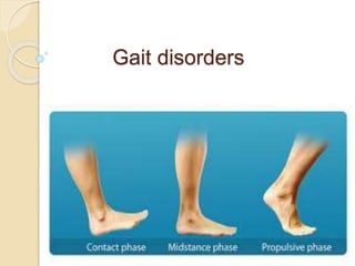
  • 10. GAIT CYCLE Period between two successive points at which the heel of the same foot strikes the ground Cadence:“the rhythm and number of steps per minute”
  • 11. Gait cycle 2 phases 8 events Phases stance swing Events 4 stance phase events 4 swing phase events
  • 12. Stance phase Swing phase Accounts for 60% gait cycle. Consists of 4 phases  initial contact  loading  mid-stance  terminal stance. Accounts for 40% gait cycle. Consists of 4 phases  Preswing  Initial swing  Mid swing  Terminal swing
  • 13. Events during stance phase 1. Heel strike:  initiates gait cycle  body’s center of gravity is at its lowest position 2. Foot flat :  plantar surface of the foot touches the ground 3. Mid stance  Swinging contra lateral foot passes the stance foot  Center of gravity at its highest position 4. Heel off  Heel looses contact with ground. 5. Toe off  terminates the stance phase as foot leaves the ground
  • 14. Events during swing phase 1. Acceleration:  begins as soon as foot leaves the ground  activation of hip flexors to accelerate leg forward 2. Mid swing:  foot passes directly beneath the body  coincident with mid stance of other foot 3. Deceleration:  describes the action of muscles as they slow  the leg and stabilize the foot in preparation for next heel strike
  • 15.
  • 16. Distance measures: Stride length ◦ distance between heels from one heel strike to the next heel strike on same side ◦ distance travelled by a person during a gait cycle Step length ◦ length between heel strike of one foot to other ◦ Rt + Lt step length = stride length Step width ◦ medio lateral distance between feet
  • 17.  A typical adult walking parameters comfortably on a level surface includes: ◦ velocity of about 80 m per minute, ◦ about 113 steps per minute, ◦ stride length of 1.41 m. ◦ About 60% of the gait cycle is spent in stance phase ◦ 40% in swing phase ◦ 10% in double limb support.  The body's center of mass is located just anterior to the S2 vertebral body.
  • 19. HISTORY  A detailed history provides the first clues to diagnosis.  note the circumstances in which walking difficulty occurs.  Ask for any associated symptoms, especially falls.  Walking over uneven ground exacerbates most walking difficulties.  Often an individual is unaware of their gait abnormality, and family or friends note altered cadence, shuffling or slowness.
  • 20. Weakness:  weakness of certain muscle groups produces characteristic difficulties during particular movements of the gait cycle. ◦ weakness of ankle dorsiflexion - foot drop ◦ Weakness of knee extension - buckling attacks ◦ Weakness of ankle plantar flexion - shallow stepped gait. ◦ ◦ proximal muscle weakness - Difficulty in climbing stairs or rising from a seated position  Catching or scraping a toe on the ground and a
  • 21. Slowness and Stiffness:  Slowness of gait is normal reaction when walking on slippery surfaces.  Similarly, when the balance was threatened any musculoskeletal or neurological disorder patient will have slowness of gait.  In Parkinson disease (PD) and other basal ganglia diseases, slowness of walking is due to shuffling with short, shallow steps.  Difficulty rising from a chair or turning in bed may be clues to loss of truncal mobility in diffuse cerebro vascular disease, hydrocephalus, and basal ganglia diseases.
  • 22.  Complaints of stiffness, heaviness in the limbs are the symptoms of a spasticity.  It is important to remember that leg muscle tone in some upper motor neuron syndromes and dystonia may be normal when the patient is examined in the supine position but is increased during walking.  In childhood, an action dystonia of the foot is a common initial symptom, walking on the toes only becoming evident after walking or running.
  • 23. Imbalance:  poor balance and unsteadiness are cardinal features of cerebellar ataxia and sensory ataxia.  cerebellar gait ataxia complains of ◦ Unsteadiness and inability to walk in a straight line and near falls. ◦ Turning and suddenly changing direction results in veering to one side. ◦ Symptoms are exacerbated by an uneven support surface.
  • 24. Sensory ataxia: ◦ presents with unsteadiness when walking in the dark. ◦ Patients complain of being uncertain of the exact position of their feet when walking. ◦ impression of walking on a spongy surface or cotton wool. Acute vestibulopathy: ◦ Is associated with vertigo and severe imbalance. Chronic vestibular dysfunction ◦ often causes veering and problems in an environment with many moving objects.
  • 25.  A wide-based unsteady gait also is a feature of frontal lobe diseases ◦ normal pressure hydrocephalus, ◦ diffuse small vessel ischemia of frontal sub cortical white matter.  Imbalance in sub cortical cerebro vascular disease commonly manifests when ◦ turning while walking, ◦ stepping backwards, ◦ bending over to pick up something.
  • 26. Falls:  Toppling Falls ◦ muscle tone is retained ◦ falling like a tree trunk ◦ due to impaired static and dynamic postural responses that control body equilibrium  collapsing falls ◦ muscle tone is lost ◦ imply a loss of consciousness characteristic of syncope or seizures  Tripping falls: ◦ many people attribute falls to tripping when in fact tripping did not occur. ◦ Tripping may be due to foot drop or shallow steps ◦ also be a consequence of secondary to inattention, dementia, or poor vision.
  • 27.  Proximal muscle weakness may result in the legs giving way and falls.  Unsteadiness and poor balance in an ataxic syndrome may lead to falls.  falls associated with postural adjustments, or falls occurring when performing multiple tasks suggest an impairment of postural responses.  Backward falls in early stages ◦ Feature of an akinetic-rigid syndromes ◦ Favors a diagnosis of MSA or PSP rather than PD ◦ Falls do occur in PD but are late feature. • In PD falls are usually forward and may also occur while turning and changing the posture.  Collapsing falls related to orthostatic hypotension, a
  • 28. Sensory Symptoms and Pain:  distribution of sensory complaints provides a clue to the site of the lesion. ◦ Ex: Distal symmetrical paresthesias of limbs S/O peripheral neuropathy.  Skeletal pain due to degenerative joint disease is aggravated by movement of the affected joints.  The normal pattern of walking is frequently modified by joint disease.  strategies to minimize pain by avoiding full weight bearing on the affected limb will produce limping.
  • 29. Urinary Incontinence:  Urinary urgency and urge incontinence seen in ◦ spinal cord lesion ◦ parkinsonism ◦ subcortical white-matter ischemia.  frontal lobe tumors, frontal lobe infarction, and hydrocephalus also produce urinary incontinence. Cognitive Changes:  Slowing of gait may be a marker of impending cognitive impairment and dementia.  executive dysfunction including inattention may produce falls.  disinhibition and impulsivity are associated with reckless gait problems and falls.
  • 30. EXAMINATION OF POSTURE AND GAIT  First observe the overall pattern of limb and body movement during walking.  Normal walking progresses in a smooth and effortless manner.  The truncal posture is upright, and the legs swing in a fluid motion with a regular stride length.  Synergistic head, trunk, and upper-limb movement flow with each step.  After the overall walking pattern is observed, the specific aspects of posture and gait should be examined.
  • 31. specific aspects of posture and gait includes: 1. Arising to stand from seated position 2. Stance and posture 3. Walking 4. Turning while walking 5. Other maneuvers ◦ Tandem walking ◦ Walking backwards ◦ Running ◦ Walking on toes, heels
  • 32. Arising from Sitting:  informs about ◦ pelvic girdle strength, ◦ control of truncal movement, ◦ coordination, and balance.  proximal muscle weakness: Inability to rise when the feet are appropriately placed under the body.  incoordination or imbalance: An abnormally wide stance base when standing.  Frontal lobe diseases: Feet are not positioned directly under the body
  • 33. Stance:  Stance base = the distance between the feet  gives an indication of balance.  Wide stance base indicates poor balance.  Wide-based gaits are ◦ typical of cerebellar or sensory ataxia ◦ diffuse cerebro vascular disease ◦ frontal lobe lesions.  Eversion of the feet to increase stability seen in diffuse cerebrovascular disease.  In mild ataxia, a widened base may only be evident with turning.  Tandem (heel to toe) walking is a good objective measure of walking stability.
  • 34. Trunk Posture:  Flexion of the trunk and a stooped posture - PD.  Upright posture with neck and trunk extension- PSP.  Slight flexion at the hips to lower the trunk - feature of cautious gait.  Neck flexion – MND Myasthenia gravis MSA Parkinsonism  Tilt of the trunk to one side - dystonia.  exaggerated lumbar lordosis ◦ stiff person syndrome[d/t Paraspinal muscle spasm] ◦ Proximal myopaties.
  • 35.
  • 36. Truncal tilt away from the side of the lesion ◦ acute vascular lesions of the thalamus( postero lateral) and basal ganglia. Truncal tilt away from the side of the lesion ◦ Acute vestibular imbalance in the lateral medullary syndrome Truncal flexion (camptocormia) ◦ paraspinal myopathies that produce weakness of trunk extension.  restricted range of spinal movement and persistence of the abnormal spinal posture when supine or during sleep are useful pointers toward a bony spinal deformity.
  • 37. Postural Responses: 1.Reactive postural responses 2. anticipatory postural responses. 3.protective postural responses. .
  • 38. pull test Reactive postural responses are examined by “the pull test”.  Examiner sharply pulls the upper trunk backward or forward while the patient is standing.  The pull should be sufficient to require the patient to step to regain their balance.  The examiner must be prepared to prevent the patient from falling.  A few short, shuffling steps backward or an impending fall backward or forward suggests impairment of righting reactions
  • 39.  Falls after postural changes such as arising from a chair or turning while walking suggest impaired anticipatory postural responses.  Falls without rescue arm movements or stepping movements to break the fall indicate loss of protective postural responses.  Injuries sustained during falls provide a clue to the loss of postural responses  A tendency to fall backward spontaneously is a sign of impaired postural reflexes in PSP and gait disorders associated with frontal lobes
  • 40. Romberg test  Patients with proprioceptive loss depends on visual cues.  the removal of vision greatly exaggerates the ataxia.  This is the basis of the Romberg test.  In which eye closure leads to a dramatic increase in unsteadiness and even falls in the patient with sensory ataxia.  When performing the Romberg test, it is important the patient is standing comfortably before eye closure and to remember that a modest increase in body sway is a normal response to eye closure.
  • 41. Walking: Initiation of Gait: start hesitation – difficulty in gait initiation. slipping clutch phenomenon ◦ few short, shuffling steps on the spot without forward progress. magnetic foot phenomenon. ◦ complete immobility with the feet seemingly glued to the floor  Patients may make exaggerated upper-body movements or alter the step pattern.
  • 42.  Start hesitation seen in ◦ frontal lobe disease, ◦ diffuse cerebrovascular disease, ◦ hydrocephalus. ◦ gait ignition failure. ◦ PD  Isolated start hesitation is seen in the syndrome of gait ignition failure.
  • 43. Stepping:  length and trajectory of each step and the rhythm of stepping should be noted.  Short, regular, shallow steps or shuffling and a slow gait - akinetic-rigid syndromes.  Freezing typically occurs when turning, when walking has been interrupted by an obstacle.  Festination (increasingly rapid, small steps) is common in PD but rare in other akinetic-rigid syndromes.
  • 44.  Unilateral loss of synergistic arm swing while walking 1. valuable sign of early PD 2. Unilateral UMN lesion 3. acute cerebellar syndrome Turning:  Requires more balance more than walking in a straight line.  Slowing on turns may be the first abnormality in walking in PD  Multiple steps on turning ◦ PD
  • 45. More Challenging Tests of Walking: Tandem walking:  Subtle degrees of cerebellar ataxia unmasked by tandem walking. Walking on toes and heels:  may bring out abnormal movements as well as deficits in the strength of dorsiflexion and plantar flexion of the ankle. Walking backward:  will sometimes reduce or abolish the dystonic foot posturing observed walking forward in action dystonia of the foot. Running:  examine the patient running to identify an action dystonia of the legs in early stages.
  • 46. Associated and Synergistic Limb Movements While Walking: Dystonic posturing of an arm or leg ◦ dystonia, ◦ parkinsonism, ◦ old hemiparesis. Choreiform movements are more prominent during walking than at rest in ◦ most chorea syndromes, ◦ levodopa-induced dyskinesia, ◦ tardive dyskinesia. Parkinsonian tremor of the dependent upper limb is often observed while walking.
  • 47. MOTOR AND SENSORY EXAMINATION:  motor and sensory function in the limbs is examined with the patient sitting or supine.  The size and length of the limbs should be measured in any child presenting with a limp.  The spinal column should be inspected for scoliosis, and the lumbar region for skin defects or hairy patches indicative of spinal dysraphism.
  • 48. DISCREPANCIES ON EXAMINATION OF GAIT:  minimal abnormal signs on physical examination of the recumbent patient, in contrast to the observed difficulty when walking. Cerebellar gait ataxia caused by a vermis lesion: ◦ perform the heel-to-shin test normally when supine but is ataxic when walking. Action dystonia: ◦ Abnormal posturing of the feet only when walking. ◦ A dystonic gait may be evident only when running or walking forward but not when walking backward.
  • 49. Frontal lobe lesion, hydrocephalus,or diffuse cerebrovascular disease : ◦ Patient totally unable to walk when standing. ◦ Gegenhalten (paratonia), with or without brisk tendon reflexes, may be the only abnormal sign in the recumbent patient ◦ Such patients perform the heel-shin test and make bicycling movements of their legs normally when lying on a bed. Leg tremor of orthostatic tremor ◦ only appears during weight bearing, especially when standing still.
  • 51. CLASSIFICATION OF GAIT PATTERNS:  A scheme based on Hughlings Jackson’s three orders of neurological function.  Lower Level Gait Disorders (simplest)  Middle Level Gait Disorders  Higher Level Gait Disorders (complex, integrative )  Each level contributes sensory and motor function, but that of higher centers is more complex and dispersed within the nervous system.
  • 52. Lower Level Gait Disorders  manifest physical signs such as weakness or sensory loss.  are due to diseases of the muscle and peripheral nerves that produce muscle weakness.  Lower level sensory gait disorders follow loss of one of the three basic senses important for gait and balance: ◦ proprioception ◦ vision ◦ vestibular sensation.
  • 53. Low level (sensory and motor) gait disorders: 1. Waddling gait - proximal weakness (myopathy) 2. high stepping gait - distal weakness (neuropathy) 3. Sensory ataxia – proprioception loss. 4. Vestibular ataxic gait -acute peripheral vestibulopathy 5. Gait difficulty due to acute visual impairment
  • 54. Myopathic Weakness and Gait:  Weakness of proximal leg and hip-girdle muscles  interferes with stabilizing the pelvis and legs on the trunk.  produces exaggerated rotation of the pelvis with each step and a waddling gait.  The hips are slightly flexed (d/t hip extensor weakness).  exaggerated lumbar lordosis.  Gower’s sign positive  myopathy is the most common cause of proximal muscle weakness,  neurogenic weakness of proximal muscles can also
  • 55.
  • 56. Neurogenic Weakness and Gait:  Muscle weakness of peripheral nerve origin.  typically affects distal leg muscles  results in a steppage gait.  The patient lifts the leg and foot high above the ground with each step because of weakness of ankle dorsiflexion and foot drop.  unilateral foot drop: ◦ common peroneal or sciatic nerve palsy or an L5 radiculopathy  Less common is foot drop caused by myopathic weakness, as in the scapuloperoneal syndromes.  Weakness of ankle plantar flexion - shallow stepped gait.
  • 57.
  • 58.  A femoral neuropathy as in diabetes mellitus: ◦ produces weakness of knee extension and buckling of the knee when walking or standing.  This may first be evident when walking down stairs.  similar weakness: ◦ Progressive muscular atrophy in MND ◦ quadriceps myopathy ◦ inclusion body myositis
  • 59. Sensory Ataxia:  wide stance base  advance cautiously  taking slow steps under visual guidance.  the feet are thrust forward with variable direction and height.  The sole of the foot strikes the floor forcibly with a slapping sound - slapping gait.  Romberg test positive.  Walking on uneven surfaces is particularly difficult.  Results from ◦ peripheral neuropathies, ◦ posterior root or dorsal root ganglionopathies, ◦ dorsal column lesions.
  • 60. Vestibular Imbalance, Vertigo, and Gait:  Acute peripheral vestibulopathy ◦ produce veering to the side of the lesion. ◦ Veering less prominent while running than walking. ◦ Patients prefer to lie stand still.  In chronic vestibular failure, ◦ gait may be normal, ◦ unsteadiness can be unmasked during eye closure and rotation of the head from side to side while walking.
  • 61. Middle-Level Gait Disorders  Related to impaired modulation of force generated by the lower level motor system include: (1) spastic gait from corticospinal tract lesions (2) ataxic gait arising from disturbances of the cerebellum and its connections. (3) hypokinetic gaits associated with parkinsonism. (4) hyperkinetic gaits associated with chorea,dystonia, and
  • 62. Spastic Gait: Spasticity of the arm and leg on one side:  The leg is slightly flexed at the hip and extended at the knee, with plantar flexion and inversion of the foot.  During the swing phase ◦ lateral flexion of the trunk toward the unaffected side. ◦ circumduction of the extended paretic leg. ◦ dragging the foot or catching the toe on the ground. ◦ Minimal associated arm swing occurs on the affected side.  Muscle tone was increased on effected side along with brisk DTR and extensor plantar response.
  • 63. Spasticity of both legs:  legs are stiffly extended at the knees, plantar flexed at the ankles, and slightly flexed at the hips.  Both legs circumduct, and the toes of the plantar flexed feet catch on the floor with each step.  There is a tendency to adduct the legs, particularly when the disorder begins in childhood. [scissoring gait]  Occasionally, bilateral leg dystonia (dystonic paraparesis) mimics a spastic paraparesis.
  • 64.  This typically occurs in doparesponsive dystonia in childhood.  may be misdiagnosed as HSP or cerebral diplegia.  Clinical differentiation between these conditions can be difficult.  Brisk tendon reflexes occur in both, and spontaneous extension of a great toe in patients with striatal disorders may be interpreted as a Babinski response.  Fanning of the toes and knee flexion suggest spastic paraplegia.  Other distinguishing features include changes in muscle tone, such as spasticity in HSP and rigidity in dystonic paraparesis.
  • 65. Cerebellar Ataxic gait:  Lesions of spino cerebellum produces gait ataxia.  wide-based stance.  Steps are taken slowly and carefully.  tendency to lurch and stagger.  Accentuated by tandem walking  Gait with lurching and staggering quality - midline cerebellar pathology.  Swaying to one side
  • 66.
  • 67. A pure truncal ataxia - midline (vermis) cerebellar syndrome. - normal examination in supine position Patients with anterior lobe atrophy: ◦ develop a 3-Hz antero posterior sway of the trunk and a rhythmic truncal and head tremor (titubation) that is superimposed on the gait ataxia. Lesions of vestibulo cerebellum:  exhibit multidirectional body sway, & dysequilibrium.
  • 68. Lesions of neo cerebellum:  produce limb ataxia  characterized by a decomposition of normal leg movement.  Steps are irregular and variable in timing (dyssynergia), length, and direction (dysmetria).  With lesions confined to one cerebellar hemisphere, ataxia is limited to the ipsilateral limbs  there is little postural instability or truncal imbalance if the vermis is not involved.  Cerebellar gait ataxia is exacerbated by the rapid postural adjustments like turning.
  • 69. Spastic Ataxic gait:  A combination of spasticity and ataxia produces a distinctive “bouncing” gait.  Gait is wide based  clonus is precipitated by standing or walking, creating a bouncing motion.  Compensatory movements, made in an effort to regain balance, set up a vicious cycle of ataxic movements, clonus, and increasing unsteadiness, rendering the patient unable to stand or walk.  Bouncing gaits must be distinguished from action myoclonus of the legs and cerebellar truncal tremors.
  • 70. Hypokinetic (Parkinsonian) Gait: Early PD: ◦ Asymmetrical reduction of arm swing and ◦ Mild slowing in gait, particularly when turning. Advanced PD: ◦ stooped posture with flexion of the shoulders, neck, trunk. ◦ little associated limb movements while walking. ◦ the arms are held immobile at the sides or slightly forward. ◦ Rest tremor of the upper limbs is often apparent when walking but leg tremor is rare during walking. ◦ Begins with start hesitation with a few rapid, short, shuffling steps. ◦ Followed by shuffling gait with small, shallow steps on a narrow base with freezing episodes ◦ In the long term, levodopa therapy may induce
  • 71.  The posture of generalized flexion of the patient with PD exaggerates the normal tendency to lean forward when walking.  To maintain balance when walking and avoid falling forward, the patient may advance with a series of rapid, small steps (festination).  The shuffling gait of PD that is responsive to levodopa characterizes the mid-level gait pattern.  As the disease progresses, dysequilibrium and falls emerge as features of a higher level gait disorder.
  • 72.
  • 73.
  • 74. Choreic Gait:  random movements of chorea - increased during walking.  The superimposition of chorea on the trunk and leg movements gives the gait a dancing quality.  Chorea can also interrupt the walking pattern, leading to a hesitant gait.  Huntington disease ◦ causes a stumbling and stuttering gait with steps forward, backward, or to one side. ◦ Variable stance but generally wide-based ◦ Excessive swaying of trunk. ◦ Balance and equilibrium are maintained until the terminal stages
  • 75.
  • 76. Dystonic Gait:  Produce bizzare gait pattetns.  classic presentation of childhood-onset primary torsion dystonia: ◦ action dystonia of a leg, with sustained abnormal posturing of the foot (typically plantar flexion and inversion) on attempting to run.  In contrast, walking backward or even running backward may be normal at an early stage. Striatal toe:  tonic extension of the great toe.  Important early sign.
  • 77.
  • 78.  A characteristic feature common to dystonic gaits is excessive flexion of the hip when walking.  Patients may hop or walk sideways in a crab-like fashion.  Hyperflexion of the hips and knee produce an attitude of general body flexion in a simian posture.  excessive flexion of the hip and knee and plantar flexion of the foot produce a birdlike (peacock) gait during the swing phase of each step.  Many patients have been thought to be hysterical
  • 79. Dopa-responsive dystonia ◦ characteristically presents in childhood with walking difficulties ◦ child walks normally in the early morning ◦ dystonic posturing of the legs increases as the day progresses or after exercise. ◦ Symptoms may be relieved by a nap (“sleep benefit”). ◦ Examination reveals dystonic plantar flexion and inversion of the foot, with brisk tendon reflexes. ◦ Some of these patients respond dramatically to levodopa. ◦ Indeed, all children presenting with a dystonic foot or leg should have a therapeutic trial of levodopa before other therapies such as anticholinergic drugs are commenced.
  • 80. Paroxysmal kinesogenic choreoathetosis: ◦ present with difficulty walking as the result of dystonic postures and involuntary movements of the legs. ◦ often appearing after standing from a seated position. ◦ These attacks are typically brief, lasting a matter of seconds.
  • 81. Mixed Movement Disorders and Gait: Athetoid cerebral palsy:  Produce motor signs reflecting abnormalities at many levels of the nervous system.  These include spasticity of the legs, truncal and gait ataxia, dystonia, and dystonic trunk and limb spasms.  DD - primary torsion dystonia.  patient with cerebral palsy usually has a ◦ history of perinatal injury or birth asphyxia. ◦ history of hypotonia. ◦ delayed developmental motor milestones. ◦ presence of spasticity and ataxia.
  • 82. Tremor of the trunk and Legs:  Leg tremor in benign essential tremor occasionally symptomatic.  Trunk and leg tremor may contribute to unsteadiness in cerebellar disease.  Orthostatic tremor: ◦ has a unique frequency (16 Hz) and distribution. ◦ rapid tremor produces an intense sensation of unsteadiness often with little obvious shaking of the legs. ◦ relieved by walking or sitting down. ◦ Patients avoid standing still (e.g., in a queue) and may shuffle on the spot in an effort to relieve the unsteadiness. ◦ Examination reveals a rippling of the quadriceps muscles during standing.
  • 83. Action Myoclonus:  Postanoxic action myoclonus of the legs is often accompanied by negative myoclonus (asterixis) that disrupts standing and walking.  Repetitive action myoclonus produces jerky movements of the legs.  Lapses of muscle activity between the jerks (negative myoclonus) cause the patient to sag toward the ground.  sequence of events gives rise to an exaggerated bouncing appearance, which is sustainable for only a few seconds before falling.
  • 84.  Difficulty walking is one of the major residual disabilities of post-anoxic myoclonus.  Stimulus sensitive cortical reflex myoclonus ◦ similar disorder of stance and gait, ◦ reflex myoclonus of the quadriceps, resulting in a bouncing posture.  acute onset negative myoclonus can occur with vascular lesions , particularly of the thalamus and frontal lobes.
  • 85. Higher Level Gait Disorders  characterized by varying combinations of ◦ dysequilibrium, ◦ Falls due to impaired postural responses ◦ short shuffling steps, and freezing.  formal neurological examination fails to reveal signs that adequately explain the gait disturbance.  Slowness of sequential leg movement and poor truncal control are often present.
  • 86.  Stepping patterns are influenced by environmental cues that induce freezing of gait.  Freezing or falling while performing multiple simultaneous tasks is common.  a variety of terms for higher level gait disorders such as ◦ apraxia of gait, ◦ magnetic gait, ◦ lower half parkinsonism, ◦ frontal gait,
  • 87. Hypokinetic Higher Level and Freezing Gait Patterns:  With progression of PD, ◦ freezing of gait, ◦ disequilibrium, ◦ loss of postural responses and falls become increasingly troublesome.  This will poorly respond to levodopa therapy.  DBS of ( STN / Gpi ) may improve or worsen gait with increased falls.  DBS in the region of the pedunculopontine nucleus is under investigation for dysequilibrium and freezing of gait.
  • 88. Akinetic rigid syndromes:  most common are MSA,PSP,CBD.  Gait patern was similar to PD except festination is less common.  Another distinguishing feature is early appearance of falls due to loss of postural and righting responses.  Falls occur in 80% of patients with progressive supranuclear palsy and can be dramatic, leading to injury.  Dramatic response to levodopa that is typical of PD does not occur in these other akinetic rigid syndromes.
  • 89. Gait in frontal lobe lesions:  Varies from an ataxic gait to an akinetic-rigid gait  Often had combination of these features.  early stages - wide stance base with turning difficulty.  There may be episodes of freezing.  Arm swing is normal or even exaggerated, giving the appearance of a “military two-step” gait.  The normal fluidity of trunk and limb motion is lost.  voluntary upper limb and hand movements are normal and there is a lively facial expression.
  • 90.  There difficulty in gait initiation [slipping clutch or magnetic feet].  Once walking is underway, steps may be better.  freezing re-emerge while attempting to turn.  Upper motor neuron signs including pseudobulbar palsy paratonic rigidity (gegenhalten) , Frontal releasing signs commonly occurs.  Postural and righting reactions are impaired and eventually lost.  Urinary incontinence and dementia frequently occur.
  • 91. Called as “lower half parkinsonism” or marche à petits pas of Dejerine or Critchley’s atherosclerotic parkinsonism.  Commonly seen in diffuse small vessel cerebrovascular disease.  upper motor neuron signs including pseudobulbar palsy, and the absence of a resting tremor distinguish this syndrome from PD.
  • 92.
  • 93. syndrome of gait ignition or gait initiation failure: ◦ Also called as “pure akinesia” or “primary progressive freezing of gait.” ◦ exhibit profound start hesitation and freezing. ◦ but step size and rhythm are normal once walking is underway. ◦ These findings are similar to those seen with walking in PD. ◦ but speech and upper limb function are normal ◦ there is no response to levodopa.
  • 94.  some elderly patients experience severe walking difficulties that resemble those described in frontal lobe disease.  The history is usually of gradual onset, without stroke- like episodes.  No identifiable lesions of the frontal lobes or in white matter on imaging.  The criteria for normal pressure hydrocephalus are not fulfilled.  there are no signs of parkinsonism and levodopa is ineffective.  There is no evidence of more generalized cerebral dysfunction. The cause of these syndromes is unknown although it is increasingly recognized that subcortical white-matter
  • 95. ELDERLY GAIT PATTERNS CAUTIOUS GAITS:  Healthy, neurologically normal elderly people ◦ Walk at slower speeds ◦ Shorter and shallower steps ◦ Stance width slightly wider than normal ◦ Synergistic associated arm movements are less vigorous. ◦ The rhythmicity of stepping is preserved.
  • 96.  These changes give the normal elderly gait a cautious or guarded appearance.  Factors contributing to a general decline in mobility of the elderly include ◦ degenerative joint disease, ◦ reducing range of limb movement, ◦ decreased cardiovascular fitness, ◦ limiting exercise capacity.
  • 97.  A cautious gait is a normal response to the perception of impaired or threatened balance.  They may be unable to walk without support.  The gait improves dramatically when support is provided.  Accordingly, a cautious gait should be interpreted as compensatory and not specific for any level of the gait classification.  A formal program of gait retraining may help restore confidence and improve the ability to walk.
  • 98. RECKLESS GAIT PATTERNS:  seen in patients with impaired postural responses and poor truncal control.  Patients do not recognize their instability and take risks that result in falls and injuries.  Such patients make inappropriate movements of the feet and trunk when sitting or standing.  The most striking examples occur in frontal dementia.  impulsivity and a failure to adapt to the precarious balance are part of the cognitive decline.
  • 99. HYSTERICAL GAIT DISORDERS The typical gait patterns encountered include: 1. transient fluctuations in posture while walking 2. knee buckling without falls 3. excessive slowness and hesitancy 4. stooped or other abnormal posture of the trunk 5. complex postural adjustments with each step 6. exaggerated body sway or excessive body motion especially brought out by tandem walking.
  • 100.  common features of psychogenic gait (and movement) disorders are: ◦ Suggestibility ◦ Variability ◦ Improvement with distraction, ◦ History of sudden onset ◦ Rapid, dramatic, and complete recovery.  Other signs like give away weakness or non physiological sensory loss will be present.  Hoover sign: ◦ As the patient lifts the normal leg, the examiner places a hand under the “paralyzed” leg and feels the presence (and strength) of synergistic hip extension.
  • 101. ASTASIA – ABASIA:  Unable to either stand or walk,  More or less normal use of their legs while in bed  Normal neurologic examination  When such patients are placed on their feet, they may take a few steps and then become unable to advance their legs.  they lurch in all directions and crumple to the floor if not assisted BEFORE LABELLING A HYSTERICAL GAIT, RULE OUT 1. Lesions of anterosuperior cerebellar vermis 2. NPH 3. Frontal lobe disease 4. Intoxications such as with phenytoin