Esophageal varices
•Transferir como PPTX, PDF•
34 gostaram•12,387 visualizações

Case Write Up - Esophageal Varices Secondary to Liver Disease

Recomendados
Mais conteúdo relacionado
Mais procurados
Mais procurados (20)
Destaque
Semelhante a Esophageal varices
Semelhante a Esophageal varices (20)
Último
Último (20)
Esophageal varices
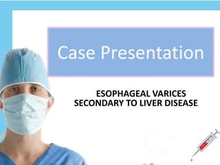
- 1. Case Presentation ESOPHAGEAL VARICES SECONDARY TO LIVER DISEASE
- 2. PATIENT’S IDENTIFICATION Name: Phuah Eng Tea Age: 59 years old Gender: Male Race: Chinese Religion: Christian Address: Klang Utama Occupation: Retired lorry driver Marital status: Married Registration No: 1564813 Ward: 4A Bed: 24 Informant: Wife
- 3. CHIEF COMPLAINT Vomiting blood, on same day of admission.
- 4. HISTORY OF PRESENTING ILLNESS Mr Phuah , 59 years old Chinese gentleman came to the Emergency Department complaining of vomiting blood (hematemesis) which is red in colour and 1 small cup full(approximately 100ml), on same day of admission. It was sudden and this was the second episode of hematemesis. This is associated with loose black stool(melena), abdominal pain, dizziness, fatigue and loss of appetite. The review of other systems were unremarkable.
- 5. PERSONALAND SOCIAL HISTORY Mr Phuah is a retired lorry driver. He is married with no children and lives in a single-storey house with his wife with basic amenities. He smokes 5-6 pieces of cigarette per day since 20 years old, he also consumes 2-3 cans of beer everyday for more than 30 years and he is a heroin drug user. He takes a normal chinese diet and undergoes sedentary lifestyle. FAMILY HISTORY There is no chronic illness or malignancies running in his family.
- 6. PAST MEDICAL HISTORY and DRUG HISTORY He has hypertension. He was admitted 2 twice before due to drug abuse and altered mental status. He was diagnosed with Hepatitis C in 2013. Currently, he is taking Covapril for his HTN. He is also a heroin drug user since young.
- 7. ANATOMICAL & PATHOLOGICAL CORRELATION Anatomical correlation Pathological correlation Esophagus Neoplasm Inflammation Stomach Inflammation Neoplasm
- 8. PHYSICAL EXAMINATION General Examination Patient was lying comfortably on bed in supine position supported with one pillow. The patient was alert, conscious and well-oriented to time, place and person. The patient was not in pain and respiratory distress. There was an ID tag on his right hand and branula attached on his dorsum of right hand which is attached to IV Metronidazole. He is moderately build, with adequate nutritional status.
- 9. Hands Hands were warm and dry No clubbing, koilonychia or leukonychia No peripheral cyanosis No palmar erythema Eyes Has conjunctival pallor No jaundice Oral cavity Tongue is coated No central cyanosis No mouth ulcers Moderate oral hygiene Neck No swelling observed Legs No pitting edema
- 10. Vital signs Blood pressure : 144/77 mmHg Pulse rate : 77 beats per minute Respiratory rate : 18 breaths per minute Temperature : 37.0 ᵒC
- 11. Local Examination Of The Abdomen On Inspection The abdomen was distended. The umbilicus was centrally located and inverted. There were no surgical scars or discoloration of the skin. All quadrants of the abdomen moved symmetrically during respiration. On palpation On superficial and deep palpation, there were no rigidity, guarding and tenderness of abdomen. The liver and spleen were not palpable.
- 12. On percussion The liver span is normal at 10cm. There was positive fluid thrill and shifting dullness On auscultation Normal bowel sound was heard(3 times per minute).
- 13. SUMMARY A 59 years old Chinese man was admitted to hospital due to hematemesis associated with loose black stool(melena), dizziness, fatigue, abdominal pain and loss of appetite. On physical examination the abdomen was distended. There was positive fluid thrill and shifting dullness.
- 14. Provisional Diagnosis Diagnosis Points to support Esophageal Varices 2⁰ to Liver Disease - Age (> 60 years old) - Past history of hematemesis and melena - History of Hepatitis C (known history of liver disease) - Intravenous drug user - Alcohol consumer and smoker - Abdominal pain - Loss of appetite - Unknown loss of weight - Loose stool (diarrhea) - Weakness & Fatigue
- 15. Differential Diagnosis DIAGNOSIS POINTS TO SUPPORT POINTS AGAINST Peptic Ulcer Disease - Nausea & vomiting - Unknown weight loss - Loss of appetite - Melena & hematemesis (severe case) - Smoking - No abdominal bloating - No burning pain before or after meal - No heartburn - No indigestion Esophageal Carcinoma - Gender - male - Unknown weight loss - Melena - Hematemesis - Vomiting - Smoking - Age (usually >65yrs) - No persistent cough - No hoarseness - No indigestion - No dyspepsia - No heartburn - No dysphagia - No chest or back pain
- 16. Gastric Carcinoma - Age (common in elderly) - Nausea or vomiting - Loss of appetite - Melena - Hematemesis - Weight loss - Diarrhea - Weakness and fatigue - No indigestion - No dysphagia - No postprandial fullness - No palpable mass on abdomen - No heartburn - No bloated feeling after eating DIAGNOSIS POINTS TO SUPPORT POINTS AGAINST
- 17. Investigations Self revised Done by hospital Full Blood Count Haemotological Test Liver Function Test Renal Function Test Renal Function Test Liver Function Test Endoscopy (OGDS) Arterial Blood Gases Coagulation Profile Chest X-Ray Chest X-Ray Abdominal X-Ray Abdominal X-Ray HIV Test Hepatitis C Screening Hepatitis C Screening Hepatitis B Screening Rapid Plasma Reagin
- 18. 1) Haemotological Test Test Patient’s Result Unit Normal Range Interpretation RBC 2.68 10^6/uL 4.5 – 6.5 Low WBC 19.6 10^3/uL 4 - 11 High HGB 8.1 g/dL 13 - 18 Low HCT 23.9 % 40 – 54 Low MCV 89.2 fL 76 – 96 Normal MCH 30.2 pg 27 – 32 Normal PLT 207 10^3/uL 150 - 400 Normal .
- 19. 2) Renal Function Test Test Result Unit Range Interpretation Urea 15.4 mmol/L 2.8 – 7.2 High Sodium 140 mmol/L 136 – 145 Normal Potassium 4.6 mmol/L 3.5 – 5.1 Normal Chloride 104 mmol/L 98 – 107 Normal Creatinine 95 umol/L 45 - 84 Normal
- 20. 3) Liver Function Test Test Patient’s Result Unit Normal Range Interpretation Total Protein 59 g/L 66 – 83 Low Albumin 28 g/L 35 – 52 Low Globulin 31 g/L 25 – 39 Normal A/G Ratio 0.9 0:9 – 1:8 Normal ALP 53 IU/L 30 – 120 Normal ALT 79 IU/L 0 – 50 High Total Bilirubin 20.1 umol/L 5 - 21 Normal
- 21. 4) Arterial Blood Gas Test Result Unit Range pH 7.382 - 7.350 – 7.450 pCO2 37.1 mmHg 35.0 – 48.0 pO2 21.3 mmHg 83.0 - 108 5) HIV Test & Rapid Plasma Reagin Test Result HIV Ag/Ab Non-Reactive Syphilis Reagin Ab Non-Reactive
- 22. 6) Hepatitis B Screening Test Result Hepatitis B e Ab Reactive Hepatitis B e Ag Non-Reactive Hepatitis B s Ag Non-Reactive 7) Hepatitis C Screening Test Result Anti HCV Reactive HCV particle Agglutination Detected
- 23. 8) Chest X-Ray Impression : Shows no abnormalities (normal)
- 24. 9) Abdominal X-Ray Impression : Shows no abnormalities (normal)
- 25. CHILD-PUGH-SCORE (SEVERITY OF CIRRHOSIS) CATEGORY 1 2 3 ENCEPHALOPATHY 0 I/II III/IV ASCITES ABSENT MILD-MODERATE SEVERE BILIRUBIN (umol/L) <34 34 – 51 >51 ALBUMIN(g/L) >35 28 – 35 <28 INR <1.3 1.3 – 1.5 >1.5 Score A : 6 or less Score B : 7-9 Score C : 10 and greater Encephalopathy Grade I : confusion, altered mood and behaviour Grade II : drowsy, inappropriate behaviour Grade III : stuporous but obeys simple commands, slurred speech, marked confusion Grade IV : unarousable coma
- 26. CATEGORY RESULT SCORE ENCEPHALOPATHY No 1 ASCITES Mild 2 BILIRUBIN (umol/L) 20 1 ALBUMIN(g/L) 28 2 INR 1.7 3 Total score : 9 Grade : Child Pugh B
- 27. RESUSCITATION • Vitals are monitored (pulse, BP, cardiac monitor, urine output) • Assessment of haemodynamic stability – severity of blood loss • IV Route - Fluid resuscitation such as normal saline • Oxygen support to prevent hypoxia of tissues – nasal cannula or facemask. • Correction of coagulopathy and thrombocytopenia • Urine output (through an indwelling catheter) • Blood transfusion is administered to patients who are shocked and are actively bleeding. Management
- 28. • Pharmacological therapy : vasoconstrictors to arrest the bleeding (terlipressin, octreotide, vasopressin, somatostatin) • Banding • Sclerotherapy • Non-selective ß-adrenergic antagonists such as propranolol and nadolol to prevent rebleeding. • Transjugular Intrahepatic Portosystemic Shunt (TIPS) • Distal Splenorenal Shunt (DSRS). • Liver transplant • Devascularization • Esophageal transection Management
- 30. TIPS DSRS
- 31. a) Currently, no treatment can prevent the development of esophageal varices in people with liver disease. b) While beta blocker drugs are effective in preventing bleeding in many people who have esophageal varices, they do not keep esophageal varices from forming. c) If one have been diagnosed with liver disease, strategies to avoid liver disease complications should be taken. - Don't drink alcohol. - Eat a healthy diet. - Reduce your risk of hepatitis. Prevention
- 32. Discussion • Portal venous pressure = portal venous flow x portal venous resistance • Hepatic venous pressure gradient (HVPG) is the difference in pressure between the portal vein (wedged hepatic venous pressure) and free hepatic vein pressure. • HVPG = surrogate for portal pressure • Increased HVPG > 5mmHg = Portal Hypertension • Clinical manifestation of portal hypertension are many included Esophageal Varices.
- 33. Increased portal pressure (HVPG > 10mmHg) Formation of varices Dilatation of varices Rupture of varices (HVPG > 12mmHg) Natural History
- 35. Anatomy of esophagus • The esophagus is a 25-cm long muscular tube that connects the pharynx to the stomach. • The esophagus extends from the lower border of the cricoid cartilage (at the level of the 6th cervical vertebra) to the cardiac orifice of the stomach at the side of the body of the 11th thoracic vertebra. • The esophagus has 3 constrictions in its vertical course. • These measurements are clinically important for endoscopy and endoscopic surgeries of the esophagus. • The esophagus has been subdivided into 3 portions: a) Cervical b) Thoracic c) Abdominal • Histologically, the esophagus has 4 concentric layers. (Mucosa, Submucosa, Muscular, Adventitia)
- 37. Blood supply • Cervical portion : inferior thyroid artery • Thoracic portion : bronchial and esophageal branches of the thoracic aorta • Abdominal portion : ascending branches of the left phrenic and left gastric arteries. Venous drainage • Venous blood from the esophagus drains into a submucosal plexus. From this plexus, blood drains to the periesophageal venous plexus. Esophageal veins arise from this plexus and drain in a segmental way similar to the arterial supply, as follows: • From the cervical esophagus, veins drain into the inferior thyroid vein • From the thoracic esophagus, veins drain into the azygos veins, hemiazygos, intercostal, and bronchial veins • From the abdominal portion, esophagus veins drain into the left gastric vein; the left gastric vein is a tributary of the portal system.
- 38. Risk Factors • High portal vein pressure. The risk of bleeding increases with the amount of pressure in the portal vein (portal hypertension). • Large varices. The larger the varices, the more likely they are to bleed. • Red marks on the varices. When viewed through an endoscope passed down your throat, some varices show long, red streaks or red spots. These marks indicate a high risk of bleeding. • Severe cirrhosis or liver failure. Most often, the more severe your liver disease, the more likely varices are to bleed. • Continued alcohol use. Your risk of variceal bleeding is far greater if you continue to drink than if you stop, especially if your disease is alcohol related. • Bacterial infection - schistosomiasis
- 39. Sign & Symptoms 1) Vomiting blood (in large amount) can cause - Dizziness - Loss consciousness - Pallor - Anaemia 2) Some patient bleed in smaller amount over a longer period, they swallow the blood rather than vomit it, so they may have : - Black, tarry or bloody stools 3) Shock and sepsis(in severe cases) 4) Low blood pressure (hypotension) 5) Rapid heart rate (tachycardia) 6) Abdominal pain 7) Confusion secondary to encephalopathy
- 40. Signs of liver disease : 1) Jaundice (yellow discoloration) 2) Spider nevi (cluster of tiny blood vessels shaped like spider) 3) Palmar erythema (Reddening on palm) 4) Splenomegaly 5) Hand deformity known as Dupuytren’s contracture 6) Testicular atrophy 7) Ascites (fluid build up) 8) Gynaecomastia 9) Hair Loss 10) Pruritus 11) Muscle loss
- 41. • Encephalopathy (sometimes called hepatic encephalopathy) • Esophageal stricture after surgery or endoscopic therapy • Hypovolemic shock • Infection (pneumonia, bloodstream infection, peritonitis) • Rebleeding after treatment • Kidney failure Possible Complications
- 42. References • Bailey & Love's Short Practice of Surgery, 25th edn • http://www.hopkinsmedicine.org • http://www.slideshare.net • http://www.nlm.nih.gov • http://emedicine.medscape.com
- 43. THANK YOU!
Notas do Editor
- Video Capsule Endoscopy (VCE) may be an alternative Coagulation profile : prothrombin time with activated partial thromboplastin time and an international normalised ratio (INR), fibrinogen level. -consumptive coagulopathy may occur with UGIB. This ass with thrombocytopenia. Platelet count < than 50 with active bleeding
- 1. Assessment of severity of blood loss :- An orthostatic decrease of 20 mm Hg in systolic blood pressure or increases in the pulse of 20 beats / min. indicate – 10% blood loss, if pt is pulsless and in shock- > 20% loss. 2. Fluid resuscitation is done by crystalloids such as normal saline or RL if hypoalbuminemia is detected use colloids. 3. Blood is also transfused when the haemoglobin concentration is less than 100 g/l Optimum resuscitation must be done before endoscopy is undertaken. Endoscopy is dangerous in the haemodynamically compromised or hypoxic patient.
- -Banding. A small rubber bands placed directly over the varices. This will stop the bleeding and get rid of the varices. -Sclerotherapy. The varices are directly injected with a blood-clotting solution instead of banding them. -TIPS A stent (a tubular device) is placed in the middle of the liver. The stent connects the hepatic vein with the portal vein. This procedure is done by placing a catheter through a vein in the neck. It is done to relieve the high BP that has built up in the liver. Distal Splenorenal Shunt (DSRS). A surgical procedure that connects the splenic vein to the left kidney vein in order to reduce pressure in the varices and control bleeding. Liver transplant This can be done in cases of end-stage liver disease. Devascularization. procedure of removing the bleeding varices. This is done when a TIPS or a surgical shunt isn't possible or unsuccessful in controlling the bleeding. Esophageal transection. A surgical procedure in which the esophagus is cut through and then stapled back together after the varicies have been tied off. Sometimes there is bleeding at the staple line
- Don't drink alcohol. Since alcohol is processed by the liver. Drinking alcohol may stress an already vulnerable liver. Eat a healthy diet. Choose a plant-based diet, whole grains and lean sources of protein. Reduce the amount of fatty and fried foods. Maintain a healthy weight. An excess amount of body fat can damage liver. Obesity is associated with a greater risk of complications of cirrhosis. Lose weight if obese or overweight. Reduce your risk of hepatitis. Sharing needles and having unprotected sex can increase your risk of hepatitis B and C.
- CONSTRICTIONS 1st : At 15 cm from the upper incisor teeth, where the esophagus commences at the cricopharyngeal sphincter; this is the narrowest portion of the esophagus and approximately corresponds to the sixth cervical vertebra. 2nd : At 23 cm from the upper incisor teeth, where it is crossed by the aortic arch and left main bronchus 3rd : At 40 cm from the upper incisor teeth, where it pierces the diaphragm; the lower esophageal sphincter (LES) is situated at this level. PORTIONS The cervical portion extends from the cricopharyngeus to the suprasternal notch The thoracic portion extends from the suprasternal notch to the diaphragm The abdominal portion extends from the diaphragm to the cardiac portion of the stomach.
- The upper esophageal sphincter (UES) is a bundle of muscles at the top of the esophagus. The muscles of the UES are under conscious control, used when breathing, eating, belching, and vomiting. They keep food and secretions from going down the windpipe. The lower esophageal sphincter (LES) is a bundle of muscles at the low end of the esophagus, where it meets the stomach. When the LES is closed, it prevents acid and stomach contents from traveling backwards from the stomach. The LES muscles are not under voluntary control.
- Although many people with advanced liver disease develop esophageal varices, most won't experience bleeding. Varices are more likely to bleed if you have:
- EV usually don’t cause S&S unless they bleed. Patient maybe suspect varices if they have any signs of liver disease :