Heart Failure Signs, Symptoms, and Pathophysiology
•Transferir como DOCX, PDF•
28 gostaram•29,328 visualizações


Recomendados
Mais conteúdo relacionado
Mais procurados
Mais procurados (20)
Destaque
Destaque (17)
Semelhante a Heart Failure Signs, Symptoms, and Pathophysiology
Semelhante a Heart Failure Signs, Symptoms, and Pathophysiology (20)
Mais de cardilogy
Mais de cardilogy (20)
Último
Último (20)
Heart Failure Signs, Symptoms, and Pathophysiology
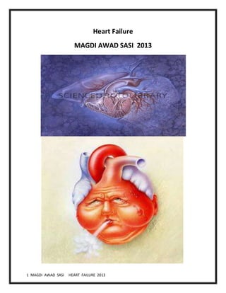
- 1. 1 MAGDI AWAD SASI HEART FAILURE 2013 Heart Failure MAGDI AWAD SASI 2013
- 2. 2 MAGDI AWAD SASI HEART FAILURE 2013 DEFINITION Heart (or cardiac) failure: pathophysiological state in which the heart is unable to pump blood at a rate commensurate with the requirements of the metabolizing tissues or can do so only from an elevated filling pressure According to AHA, HF is a clinical syndrome including circulatory congestion or inadequate tissue perfusion , due to abnormal heart function and associated neurohormonal abnormalities. An inadequate or decreased cardiac output which causes an increase in the blood volume within the vascular system. The resulting congestion within the venous system interferes with the movement of body fluids, resulting in fluid accumulation in the tissue spaces, causing edema Frequency : United States • More than 3 million people have congestive heart failure (CHF), and more than 400,000 new patients present yearly. The prevalence rate is 1-2%. Race Blacks are 1.5 times more likely to die of CHF than whites are. Nevertheless, black patients appear to have similar or lower in-hospital mortality rates than white patients. Sex Prevalence is greater in males than in females in patients aged 40-75 yrs. No sex predilection is noted among patients older than 75 years. Age Prevalence of CHF increases with increasing age and affects about 10% of the population older than 75 years
- 3. 3 MAGDI AWAD SASI HEART FAILURE 2013 Mortality/Morbidity Approximately 30-40% of patients with congestive heart failure (CHF) are hospitalized every year. CHF is the leading diagnosis-related group (DRG) among hospitalized patients older than 65 years. 35% will die within one year of diagnosis. Less than 50% of patients with HF have typical physical signs. Less than 50% of patients being correctly identified during the initial consultation. 50% readmission rate within 6 months 50% of HF patients will die 5 years after the diagnosis The most common cause of death is progressive heart failure, but sudden death may account for up to 45% of all deaths. Patients with coexisting insulin-dependent diabetes mellitus have a significantly increased mortality rate. Cardiac Physiology (remember this?) • CO = SV x HR • HR: parasympathetic and sympathetic tone • SV: preload, afterload, contractility Preload • Def: Passive stretch of muscle prior to contraction • Measurement: Swan-Ganz – LVEDP( venous return) • Really a function of diastole
- 4. 4 MAGDI AWAD SASI HEART FAILURE 2013 • Affected by compliance – Low compliance = higher LVEDP with lower LVEDV Afterload • Def: Force opposing/stretching muscle after contraction begins • Measurement: SVR Contractility • Def: Normal ability of the muscle to contract at a given force for a given stretch, independent of preload or afterload forces • In other words: – How healthy is your heart muscle? • Ischemia,infarction, Hypertrophy (?), Muscle loss Pathophysiology • Hemodynamic changes • Neurohormonal changes • Cellular changes 1.Hemodynamic changes: • HF can be secondary to systolic dysfunction or diastolic dysfunction • 2. Cellular changes • Changes in Ca+2 handling • Changes in adrenergic receptors: Slight in α1 receptors
- 5. 5 MAGDI AWAD SASI HEART FAILURE 2013 β1 receptors desensitization followed by down regulation • Changes in contractile proteins • Program cell death (Apoptosis) • Increase amount of fibrous tissue • 3. Neurohormonal changes Neurohormonal changes N/H changes Favorable effect Unfavorable effect Sympathetic activity HR , contractility, vasoconst. Venous return, filling Arteriolar constriction After load workload O2 consumption Renin-Angiotensin – Aldosterone Salt & water retention Venous Return Vasoconstriction after load Vasopressin Same effect Same effect interleukins &TNF- May have roles in myocyte hypertrophy Apoptosis Endothelin Vasoconstriction VR After load Classifying Heart Failure • Forward Vs Backward • Rt. Vs Lt. sided HF • Acute Vs Chronic HF • Low Vs High output HF • Systolic Vs Diastolic HF
- 6. 6 MAGDI AWAD SASI HEART FAILURE 2013
- 7. 7 MAGDI AWAD SASI HEART FAILURE 2013 • A etiology It is a common end point for many diseases of cardiovascular system 1. Vascular – Ischemic heart disease, myocardial infarction 2. Valvular – stenosis/regurgitation( RHD, infective endocarditis) 3. Preesure- hypertension 4.Muscle- cardiomyopathy 5. Rhythm- atrial fibrillation Symptoms: Left ventricular failure- The patient present with chest symptoms which may delay the diagnosis and keep the patient to seek chest consultants advice. Failure of forward flow from left ventricle into the aorta result into pulmonary congestion with chest symptoms. Since the left ventricle does not empty completely, it cannot accept blood returning from the lungs via the pulmonary veins. The pulmonary veins become engorged and fluid seeps out through the veins and collects in the lungs. LVF can be acute or chronic depending on the underlying cause of heart disease LVF= CHEST SYMTOMS= D/D OF ALL LUNG DISEASES= CHEST FINDING
- 8. 8 MAGDI AWAD SASI HEART FAILURE 2013 LVF symptoms are: Dyspnea at rest Dyspnea upon exertion: This has been found to be the most sensitive symptom reported, yet the specificity for dyspnea is less than 60%. Orthopnea and paroxysmal nocturnal dyspnea (PND): These symptoms are observed; however, the sensitivity for orthopnea and PND is only 20- 30%. Cough: Cough that produces pink, frothy sputum is highly suggestive of congestive heart failure (CHF).The sputum is whitish , soap like , watery. Wheezing= new onset of wheezing above age of 60 years or wheezing in elder diabetic or wheezing all of a sudden in HTN patient is cardiac asthma until prove other wise. Palpitation in arrhythmia Chest pain or tightness especially if the cause is vascular(IHD/ACS). Non specific symptoms of low COP Malaise, Weakness,Light headedness, fatigue, dizziness ,sweating ,lose of effort.
- 9. 9 MAGDI AWAD SASI HEART FAILURE 2013 Right ventricular heart failure The patient present with abdominal symptoms which may delay the diagnosis and keep the patient to seek gastroenterologist advice. Failure of forward flow from right ventricle into the pulmonary artery results into systemic congestion with abdominal symptoms. Failure of the right ventricle to maintain a normal output of blood. Since the right ventricle does not empty completely, it cannot fully accept blood returning from the body. Pressure builds in the veins of the body causing fluid to seep out and collect in the cells of the body – especially the extremities. Engorgement of the systemic veins produces pitting edema, enlargement of the liver, and ascites.
- 10. 10 MAGDI AWAD SASI HEART FAILURE 2013 RVF can be acute or chronic depending on the underlying cause of heart disease RVF= ABDOMENAL SYMTOMS= D/DOF ABDOMENAL DISEASES= ABD FINDING. Right-sided or right ventricular (RV) heart failure usually occurs as a result of left-sided failure. When the left ventricle fails, increased fluid pressure is, in effect, transferred back through the lungs, ultimately damaging the heart's right side. When the right side loses pumping power, blood backs up in the body's veins. This usually causes swelling in the legs and ankles. RVF symptoms are: Abdominal distention Right hypochondrial pain Epigastric fullness, early satiety , nausea , vomiting(stomach congestion) Change of bowel habit –constipation, diarrhea, malabsorption Bilateral leg swelling Yellowish coloration of sclera Change of urine frequency and colour These symptoms are caused by internal abdominal organs congestion And can be mistaken for local abdominal pathology. RVF is caused by: 1. Left ventricular failure - most common cause- mitral stenosis 2. Pulmonary hypertension caused by chronic lung disease (cor pulmonale) 3. Cardimyopathies and myocarditis
- 11. 11 MAGDI AWAD SASI HEART FAILURE 2013 SIGNS: General appearance Patients with mild heart failure appear to be in no distress after a few minutes of rest, but they may be obviously dyspneic during and immediately after moderate activity. Patients with LV failure may be dyspneic when lying flat without elevation of the head for more than a few minutes. Those with severe heart failure appear anxious and may exhibit signs of air hunger in this position. Patients with recent onset of heart failure are generally well nourished, but those with chronic severe heart failure are often malnourished and sometimes even cachectic. Chronic marked elevation of systemic venous pressure may produce exophthalmos and severe tricuspid regurgitation and may lead to visible pulsation of the eyes and of the neck veins. Central cyanosis, icterus, and malar flush may be evident in patients with severe heart failure. In mild or moderate heart failure, stroke volume is normal at rest; in severe heart failure, it is reduced, as reflected by a diminished pulse pressure and a dusky discoloration of the skin. With very severe heart failure, particularly if cardiac output has declined acutely, systolic arterial pressure may be reduced. The pulse may be weak, rapid, and thready; the proportional pulse pressure (pulse pressure/systolic pressure) may be markedly reduced. The proportional pulse pressure correlates reasonably well with cardiac output. Evidence of increased adrenergic activity Increased adrenergic activity is manifested by tachycardia, diaphoresis, pallor, peripheral cyanosis with pallor and coldness of the extremities, and obvious distention of the peripheral veins secondary to venoconstriction. Diastolic arterial pressure may be slightly elevated. Pulmonary rales =LVF Rales heard over the lung bases( bilateral basal end inspiratory) are characteristic of heart failure of at least moderate severity. The absence of rales certainly does not exclude elevation of pulmonary capillary pressure due to LV failure.
- 12. 12 MAGDI AWAD SASI HEART FAILURE 2013 Protodiastolic (S3) gallop: This is the earliest cardiac physical finding in decompensated heart failure in the absence of severe mitral or tricuspid regurgitation or left-to-right shunts. LVF=CHEST-B/L BASAL INSPIRATORY CREPITATION + HEART-S3 Systemic venous hypertension: This is manifested by jugular venous distention. Normally, jugular venous pressure declines with respiration; however, it increases in patients with heart failure, a finding known as the Kussmaul sign. This reflects an increase in right atrial pressure and therefore right-sided heart failure. Hepatojugular reflux: This represents distension of the jugular vein induced by applying manual pressure over the liver. The patient's body should be positioned at a 45 º angle. This is found in patients with elevated left-sided filling pressures and reflects elevated capillary wedge pressure and left-sided heart failure. Edema Bilateral pitting pedal odema. Usually, a substantial gain of extracellular fluid volume (ie, a minimum of 5 L in adults) must occur before peripheral edema is manifested. Edema, in the absence of dyspnea or other signs of LV or RV failure, is not solely indicative of heart failure and can be observed in many other conditions, including chronic venous insufficiency, nephrotic syndrome, or other syndromes of hypoproteinemia or osmotic imbalance. Hepatomegaly Hepatomegaly is prominent in patients with chronic right-sided heart failure, but it may occur rapidly in acute heart failure. When occurring acutely, the liver is usually tender. In patients with considerable tricuspid regurgitation, a prominent systolic pulsation of the liver, attributable to an enlarged right atrial V wave, is often noted. A presystolic pulsation of the liver, attributable to an enlarged right atrial A wave, can occur in tricuspid stenosis, constrictive pericarditis, restrictive cardiomyopathy involving the RV, and pulmonary hypertension (primary or secondary).
- 13. 13 MAGDI AWAD SASI HEART FAILURE 2013 Hydrothorax (pleural effusion) Hydrothorax is most commonly observed in patients with hypertension involving both systemic and pulmonary systems. Hydrothorax is usually bilateral, although when unilateral, it is usually confined to the right side of the chest. When hydrothorax develops, dyspnea usually intensifies because of further reductions in vital capacity. Ascites This finding occurs in patients with increased pressure in the hepatic veins and in the veins draining into the peritoneum. Ascites usually reflects long-standing systemic venous hypertension. Pulsus alternans ( one strong and one weak beat) Pulsus alternans occurs most commonly in heart failure due to increased resistance to LV ejection, as occurs in hypertension, aortic stenosis, coronary atherosclerosis, and dilated cardiomyopathy. It is usually associated with an S3 gallop, signifies advanced myocardial disease, and often disappears with treatment of heart failure. Accentuation of P2 heart sound, S3 gallop, and systolic murmurs This accentuation is a cardinal sign of increased pulmonary artery pressure. It disappears or improves after treatment of heart failure. Mitral and tricuspid regurgitation murmurs are often present in patients with decompensated heart failure because of ventricular dilatation. These murmurs often disappear or diminish when compensation is restored. Note that correlation between the intensity of the murmur of mitral regurgitation and its significance in patients with heart failure is poor. Severe mitral regurgitation may be accompanied by soft murmur. The presence of an S3 gallop in adults is important, pathologic, and often the most apparent finding on cardiac auscultation in patients with significant heart failure. Cardiac cachexia Cardiac cachexia is found in long-standing heart failure, particularly of the RV, because of anorexia from hepatic and intestinal congestion and sometimes because of digitalis toxicity. Occasionally, impaired intestinal absorption of fat and (rarely) protein-losing enteropathy occur.
- 14. 14 MAGDI AWAD SASI HEART FAILURE 2013 Left heart failure IHD, Myocarditis, Valvular heart diseases Forward failure Backward failure ↓ cardiac output Tissue anoxia ↓ renal perfusion Activation of RAAS PULMONARY CONGESTION and OEDEMA Na+, H2O retention Residual blood in left ventricle Left atrial pressure and volume Pressure in pulmonary venous circulation Pulmonary arterial hypertension Right ventricular pressure SYSTEMIC VENOUS CONGESTION and PERIPHERAL OEDEMA Right heart failure Right side valvular disease Rt side myocardial disease Pulmonary hypertension
- 15. 15 MAGDI AWAD SASI HEART FAILURE 2013 Precipitating Factors • Infection Sodium Intake • Medications!!! Anemia • Thyroid disorders Endocarditis • Pulm Embolus Noncompliance • Arrhythmia Myocardial Infarction • Stress reaction Diagnosis of CHF / Routine Tests: CBC count INVESIVE Electrolytes 1. Exercise stress test Renal function tests 2. Cardiac catheterization Liver function tests 3. Holter monitor B-type natriuretic peptide ECG Chest x-ray Echocardiogram CXR Cardiomegaly Vascular redistribution Kerley B lines Interstitial edema Peri-bronchial “cuffing” Effusions
- 16. 16 MAGDI AWAD SASI HEART FAILURE 2013
- 17. 17 MAGDI AWAD SASI HEART FAILURE 2013 Chest radiographs in patients with abrupt onset are usually helpful but can be limited because a delay of as long as 12 hours is possible from the onset of dyspnea due to acute heart failure to the development of classic abnormal findings on radiographs. Classic radiographic findings demonstrate cardiomegaly (in patients with underlying CHF) and alveolar edema with pleural effusions and bilateral infiltrates in a butterfly pattern. The other signs are loss of sharp definition of pulmonary vasculature, haziness of hilar shadows, and thickening of interlobular septa (Kerley B lines). In long standing biventricular chronic heart failure, chest radiographs may only show cardiomegaly without alveolar edema or pleural effusions due to adaptive lung mechanism with increased arterial vasoconstriction and lymphatic drainage.
- 18. 18 MAGDI AWAD SASI HEART FAILURE 2013 Electrocardiography o The presence of left atrial enlargement and LV hypertrophy is sensitive (although nonspecific) for chronic LV dysfunction. o ECG may suggest an acute tachyarrhythmia or bradyarrhythmia. o ECG may aid in the diagnosis of acute myocardial ischemia or infarction as the cause of heart failure or may suggest the likelihood of prior myocardial infarction or presence of coronary artery disease as the cause of heart failure. o ECG is of limited help when an acute valvular abnormality or LV systolic dysfunction is considered to be the cause of heart failure; however, the presence of left bundle branch block (LBBB) on an ECG is a strong marker for diminished LV systolic function Echocardiogram Chamber enlargement Wall motion abnormalities Diminished ejection fraction Possible LVH Possible valvular problems Assess diastolic dysfunction
- 19. 19 MAGDI AWAD SASI HEART FAILURE 2013 Heart disease (any) Hypertension Diabetes, Hypercholesterolemia Asymptomatic LV dysfunction Systolic / Diastolic Marked symptoms at rest despite max. therapy Dyspnea, Fatigue Reduced exercise tolerance Stages in the Evolution of Heart Failure A B C D AHA guidelines 2001 Treatment Goals • Improve symptoms A. Enhance well-being and quality of life B. Increase exercise tolerance • Improve survival A. Prevent progressive heart failure B. Prevent sudden death C. Prevent thromboembolic episodes
- 20. 20 MAGDI AWAD SASI HEART FAILURE 2013 • Treatment :: • Oxygen – nasal, BiPAP, intubation • Fluid Balance Restrict fluid- 1000ml daily/ salt intake Monitor Input/Outputs and daily weight Dialysis if needed -Critical renal failure patients Aspirin 75mg Promoting Rest and Activity Bed rest or limited activity may be necessary during the acute phase Provide an overbed table close to the patient to allow resting the head and arms Use pillows for added support. Gradual ambulation is encouraged to prevent risk of venous thrombosis and embolism due to prolonged immobility Activities should progress through dangling, sitting up on a chair and then walking in increased distances under close supervision. Assess for signs of activity intolerance (dyspnea, fatigue and increased pulse rate that does not stabilize readily) Providing Skin Care Edematous skin is poorly nourished and susceptible to pressure sores Change position at frequent intervals Assess the sacral area regularly Use protective devices to prevent pressure sores Promoting Elimination Advise to avoid straining at defecation which involves Valsalva manoeuvre. Administer laxative as ordered Encourage use of bedside commode
- 21. 21 MAGDI AWAD SASI HEART FAILURE 2013 Preload Reduction – Loop diuretics Lasix 20-200mg IV (q 6-8 hours) – Nitrates -Nitroglycerin IV:10-200 mcg/min – ACEi / ARB Captopril 6.25-50mg PO q8h Enalapril 2.5-20mg PO BID Afterload Reduction – IV NTG, Nitroprusside – Hydralazine 10-100mg PO q6-8 h – ACE-I / ARB Ionotropic Support – Dopamine / Dobutamine 500mg in 250cc D5W 3-10 cg/kg/min
- 22. 22 MAGDI AWAD SASI HEART FAILURE 2013 – Amrinone / Milrinone n Digoxin (chronic) – Mechanical (ABP) Cardiogenic shock unresponsive to above tx A.-----------Diuretics • volume overload , sodium overload , preload reduction Advantages • Highly effective in most classes • Essential with fluid retention • Well tolerated, simple to use Disadvantages • Electrolyte abnormalities Na, k ,Ca ,Mg • Hypovolemia, hypotension, renal dysfunction • Activation of neurohormaonal system AIM----Elimination of symptoms and/or signs of congestion • Avoid volume depletion A. Postural hypotension B. Increase in heart rate C. Increase in BUN/Cr D. Neuroendocrine activation Thiazide Diuretics-----Hydrochlorothiazide, Chlorthalidone , Metolazone Loop Diuretics----------Furosemide , Torsemide Potassium Sparing Diuretics---Spironolactone , Triamterene , Amiloride Spironolactone : Aldosterone inhibition minimize potassium loss, prevent sodium and water retention, endothelial dysfunction and myocardial fibrosis.
- 23. 23 MAGDI AWAD SASI HEART FAILURE 2013 B-------------ACE Inhibitors ACE inhibitors should be the initial treatment for heart failure • Improve hemodynamic status • Attenuate neurohumoral abnormalities • Improve symptoms, left ventricular ejection fraction . • Reduce incidence of hospitization • Slow progression • Reduce mortality Neurohormonal Changes • Decreased angiotensin II. • Reduction in arterial resistance (afterload).
- 24. 24 MAGDI AWAD SASI HEART FAILURE 2013 • Reduction in venous tension (preload). • Inhibition of cardiac and vascular remodeling . • Increased bradykinin • Decreased or no change in aldosterone • Decreased norepinephrine Reduction in Sudden Death/Potential Mechanisms • Increase in serum/total body potassium • Decreased adrenergic stimulation • Reduced heart size and decrease in ventricular hypertrophy • Prevention of myocardial ischemia • Prevention of progressive myocardial damage FDA Approved • Captopril ,Enalapril ,Lisinopril ,Quinapril ,Trandolapril ,Fosinopril Adverse effects : Dry irritating persistent cough Hyperkalemia Angioedema Fetal toxicity
- 25. 25 MAGDI AWAD SASI HEART FAILURE 2013 CHF: ACE Inhibitor Doseages Captopril Enalapril Lisinipril Quinapril • Start: 6.25 bid/tid • Usual: 6.25-50 bid/tid • Start: 2.5 qd/bid • Usual: 2.5-10 bid • Start: 2.5-5 qd • Usual: 5-20 qd • Start: 5 bid • Usual 10-20 bid C---------------Beta Blockers • Primary mechanism is inhibition of down regulation of beta receptors • Additional mechanisms A. Restore receptor density B.Protect against cardiotoxicity of catecholeamines C. Improve systolic/diastolic function in ischemic myocardium • First Generation: Beta 1 and Beta 2 Propranolol/Timolol • Second Generation: Beta 1 Metoprolol/Atenolol • Third Generation: Vasodilating Properties Carvedilol Improve symptoms and clinical class
- 26. 26 MAGDI AWAD SASI HEART FAILURE 2013 • Degree of benefit appears to relate to degree of disability before treatment • Should be used in all stable Class II/III patients unless contraindicated • Treatment should not be initiated in patients with acutely decompensated CHF • Clinical response may take 2 to 3 months Risks of Treatment • Hypotension Fluid retention and worsening CHF • Bradycardia and heart block
- 27. 27 MAGDI AWAD SASI HEART FAILURE 2013 CHF: Beta Blockers • Carvedilol • Metoprolol • Bisoprolol • Start: 3.125 mg bid • Usual: 25 mg bid • Start: 12.5-25 mg qd • Usual: 50-100 mg bid • Start: 1.25 mg qd • Usual: 5-10 mg qd Start at low dose and monitor for bradycardia Carvedilol and Metoprolol are the most commonly used for CCF amongst beta blockers D------------Angiotensin Receptor AT-1 blockers (ARB) : Losartan, Irbesartan, Candesartan Competitive antagonists of Angiotensin II (AT-1). No inhibition of ACE or Cough.
- 28. 28 MAGDI AWAD SASI HEART FAILURE 2013 E--------------Vasodilators : Isosorbide dinitrate and hydralazine also used specially in patients who cannot tolerate ACE inhibitors. Amlodipine and prazosin are other vasodilators can be used in CCF.
- 29. 29 MAGDI AWAD SASI HEART FAILURE 2013 F-------Cardiac glycosides : Digoxin : Inhibition of Na/K ATPase pump increase intracellular sodium concentration – eventually increase cytosolic calcium. It restores the vagal tone and abolishes the sympathetic over activity. Has positive inotropic (strengthens force of cardiac contractility) and negative chronotropic effects (decreases heart rate. Increase the refractoriness of AV node thus decrease ventricular response to atrial rate.
- 30. 30 MAGDI AWAD SASI HEART FAILURE 2013 Digoxin is used as a first-line drug in patients with congestive heart failure who are in atrial fibrillation. Adverse effects / Precautions : Nausea, vomiting, gynecomastia, visual disturbances and psychosis. Ventricular bigeminy, AV block and bradycardia. Antidote for Toxicity: Digibind Nursing Responsibilities Assess heart rate before administration; if below 60 bpm or above 120 bpm, withhold the drug. Monitor serum potassium Assess for signs of Digitalis toxicity - Bradycardia - GI manifestations (anorexia, nausea, vomiting and diarrhea) - Dysrrhythmias - Altered visual perceptions - In males: gynecomastia, decreased libido and impotence Amiodarone and verapamil can increase the plasma concentration of digoxin by inhibiting its excretion IN DIASTOLIC FAILURE; USE B-BLOCKER, ACE-I , CALCIUM CHANNEL BLOCKER RATE LIMITING • Difficult to treat • Diuretics for volume overload. Avoid volume depletion • Prevent tachycardia • Rate-limiting calcium channel blockers first choice • Beta 1 beta blockers second choice
- 31. 31 MAGDI AWAD SASI HEART FAILURE 2013 ADVERSE PROGNOSTIC MARKERS IN CHRONIC HEART FAILURE • Old Age, • Severity of heart failure (NYHA class) • Left ventricular dysfunction, • Diabetes Mellitus, • Raised creatinine, • Hyponatremia , Hypoalbuminaemia,Anaemia • Presence of arrhythmia : AF / VT Causes of Mortality in Heart Failure • Pump failure • Arrhythmia • Severe Anaemia • Associated serious co-morbidities i.e. Renal failure Cardiac Inotropes = • Dopamine acts at a variety of receptors (dose dependant) • Rapid elimination- can only be administered as a continuous infusion Dobutamine • Stimulates beta-adrenergic receptors and produces a positive inotropic response • Unlike the vasoconstriction seen with high doses of dopamine, dobutamine produces a mild vasodilatation .
- 32. 32 MAGDI AWAD SASI HEART FAILURE 2013 PDE Phosphodiesterase inhibitors • Inamrinone (amrinone) and Milrinone (bipyridines) • Acts by inhibiting the enzyme Phosphodiesterase • Thus lead to increase of intracellular concentrations of cAMP • cAMP is responsible for the conversion of inactive protein kinase to active form • Protein kinases are responsible for phosphorylation of Ca channels • Thus causing increased Ca entry into the cell. MECHANISM OF ACTION: • Increase myocardial contractility by increasing the Ca influx during AP • Also have vasodilating effect • Selective for PDE isoenzyme-3 (found in cardiac and smooth muscle) ADVERSE RECTIONS • Inamrinone: nausea, vomiting, arrhythmias, thrombocytopenia and liver enzyme changes • Withdrawn in some countries • Milrinone: arrhythmias, less likely to cause other ADR Niseritide • Brain (B-type) natriuretic peptide (BNP) is secreted constitutively by ventricular myocytes in response to stretch • BNP binds to receptors in the vasculature, kidney, and other organs, producing potent vasodilation with rapid onset and offset of action by increasing levels of cGMP
- 33. 33 MAGDI AWAD SASI HEART FAILURE 2013 • Niseritide is recombinant human BNP approved for treatment of acute decompensated CHF. • It reduces systemic and pulmonary vascular resistances, causing an indirect increase in cardiac output and diuresis. • Effective in HF because cause reduction in preload and afterload • ADR- hypotension OTHERS; Implantable cardioverter-defibrillator Pacemakers, Biventricular pacemaker Heart transplant Cardiac Resynchronisation Therapy
- 34. 34 MAGDI AWAD SASI HEART FAILURE 2013