Nikon Small World 2016: Winners & Honorable Mentions
•Transferir como PPSX, PDF•
5 gostaram•505 visualizações

Nikon Small World 2016: Winners & Honorable Mentions

Recomendados
Mais conteúdo relacionado
Semelhante a Nikon Small World 2016: Winners & Honorable Mentions
Semelhante a Nikon Small World 2016: Winners & Honorable Mentions (20)
Mais de maditabalnco
Mais de maditabalnco (20)
Último
Último (20)
Nikon Small World 2016: Winners & Honorable Mentions
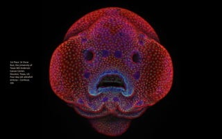
- 1. 1st Place: Dr Oscar Ruiz, the University of Texas MD Anderson Cancer Center, Houston, Texas, US: Four-day-old zebrafish embryo – Confocal, 10x
- 2. 2nd Place: Douglas L Moore, University of Wisconsin – Stevens Point Museum of Natural History, Stevens Point, Wisconsin, USA: Polished slab of Teepee Canyon agate – Stereomicroscopy, 90x
- 3. 3rd Place: Rebecca Nutbrown University of Oxford, Nuffield Department of Clinical Neurosciences Oxford, United Kingdom: Culture of neurons (stained green) derived from human skin cells, and Schwann cells, a second type of brain cell (stained red) – Confocal/Immunofluoresce nce/iPSCs, 20x
- 4. 4th Place: Jochen Schroeder, Chiang Mai, Thailand: Butterfly proboscis – Image Stacking, 6.3x
- 5. 5th Place: Dr Igor Siwanowicz, Howard Hughes Medical Institute (HHMI), Janelia Research Campus, Ashburn, Virginia, USA: Front foot (tarsus) of a male diving beetle – Confocal, 100
- 6. 6th Place: Marek Mis, Podlaskie, Poland: Air bubbles formed from melted ascorbic acid crystals – Polarized Light, 50x
- 7. 7th Place: Dr David Maitland, Feltwell, United Kingdom: Leaves of Selaginella (lesser club moss) – Differential Interference Contrast, 40x
- 8. 8th Place: Samuel Silberman, Monoson Yahud, Israel: Wildflower stamens – Fiber Optic Illumination, 40x
- 9. 9th Place: Vin Kitayama and Sanae Kitayama, Vinsanchi Art Museum, Azumino, Nagano, Japan Espresso coffee crystals – Polarized Light
- 10. 10th Place: Rogelio Moreno Gill, Panama, Panama: Frontonia (single-celled organism) showing ingested food, cilia, mouth and trichocysts – Differential Interference Contrast, 200x
- 11. 11th Place: Francis Sneyers Brecht, Belgium: Scales of a butterfly wing underside (Vanessa atalanta) – Macroscopy, 10x
- 12. 12th Place: Dr Dylan Burnette, Vanderbilt University School of Medicine Nashville, Tennessee, USA – Human HeLa cell undergoing cell division (cytokinesis). DNA (yellow), myosin II (blue) and actin filaments (red) – Structured Illumination, 9x
- 13. 13th Place: Walter Piorkowski, South Beloit, Illinois, USA: Poison fangs of a centipede (Lithobius erythrocephalus) – Fibre Optic Illumination/Image Stacking, 16x
- 14. 14th Place: Dr Keunyoung Kim, University of California, San Diego, National Center for Microscopy and Imaging Research, La Jolla, California, USA: Mouse retinal ganglion cells – Fluorescence/Confocal 40x
- 15. 15th Place: Geir Drange, Asker, Norway: Head section of an orange ladybird (Halyzia sedecimguttata) – Reflected Light/Focus Stacking, 10x
- 16. 16th Place: Stefano Barone, Diatom Shop, Palazzo Pignano, Italy: 65 fossil Radiolarians (zooplankton) carefully arranged by hand in Victorian style – Darkfield, 100x
- 17. 17th Place: Jose Almodovar, University of Puerto Rico, Mayaguez Campus, Biology Department, Mayaguez, Puerto Rico: Slime mould (Mixomicete) – Image Stacking/Reflected Light, 5x
- 18. 18th Place: Pia Scanlon, Department of Agriculture and Food, Western Australia: Parts of wing-cover (elytron), abdominal segments and hind leg of a broad-shouldered leaf beetle (Oreina cacaliae) – Stereomicroscopy, Image Stacking, 40x
- 19. 19th Place: Dr Gist F Croft, Lauren Pietilla, Stephanie Tse, Dr. Szilvia Galgoczi, Maria Fenner, Dr Ali H. Brivanlou, Rockefeller University, Brivanlou Laboratory New York, New York, USA: Human neural rosette primordial brain cells, differentiated from embryonic stem cells – Confocal, 10x
- 20. 20th Place: Michael Crutchley, Haverfordwest, Pembrokeshire, United Kingdom: Fungi on cow dung – Darkfield, 30x
- 22. Zebrafish fin with cylindrical bone segments and rows of pigmented epithelial cells (50x).Dr. Leonardo Andrade
- 23. Scales of a butterfly wing (10x). Evan Darling
- 24. Dentate gyrus of a optically-cleared transgenic mouse brainin 3D (10x).Hei Ming Lai & Dr. Wutian Wu
- 25. Retinal ganglion cellsin the whole-mounted mouse retina (20x). Dr. Keunyoung Kim
- 26. Micrasterias thomasiana (algae) (400x). Jacek Myslowski
- 27. Tail of a a small shrimp (40x). Dr. Charles Krebs
- 28. Seeds of an Indian Paintbrush wildflower (Castilleja indivisa) (4x). David Millard
- 29. Curvepod Fumewort (Corydalis curvisiliqua) seed (4.5x). David Millard
- 30. Leg of a water boatman (Corixidae) (25x). Marek Miś
- 31. Interior of a bisected trap of a humped bladderwort (Urticularia gibba), a fresh water carnivorous plant (100x). Dr. Igor Siwanowicz
- 32. Trumpet animalcule containing endosymbionts (160x). Wim van Egmond
- 33. Wildflower stamens (40x). Samuel Silberman
- 34. Caudal gill of a dragonfly larva (25x). Marek Miś
- 35. Gears coupling hind legs of a planthopper nymph (250x).Dr. Igor Siwanowicz