
Recomendados
Mais conteúdo relacionado
Mais procurados
Mais procurados (20)
Destaque
Destaque (20)
Semelhante a Gel electrophoresis native, denaturing&reducing
Semelhante a Gel electrophoresis native, denaturing&reducing (20)
Mais de Lovnish Thakur
Mais de Lovnish Thakur (20)
Último
Último (20)
Gel electrophoresis native, denaturing&reducing
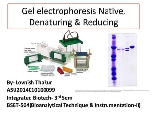
- 1. Gel electrophoresis Native, Denaturing & Reducing By- Lovnish Thakur ASU2014010100099 Integrated Biotech- 3rd Sem BSBT-504(Bioanalytical Technique & Instrumentation-II) http://www.google.co.in/imgres?imgurl=http://genotech.ir/ckfinder/userfiles/images/proteanmini(1).jpg&imgrefurl=http://genotech.ir/Training/Details.aspx?ID%3D15&h=307&w=440&tbnid=3yosiKz1AOFetM: &docid=0KtNzihlsekF6M&ei=JEEvVuT8C4WwmAWut7wg&tbm=isch&ved=0CGwQMyhJMElqFQoTCOSe6t2o4sgCFQUYpgodrhsPBA
- 2. Gel electrophoresis Electrophoresis is a technique used to separate and sometimes purify macromolecules - especially proteins and nucleic acids - that differ in size, charge or conformation.
- 4. SDS-PAGE(Denaturing gel electrophoresis) . Denaturing the proteins nullifies structural effects on mobility, allowing separation on a true charge/mass ratio basis. The most commonly used denaturant is sodium dodecyl sulfate (SDS). Proteins solubilized in SDS bind the detergent uniformly along their length to a level of 1.4 g SDS/g protein. This creates a charge/mass ratio which is consistent between proteins. For this reason, separation on a polyacrylamide gel in the presence of SDS occurs by mass alone..
- 6. Chemical Requirement Acrylamide solutions Ammonium persulfate is used as a catalyst for the copolymerization of acrylamide and bisacrylamide gels. SDS (0.1%) Protein standard molecular-weight markers Protein samples to be resolved SDS stock solution for resolving and stacking gels 1× SDS gel-loading buffer 0.2% (w/v) bromophenol blue 20% (v/v) glycerol 200 mM β-mercaptoethanol TEMED (electrophoresis grade) Tris-glycine buffers for SDS-polyacrylamide gels.
- 7. Procedure Gel casting • Assemble the glass plates according to the manufacturer’s instructions. • Make gel using desired concentration of acrylamide and bisacrylamide and Ammonium persulfate as a catalyst for copolymerization. • Pour the acrylamide solution into the gap between the glass plates. • After polymerization of resolving gel is complete (30 minutes), pour off the overlay and wash the top of the gel several times with de- ionized H2O to remove any unpolymerized acrylamide. • Pour the stacking gel solution directly onto the surface of the polymerized resolving gel. • Immediately insert a clean Teflon comb into the stacking gel solution.
- 8. Loading • After polymerization is complete (30 minutes), remove the Teflon comb carefully. • Mount the gel in the electrophoresis apparatus. Add Tris-glycine electrophoresis buffer to the top and bottom reservoirs. • Load up to 15 μl of each of the samples in a predetermined order into the bottom of the wells.
- 9. Run gel Attach the electrophoresis apparatus to an electric power supply • Apply a voltage • Run the gel until the bromophenol blue reaches the bottom of the resolving gel. • Turn off the power supply. • Remove the glass plates from the electrophoresis apparatus and place them on a paper towel. • At this stage, the gel can be fixed, stained with Coomassie Brilliant Blue or silver salts.
- 11. Reducing • In Reducing gel electrophoresis mercaptoetanol is used which assist SDS in denaturing by reducing the disulphide bond in protein that is the reason it is called reducing electrophoresis. • The formation of nonlinear species can effect mobility - ie, if you have an intra chain disulfide bond in a monomer, it may run differently than the reduced monomer.
- 12. Native PAGE • Sometime, we need to separate protein in non- denaturing conditions. This type of polyacrylamide gel electrophoresis is also called native gel electrophoresis because protein remains in native form even after electrophoresis Difference between Native & SDS PAGE • The basic difference in the native gel electrophoresis (native-PAGE) is the electrophoresis buffer does not contain SDS. • Also, loading buffer does not have SDS and reducing agents and samples are not boiled. • Rest of the things are similar to SDS- PAGE gel electrophoresis
- 13. ADVANTAGE • Biological activity of protein remain intact • Native PAGE is used for separation of enzymes/isozymes.
- 14. APPLICATIONS
- 15. Quantification of muscle mitochondria oxidative phosphorylation enzyme via histochemical staining of blue native polyacrylamide gel Summary- In this blue-Native Polyacryamide gel electrophoresis is used to separate intact multi-subunit complex of enzyme. For particular enzyme separation require denaturing PAGE. In this they demonstrate that they can study enzyme activity using these technique. And compare the number of mitochondria in normal and heart cell by quantification of phosphorylation enzyme.
- 16. Separation and identification of hen egg protein isoforms using SDS–PAGE and 2D gel electrophoresis with MALDI-TOF mass spectrometry Summary- High-resolution techniques for proteome analysis, including SDS–PAGE and 2-dimensional (2D) gel electrophoresis, combined with mass- spectrometry, were employed to separate and identify several protein components in hens egg. An advanced and sensitive glycoprotein staining kit was used to detect the presence of glycosylated proteins in the egg samples. Numerous spots were revealed when a mixture of egg white and yolk was subjected to 2D gel electrophoresis. Several of the already known egg proteins were identified. Result- Isoforms of ovalbumin and conalbumin were visualised.
- 17. Reference • http://www.ispybio.com/search/protocols/sds%2 0protocol29.pdf • http://www.protocol-online.org/biology- forums/posts/12890.html • https://classes.soe.ucsc.edu/bme220l/Spring11/ Reading/SDS-PAGE-theory.pdf • http://www.researchgate.net/publication/22338 5062_Separation_and_identification_of_hen_egg _protein_isoforms_using_SDS-PAGE_and_2- D_gel_electrophoresis_with_MALDI- TOF_mass_spectrometry._Food_Chem
- 18. THANK YOU