Hereditary Hemochromatosis
•Transferir como PPTX, PDF•
30 gostaram•9,243 visualizações

celtic curse

Recomendados
Mais conteúdo relacionado
Mais procurados
Mais procurados (20)
Semelhante a Hereditary Hemochromatosis
Semelhante a Hereditary Hemochromatosis (20)
Último
Último (20)
Hereditary Hemochromatosis
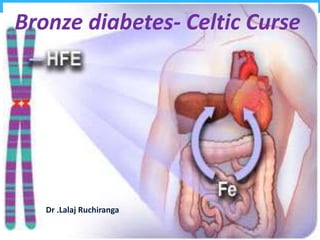
- 1. Bronze diabetes- Celtic Curse Dr .Lalaj Ruchiranga
- 2. Introduction • “Celtic Curse”- also known as “Bronze diabetes” • Hemochromatosis is the abnormal accumulation of iron in parenchymal organs, such as the liver, pancreas, and heart leading to organ toxicity. • Most common autosomal recessive genetic disorder
- 3. Epidemiology • Hereditary hemochromatosis (HH) remains the most common genetic disorder in Caucasians. • Women typically presented approximately 10 years later than men • Female: male- 1 : 10 • Population screening has shown prevalance of heterozygotes is about 10 %. • Prevalence of HH in the USA is 1 in 200-500 individuals.
- 5. Genetic basis • Principal HFE gene defect was first described in 1996, • Tightly linked to the HLA-A locus on chromosome 6p • G-to-a missense mutation ; • Cysteine tyrosine at 282 (C282Y) • C282Y homozygotes account for 80%-85% • Histidine aspartate at 63 (h63d) • Serine cysteine at 65 (s65c)
- 6. Schematic representation of the protein product of HFE
- 7. Pathophysiology (1) Increased absorption of dietary iron in the upper intestine, (2) Decreased expression of the iron-regulatory hormone hepcidin, (3) Altered function of HFE protein, (4) Tissue injury and fibrogenesis induced by iron.
- 8. Hepcidin Interactions between duodenal enterocytes, hepatocytes, and macrophages in iron homeostasis regulated by hepcidin.
- 9. Classification of iron overload syndromes Hereditary Hemochromatosis • HFE-related • C282Y/C282Y • C282Y/H63D • C282Y/S65C • Non–HFE-related • Hemojuvelin (HJV) • Transferrin receptor-2 (TfR2) • Ferroportin (SLC40A1) • Hepcidin (HAMP) • African iron overload
- 10. Classification of iron overload syndromes cont.; • Secondary Iron Overload • Iron-loading anemia • Thalassemia major • Sideroblastic anemia • Chronic hemolytic anemia • Aplastic anemia • Parenteral iron overload • Red blood cell transfusions • Iron–dextran injections • Long-term hemodialysis • Chronic liver disease • Porphyria cutanea tarda • Hepatitis C • Hepatitis B • Alcoholic liver disease • Nonalcoholic fatty liver disease
- 11. It’s a spectrum of disease • Phenotypic expression only occurs in approximately 70% of C282Y homozygotes, • Fewer than 10% of C282Y homozygotes will develop severe iron overload accompanied by organ damage and clinical manifestations of hemochromatosis
- 12. EuropeanAssociationfor the Study of Liver Diseases staging system • Stage 1 -genetic disorder +, no increase in iron stores ‘‘genetic susceptibility.’’ • Stage 2- genetic disorder +, phenotypic evidence of iron overload without tissue or organ damage. • Stage 3 genetic disorder +, with iron overload with tissue and organ damage.
- 13. Natural progression of disease
- 14. CLINICAL MANIFESTATIONS • Liver function abnormalities — 75 % • Weakness and lethargy — 74 % • Skin hyperpigmentation — 70 % • Diabetes mellitus — 48 % • Arthralgia — 44 % • Impotence in males — 45% • Electrocardiographic abnormalities — 31%
- 15. Hepatic manifestations • Liver is usually the first organ to be affected • Hepatomegaly in >95% of symptomatic patients. • Portal hypertension and esophageal varices occur less commonly than in cirrhotic. • Hepatocellular carcinoma develops in about 30% of patients with cirrhosis. • Incidence increases with age, common in men, almost exclusively in cirrhotic patients
- 16. Skin • Skin pigmentation -characteristic metallic or slate-gray hue. • Results from increased melanin and iron in the dermis. • Pigmentation usually is generalized, More pronounced on; • The face, neck, • Extensor aspects of the lower forearms, • Dorsa of the hands, lower legs, • Genital regions, • In scars.
- 17. Diabetes • Diabetes mellitus occurs in about 65% • More likely to develop in those with a family history of diabetes, • Insulin resistance is more common in association with hemochromatosis
- 18. Arthropathy • Arthropathy develops in 25–50% of symptomatic patients • Usually occurs after age 50. • 2nd and 3rd mcp joints, are usually the first joints involved. • Calcium pyrophosphate (chondrocalcinosis or pseudogout), mainly in the knee.
- 19. Calcium pyrophosphate deposition disease, atypical osteoarthritis involving the 2nd & 3rd MCP joints & KJ
- 20. Cardiac involvement • Presenting manifestation in about 15%. • Most common manifestation is congestive heart failure. • Cardiac arrhythmias ; • premature supraventricular beats, • Paroxysmal tachyarrhythmia's, • Atrial flutter, • Atrial fibrillation, • Varying degrees of AV block.
- 21. Hypogonadism •Occurs in both sexes. •Impairment of hypothalamic-pituitary function by iron deposition • Loss of libido, • Impotence, • Amenorrhea, • Testicular atrophy, • Gynecomastia, • sparse body hair.
- 22. Screening for HH • High risk groups; • Family history of HH (1st degree) • Those with suspected organ involvement • Those with chance detection of biochemical and/or radiological abnormalities • Optimal timing for screening family members is between the ages of 18 and 30, • Generally recommended that all patients with abnormal liver function have iron studies done at some point
- 23. Diagnosis 1. Transferrin saturation — • If transferrin saturation >45% • the presence of the C282Y or H63D mutation confirm the diagnosis of hemochromatosis 2.Plasma ferritin — normal 40 to 200 ng/ml • >200 mcg/L in premenopausal women • > 300 mcg/L in men and postmenopausal women indicate primary iron overload • False +ve elevations related to inflammation. • In the absence of increased iron stores in patients With necroinflammatory liver disease • Serum ferritin levels have an additional value as a predictor of advanced fibrosis and cirrhosis in confirmed HH
- 25. Liver biopsy Liver biopsy should be considered; •For the purpose of determining the presence or absence of advanced fibrosis or cirrhosis. •Screening for hcc •For measurement of HIC.
- 26. Treatment of Hemochromatosis • Phlebotomy remains the sole recommended treatment - simple, inexpensive, and safe. • Each 500 mL of whole blood removed contains 200 to 250 mg of iron. Induction phase • One phlebotomy (500 mL) one to two per week. • Check hematocrit (Hct) prior to each phlebotomy; • do not allow Hct to fall by more than 20 percent of prior level • Check serum ferritin every 10 to 12 phlebotomies
- 27. Maintenance phase • The phlebotomy should be performed every 2-4 months. • The interval between procedures is determined by the level of ferritin, which should be 50 - 100mcg/ml. • Dietary adjustments are unnecessary. • Vitamin c supplements and iron supplements should be avoided. • Check hematocrit/hemoglobin prior to each phlebotomy. • Allow hematocrit/hemoglobin to fall by no more than 20% of prior level
- 28. Response to phlebotomy treatment in patients with HH • Improved survival if diagnosis and treatment before Development of cirrhosis and diabetes • Improved sense of well-being, energy level Cardiac function Control of diabetes • Reduction of tissue iron stores to normal Of portal hypertension in patients with cirrhosis In skin pigmentation • Reversal of hepatic fibrosis (in approximately 30% of cases) • No reversal of established cirrhosis or diabetes Arthropathy Testicular atrophy • Elimination of risk of hh-related HCC if iron removal is Achieved before development of cirrhosis
- 29. ChelationTherapy • Treatment with iron chelation agents is recommended; • (heart disease, anemia, poor venous access) • Deferoxamine intravenously or subcutaneously (25 to 40 mg/kg) • IV 8-10 hours 5 nights per week. • subcutaneous bolus injections B.d • Deferasirox (exjade) orally • 100 mg/kg administered once daily 5 times a week. • Very efficient in liver iron removal. • Less effective in the splenic & pancreatic iron. • Renal functions should be monitored.
- 30. • We recommend that patients with abnormal Iron studies should be evaluated as patients with Hemochromatosis, even in the absence of symptoms.(A) • All patients with evidence of liver disease should be evaluated for hemochromatosis. (1b) AASLD Recommendations
- 31. • In a patient with suggestive symptoms, physical findings, or family history, a combination of TS and ferritin should be obtained rather than relying on a single test. (1B) if either is abnormal (TS 45% or ferritin above the upper limit of normal), then HFE mutation analysis should be performed. (1b) • We recommend screening (iron studies and hfe mutation analysis) of first-degree relatives of patients with hfe- related hh to detect early disease and prevent complications. (1a)
- 32. • Patients with hemochromatosis and iron overload should undergo therapeutic phlebotomy weekly (as tolerated). (1a) • Target levels of phlebotomy should be a Ferritin level of 50-100 lg/L. (1b) • Iron chelation with either deferoxamine mesylate or deferasirox is recommended in iron overloaded patients with dyserythropoietic syndromes or chronic hemolytic anemia. (1a)
- 33. References Diagnosis and Management of Hemochromatosis: 2011 Practice Guideline by the American Association for the Study of Liver Diseases(AASLD). UpToDate 19.3 version