

Recomendados
Mais conteúdo relacionado
Mais procurados
Mais procurados (20)
Semelhante a Nasopharngeal angiofibroma
Semelhante a Nasopharngeal angiofibroma (20)
Último
Último (20)
Nasopharngeal angiofibroma
- 1. NASOPHARYNGEAL ANGIOFIBROMA PRESENTED BY KOMAL SOOMRO 4TH YEAR MBBS ENT
- 3. INTRODUCTION: • It is a benign but locally aggressive tumor. • It is a rare tumor, though it is the commonest of all the benign tumors of nasopharynx. • The exact cause is unknown but it occurs mostly in adolescent males. it is thought to be testosterone dependant. • These patients have hamartomatous nidus of vascular tissue which get activated to form angiofibroma when male sex hormone is released.
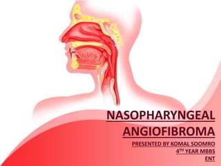
- 4. ANATOMY OF NASOPHARYNX • It opens anteriorly into the nasal cavity.
- 5. SITE OF ORIGIN AND GROWTH: It arises from the posterior part of the nasal cavity close to the superior margin of sphenopalatine foramen from here tumor grows into the nasal cavity, nasopahrynx and into the pterygopalatine fossa running behind the posterior wall of maxillary sinus. Laterally tumor extends into the pterygomaxillary fossa and thence to infratemporal fossa and cheek.
- 7. PATHOLOGY: The exact aetiology of tumor is unknown but it tends to develop in males between 10 and 25 years old. Histologically it is composed of fibrous connective tissues interspersed with variable proportion of endothelium lined blood spaces. The vessels are just endothelium lined spaces with no muscle coat therefore severe bleeding may occur on taking biopsy and surgical removal as these vessels can not contract to stop bleeding.
- 9. EXTENSIONS OF THE TUMOUR: Nasopharyngeal angiofibroma is a benign tumour but locally invasive and destroys the adjoining structures. it may extend into: 1. Nasal cavity 2. Paranasal sinuses 3. Pterygomaxillary fossa 4. Orbits 5. Cranial cavity (middle cranial fossa)
- 11. CLINICAL FEATURES: Profuse and recurrent epistaxis. Progressive nasal obstruction and hyponasal voice. Conductive hear loss and middle ear effusion. Extension of tumor in different directions produces symptoms like facial swelling, proptosis, diplopia,broadening of nasal bridge, palatal buldge and cranial nerve palsies. On examination a pink or purplish lobulated soft mass is seen. The mass may bleed on touch
- 13. INVESTIGATION: • IMAGING STUDIES: • Plain x-rays of the nasopharynx (lateral view) and paranasal sinuses (occipito-mental view) will show the presence of soft tissues mass. • CT scan is particularly helpful to find the extent of the tumour. In addition CT scan with contrast will show the vascularity of the tumour. • MRI is also helpful especially to see the extension of the soft tissue tumour into the cranium, orbit and infra-temporal fossa.
- 15. • ANGIOGRAPHY: • Carotid or four vessel angiography (two carotids and two vertebral) will show the vascular nature of the tumour, its feeding vessels and extension of the tumour. In addition during angiography embolization of the feeding vessel with gelfoam can be done pre-operatively to shrink the tumour and reduce bleeding during surgery. • BIOPSY: • It is contraindicated in suspected cases of angiofibroma because it will cause profuse bleeding (as the muscular coat of the vessel is absent).
- 17. TREATMENT: Surgical excision is the treatment of choice. various surgical approaches to angiofibroma depending on its origin and extension are listed below: 1. Trans-antral 2. Trans-palatal 3. Trans-mandibular 4. Lateral rhinotomy 5. Lateral pharyngeal 6. Mid facial degloving 7. Endoscopic 8. transplatine+sublabial (sardana’s approach) 9. Transmaxillary (le fort I approach)
- 18. Profuse bleeding during surgery is the main problem in removal of tumour so different methods are described to reduce the bleeding: 1. External carotid artery ligation was employed before surgery to reduce bleeding. 2. Estrogen therapy for three weeks before surgery is also done to reduce the vascularity. 3. Now super selective embolization is done prior to surgery in which after angiography embolization of the feeding vessels is done by gelfoam. Surgery is performed usually within 24 to 48 hours after embolization.
- 19. RADIOTHERAPY: It has been used as a primary mode of treatment. A dose of 3000 to 3500 cGy in 15-18 fractions is delivered in 3 weeks. Tumour regresses slowly in about a year sometimes even upto 3 years. CHEMOTHERAPY: Recurrent and residual lesions have been treated by chemotherapy, doxorubicin, vincristine, and dacarbazine in combination. HARMONAL: Diethylstilboestrol and flutamide have been used as tumour occurs in males at puberty.