
Recomendados
Mais conteúdo relacionado
Mais procurados
Mais procurados (20)
Semelhante a Bacteria pdf
Semelhante a Bacteria pdf (20)
Último
Último (20)
Bacteria pdf
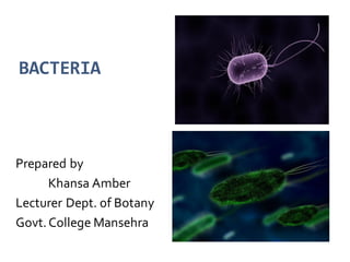
- 1. BACTERIA Prepared by Khansa Amber Lecturer Dept. of Botany Govt.College Mansehra
- 2. Contents Brief introduction History Characteristics Morphology Classification Nutrition Structure Reproduction Economic importance of bacteria
- 3. INTRODUCTION TO BACTERIA Bacteria are microscopic organisms. ‘germs’ and ‘microbes’. Smallest and simplest cellular organism. Bacteria + Cyanobacteria (Blue-Green algae) placed in separate kingdom Monera. Prokaryotic.
- 4. History of bacteria 1976 – Leawon hook (Father of microbiology) ---- -Microorganisms Louis Pasture---confirmed---presence. Robert kotch----discovered---1st bacteria
- 5. CHARACTERISTICS OF BACTERIA Unicellular Colonial Diameter between 1-5 μm Prokaryotic Cosmopolitan While several bacterial species are pathogenic (capable of causing disease) Most are non-infectious.
- 6. Many have critical roles in Decay Fermentation Nutrient recycling Nitrogen fixation. Bacteria are usually classified as gram positive or gram-negative based on a basic microbiological staining procedure called the gram stain. They come in a variety of shapes and sizes.
- 7. MORPHOLOGY Bacteria display a wide diversity of shapes and sizes called morphologies Cannot be seen with human eyes (microscopic) Their presence was only first recognized in 1677, when the Dutch naturalist Antonie van Leeuwenhoek saw microscopic organisms in a variety of substances with the aid of primitive microscopes. Now bacteria are usually examined under light microscopes capable of more than 1,000-fold magnification Details of their internal structure can be observed only with the aid of much more powerful transmission electron microscopes. Unless special phase-contrast microscopes are used, bacteria have to be stained with a coloured dye so that they will stand out from their background.
- 8. CLASSIFICATION OF BACTERIA With over millions of bacteria present in the planet, it is not an easy job to identify, isolate and study a particular species or particular bacteria as such. • Microbiologists categorized bacteria based on basic and important factors making all the bacteria fall under any one of the categories and thus making the process of isolation and identification much easier. • Bacteria are classified based on various factors like shape (morphology), Cell wall structure, Respiration (metabolism), type of nutritional source, characteristic and environmental factor. Bacteria are classifiedbased on various factors • shape (morphology) • Cell wall structure • Respiration (metabolism) • type of nutritional source • characteristic • environmental factor etc.
- 9. Classification on the basis of Stainning method Capsule Gram positive bacteria - take up crystal violet dye andretain their blue or violet color. Gram negative bacteria - do not take up crystal violet dye, and thus appear red or pink. •Capsulated • Encapsulated
- 10. Classification Based on Spore Formation Classification Based on their association Spore forming Non-sporeforming With host Beneficial Pathogenic Harmless
- 11. Respiration AerobicRespiration : Sugars are broken downin the presence of oxygen to produce carbondioxide, water, and energy. Anaerobic Respiration : breaks downsugars and releases energy in theabsence of oxygen. Slower and less efficient than aerobicrespiration. Itinvolves chemicalsotherthan oxygen and carbondioxide. FacultativeAnaerobic Respiration: Ableto perform either aerobic / anaerobicrespiration depending on theoxygen contentof their environment. E.g : Coliformbacteria Microaerophiles: Sugars are broken downin the presence of minute amountsof oxygen to produceenergy.
- 12. Classification Based on Environment Mesophiles- which require moderate temp to survive. Neutrophiles- require moderate conditions to survive. Extremophiles- can survive in extreme conditions. Acidophiles- which can tolerate low pH conditions. Alkaliphiles- which can tolerate high pH conditions. Thermophiles- which can resist high temperature. Psychrophiles- can survive extremely cold conditions. Halophiles- can survive in highlysaline conditions. Osmophiles- can survive in high sugar osmotic conditions.
- 13. Shape Flagella Colonization Cocci Bacilli Spirals Spirillum Spirochete Atrichous Monotrichous Amphitrichous Lopotrichous Peritrichous Cocci Monococcuss Diplococcuss Streptococcus Staphylococcus Tetracoccus Sarcinae Bacilli Monobacillus Diplobacillus Triplobacillus Spirals vibrios Spirilla Spirocheates
- 14. On the basis of shape Coccus: Spherical (round) Bacillus: Rod shaped Vibrio:Comma shaped with flagella Spirillum: Spiral shape Spirochete: wormlike spiral shape
- 15. On the basis of Flagella Atrichous Withoutflagella Monotrichous a single flagellumatone pole. Amphitrichous 2 or more than 2 flagella onbothends Lopotrichous Tuffofflagella on one side Peritrichous Flagella throughoutthe surface
- 16. On the basis of colonization Cocci Monococcuss (One) Diplococcuss Streptococcus Staphylococcus Tetracoccus Sarcinae Bacilli Monobacillus Diplobacillus Triplobacillus Spirals vibrios Spirilla Spirocheates
- 17. Nutritional Source 1. Autotrophs: Use inorganic source and synthesize organic compounds. E.g Obtainthe carbon it requires from carbon dioxide Photoautotrophs: Directlyuse sunlight in order to produce sugarfrom carbondioxide Chemoautotrophs : depend on various chemicalreactions. Use inorganicenergysources, such as hydrogensulfide,elementalsulfur, ferrous iron,molecularhydrogen, and ammonia. 2. Heterotrophs : Rely on organic sources e.g lipids, Carbohydrates etc. 3 types Saprotrophic Bacteria Dead organicmatter Decomposeandget food. Parasitic bacteria Host May be Facultative parasites (not dependent completelyon host) Obligate parasites. (host dependent)
- 18. STRUCTURE OF BACTERIAL CELL Capsule / Glycocalyx Pilli/ Fimbriae Flagella Cell wall Cell/ Plasma membrane Cytoplasm Chromosomes Ribosomes
- 19. Capsule / Glycocalyx Outside cellwall sugarcoatirregularseceretion ofpolysaccharides, capsule: tightly bound regulardistribution Protects and prevents from drying,also protects from phagocytes *Slim layer:thin secretion ofpolysaccharides,and often a significant componentof“biofilms” 2.Polysaccharidesfirmly attached to the cell wall. Capsules adhere to solid surfaces and to nutrients in the environment. Adhesive powerofcapsules is a major factor in the initiation of some bacterialdiseases. Capsulealso protectbacteria from being phagocitized bycells of the hosts immune system. Sticky layer Used to Adhere with With Substrate To form colonies ResistAntibiotics Shield pathogenic bacteriafrom attack by Host’s Immune system
- 20. Pilli / Fimbriae Hollow filamentous rods Motile& non motile. Shortprotein appendages Smaller than flagella Fimbriae Bristlelike small/short fibres andnumerous. Present on bothGram positiveandGram negativebacteria. Cell to cell attachments Adhere bacteria to surfaces(hostepithelium) Fimbriaecan also detect chemical signals. Fimbriaealso act as receptors for bacteriophages. Pilli long hair like tubularmicrofibres like structures. Even thoughpiliarise from plasmamembrane they are not consideredpart of theplasmamembrane. present only on someGram negative bacteria. F pillus /Sex pillus –bacterial mating—sexualreproduction. Used in conjugationfor Exchange ofgeneticinformation Aid Flotation by increasingbuoyancy
- 21. Flagella Motile Hair like, helical cytoplasmic appendages. ProkaryoticFlagella are much thinner than eukaryotic Flagella and they lack the typical 9+2arrangement of microtubules. Theyare approximately 3-2μm long and end in a square tip. Thebacterial flagella is non-contractile, compose of single type of proteins subunits called flagellin. With the help of Flagella bacteria can move at average speed of 50 μm/sec. Capable of “Taxis” Positive chemotaxis Movetowards nutrients Negative chemotaxis Moveaway from toxic substances Somebacteria are motile and some are not. Almost all motile bacteria possess flagella as the organ of locomotion. Such bacteria tend to move towards or away fromthe source of stimulus .these stimuli can be chemicals, light, air or magnetism.
- 22. CELL WALL Cell wall – Murein (Peptidoglycan) 5 amino acids N-acetyl glucosamine N-acetyl Muramic acid Rigidstructuregive shape to the cell. Necessary for the growth and development Maintains cell shape. Protect cell from osmotyic lysis Act as a barrier Attachment site for flagella Site of action of certain antimicrobial agents
- 24. Types of bacteria on the basis of cell wall
- 25. Plasma membrane Thin n elastic , can be only seen with electron microscope Phospholipids (20-30%) and protein (60-70%). 5-10 nm in thickness. It lies below the peptidoglycan layer of the cell wall and encloses the cytoplasm. The arrangement of a lipid and protein to form a layer is called a It is a phospholipid bilayer with polar heads on rather side of the membrane . Phospholipid molecules oriented so that hydrophilic,water-loving heads directed outward and hydrophobic ,water-hating tails directed inward. Proteins embedded in two layers of lipids (lipid bilayayer) (fluid mosaic model). Specialized structures called as mesomers or chondroids are present.
- 26. Functionof plasma membrane Housing enzymes for cell wall, outer membrane synthesis, assembly n secretion of extractoplasmic n extracellular substances Semipermeable membrane Generation of ATP Cell motility Mediation of chromosomal segragation during replication Site of energy Production Attachment with bacterial DNA A selectively permeable barrier Integral proteins form channels Peripheral proteins can act as a receptors Excretion of hydrolytic enzymes Site for the initiation of cell wall synthesis Site of the attachment of the chromosome Site of synthesis of phospholipids Bear receptors and proteins of sensory transduction
- 27. CYTOPLASM The cytoplasm or protoplasm is the portion of the cell that lies within the cytoplasmic membrane . It is a gel like structure and includes the prokaryotic chromosomes and ribosomes. Constituents of cytoplasm include proteins, vitamins, ions, nucleic acids and their precursors, amino acids and their precursors, carbohydrates and their derivatives, fatty acids and their derivatives. The cytoplasm does not exhibit any internal mobility. Cytoplasm also lacks organelles such as mitochondria, Golgi apparatus and endoplasmic reticulum. Recent studies suggest that some bacteria possess cytoskeleton.
- 28. RIBOSOMES Bacterial cells can contain thousands of ribosomes, which are the sites of protein synthesis. The distinct granular appearance of prokaryotic cytoplasm is due to the presence and distribution of ribosomes. They often aggregate to form structures known as polysomes. Bacterial ribosomes are termed 70s and eukaryotic ribosomes are termed as 80s. The difference between the bacterial and eukaryotic ribosomes is often exploited during antibiotic therapy.
- 29. SPORE In poor growth conditions some bacteria produce resistant survival forms termed endospores. This process is known as sporulation. They are resistant to extreme environmental conditions such as higher temperature, dryness, toxic chemicals and UV radiations. Once the endospores are formed the vegetative portion of the bacterium is degraded and the dormant endospore is released. The endospore is able to survive for long periods of time until the environmental conditions again become favorable for growth. Endospore forming bacteria Clostridiumbotulinum, Bacillusbrevis, Bacillus thuringiensis
- 30. PROTOPLAST AND SPHEROPLAST When bacteria are treated with enzymes that hydrolyze the cell wall are antibiotics that interfere with biosynthesis of peptidoglycan, wall-less bacteria are often produced. Such a treatment of bacteria in osmotically protective medium liberates protoplast from gram positive bacteria and Spheroplast from gram negative bacteria. Spheroplast retains the outer membrane. They are produced more readily with penicillin than with lysozyme.
- 31. Specialized membrane Have infoldings to form complex internal structure i.e mesosomes and photosynthetic membranes. Mesosomes Infolding. Associated to dna during cell division. It help in seperation of two daughter cells Provide surface area to cell membrane for cellular respiration.
- 32. Photosynthetic membrane In photosynthetic bacteria. Tubular sheet like infoldings. Resembles thylakoid in cyanobacteria. Have photosynthetic pigment called Bacteriochlorophyll (Site of photosynthesis)
- 33. Cytoplasm a) Cytosol b) Ribosomes c) Chromosomes a) Cytosol Complex of inorganic acid, a.a, proteins, peptides, nitrogenous bases, vitamin, enzymes, co enzymes which provide chemical environment for metabolic activities, cellular activities.
- 34. b) Ribosomes RNA bodies Freely in cytoplasm Protein synthesis. 2 subunits( 50 S + 30S) Consists of RNA and protein Abundant in cytoplasm Often grouped in long chains called polyribosomes. Smaller than the ribosomes in eukaryotic cells but have a similar function.
- 35. They translate the genetic code from the molecular language of nucleic acid to that of amino acids—the building blocks of proteins. Bacterial ribosomes are similar to those of eukaryotes, but are smaller and have a slightly different composition and molecular structure. Bacterial ribosomes are never bound to other organelles as they sometimes are bound to the endoplasmic reticulum in eukaryotes, but are free-standing structures distributed throughout the cytoplasm. There are sufficient differences between bacterial ribosomes and eukaryotic ribosomes that some antibiotics will inhibit the functioning of bacterial ribosomes, but not a eukaryote's, thus killing bacteria but not the eukaryotic organisms they are infecting. Streptomycin binds 70S ribosome and stops protein synthesis but it can not bind 80S
- 36. c) Chromosome The chromosome in bacteria is typically a single,closed circle DNA that is concentratedin a nucleoid region. Mitotic apparatus and nuclearmembraneare completelylacking. As in all organisms,bacterialDNA containsthe four nitrogenous bases A,G,C,T. The amount of DNA in bacterial chromosomes ranges from 580,000 base pairs in Mycoplasma gallinarum to 4,700,000base pairs in E. coli to 9,140,000 basepairs in Myxococcus xanthus. Unlike the DNA in eukaryotic cells, which resides in the nucleus, DNA in bacterial cells is not sequestered in a membrane-bound organelle but appears as a long coil distributed through the cytoplasm. In many bacteria the DNA is present as a single, circular chromosome and in some cases the DNA is linearratherthan circular.some bacteria may contain two chromosomes The bacterialchromosomeconsists ofa single,circle of deoxyribonucleic acid. DNA is double stranded- two strands line up antiparrallel to each other and the bases are linked togetherwith hydrogenbondswhich includesmostof the genetic materialof the organism .
- 37. Plasmids Extra chromosomal circular DNA. • contain genes for antibiotic resistance or virulence. • Structure Similar to most bacterial chromosomes, but considerably smaller.. Size : Chromosomal DNA is typically about 4000 kb, plasmid DNA ranges from 1-200 kb. Number of plasmids: 1-700 copies of plasmid in a cell. Plasmids and the associated traits can be transferred between bacteria, even from one bacterial species to another. Plasmids are not involved in reproduction. Plasmids replicate independently of the chromosome. Plasmids are passed to other bacteria by two means. For most plasmid types, copies in the cytoplasm are passed on to daughter cells during binary fission. Consisted of few genes. Self replictiong. Certain plasmids provideresistance to antibiotics and disinfectants . • Extra-chromosomal circular DNA • multiple copy number • coding - pathogenesis factors - antibiotic resistance factors • bacterial replication
- 38. PlasmidFunction Thefunctionof plasmidsis not always known, but they are not normally essential for survival of host,although theirpresence generally gives thehostsome advantage. Antibioticresistance- Some plasmids code for proteins thatdegrade antibiotics-abig advantage forpathogens. Someencodefor proteins which confer virulence factorsonthehost.Forexample- E. coli plasmid Ent P307codes foran enterotoxin whichmakes E. coli pathogenic. Conjugativeplasmids – Theseallow exchangeof DNA between bacterial cells.
- 39. Spores and cysts Bacteria produces endospores e.g bacillus spp. They can resist heat, desiccation and radiation to overcome unfavorable conditions Gas vacuole Hollow, rigid cylinders. Impermeable to water. In some aquatic species hey provide buoyancy.
- 40. Inclusion Composition Glycogen poly-glucose Reservecarbonand energy source Poly-betahydroxybutyric acid (PHB) lipid Reservecarbonand energy source Poly-phosphates polymers ofPO4 Reservephosphate,possibly high-energyPO4 Sulfurglobules elementalS Reserveenergyand orelectrons Magnetosomes magnetite (iron oxide) Provide orientationin magnetic field Gas vesicles protein shells inflated with gases Provide buoyancyin aquatic environments Parasporalcrystals protein Produced by endospore-forming Bacilli- toxic to insects
- 41. Reproduction in bacteria • Cell growth and reproduction by cell division are tightly linked in unicellular organisms. Bacteria grow to a fixed size and then reproduce through binary fission, a form of asexual reproduction Under optimal conditions, bacteria can grow and divide extremely rapidly, and bacterial populations can double as quickly as every 9.8 minutes. In cell division, two identical clone daughter cells are produced. Budding involves a cell forming a protrusion that breaks away and produces a daughter cell
- 42. Binary fission Most prokaryotes reproduce by a process of binary fission, in which the cell grows in volume until it divides in half to yield two identical daughter cells. Each daughter cell can continue to grow at the same rate as its parent. For this process to occur, the cell must grow over its entire surface until the time of cell division, when a new hemispherical pole forms at the division septum in the middle of the cell. The septum grows inward from the plasma membrane along the midpoint and forms as the side wall which pinches inward, dividing the cell in two. In order for the cell to divide in half, the peptidoglycan structure must be different in the hemispherical cap than in the straight portion of the cell wall, and different wall-cross-linking enzymes must be active at the septum than elsewhere.
- 43. Budding A group of environmental bacteria reproduces by budding. In this process a small bud forms at one end of the mother cell As growth proceeds, the size of the mother cell remains about constant, but the bud enlarges. When the bud is about the same size as the mother cell, it separates. This type of reproduction is analogous to that in budding fungi, such as brewer’s yeast (Saccharomyces cerevisiae). One difference between fission and budding is that, in the latter, the mother cell often has different properties from the offspring. E.g : In some strains, mother cells have a flagellum and are motile, whereas the daughter buds lack flagella.
- 44. BACTERIAL RECOMBINATION Three mechanisms of genetic recombination •Conjugation •Transformation •Transduction
- 45. CONJUGATION Two bacterial cells come together and mate such that a gene transfer occurs between them. Can only occur between cells of opposite mating types. – The donor (or "male") carries a fertility factor (F+). – The recipient ("female") does not (F−). One cell, the donor cell (F+), gives up DNA; and another cell, the recipient cell (F−), receives the DNA. The transfer is nonreciprocal, and a special pilus called the sex pilus joins the donor and recipient during the transfer. The channel for transfer is usually a special conjugation tubeformed during contact between the two cells. The DNA most often transferred is a copy of the F factor plasmid. The factor moves to the recipient, and when it enters the recipient, it is copied to produce a double-stranded DNA for integration.
- 46. BACTERIAL TRANSFORMATION • Discovered by Frederick Griffith in 1928. • Many bacteria can acquire new genes by taking up DNA molecules (ex: plasmid) from their surroundings. • When bacteria undergo lysis, they release considerable amounts of DNA into the environment. • This DNA may be picked up by a competent cell- one capable of taking up the DNA and undergoing a transformation. • To be competent, bacteria must be in the logarithmic stage of growth, and a competence factor needed for the transformation must be present.
- 47. BACTERIAL TRANSDUCTION Bacterial viruses ( bacteriophages) transfer DNA fragments from one bacterium (the donor) to another bacterium (the recipient). The viruses involved contain a strand of DNA enclosed in an outer coat of protein.
- 48. After a bacteriophage enters a bacterium, it may encourage the bacterium to make copies of the phage. At the conclusion of the process, the host bacterium undergoes lysis and releases new phages. This cycle is called the lytic cycle. Under other circumstances, the virus may attach to the bacterial chromosome and integrate its DNA into the bacterial DNA. It may remain here for a period of time before detaching and continuing its replicative process. This cycle is known as the lysogenic cycle. Under these conditions, the virus does not destroy the host bacterium, but remains in a lysogenic condition with it. The virus is called a temperate phage, also known as a prophage. At a later time, the virus can detach, and the lytic cycle will ensue. It will express not only its genes, but also the genes acquired from the donor bacterium.
- 49. THANKYOU !