
Recomendados
Mais conteúdo relacionado
Mais procurados
Mais procurados (20)
Destaque
Semelhante a Chap18 blood
Semelhante a Chap18 blood (20)
Mais de kevperrino
Mais de kevperrino (20)
Último
Último (20)
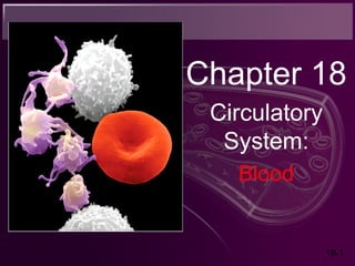
Chap18 blood
- 2. Function of Circulatory System Fundamental purpose of the circulatory system is to transport substances from one place to another in the blood. Functions of Circulatory System: 1. Transportation -O2, CO2, nutrients, wastes, hormones, and heat 2. Protection - role in inflammation = ↓ spread of infection - white blood cells, antibodies, platelets; 3. Regulation - Stabilize fluid distribution and pH
- 3. 18-3 Components & General Properties of Blood • Adults have 4-6 L of blood – Contains two FORMED elements: 1. Plasma - a clear extracellular fluid 2. Formed Elements – cells and cell fragments
- 4. 18-4 Formed Elements • Classified as: – Erythrocytes (RBCs) – Platelets – Leukocytes (WBCs) • Granulocytes – Neutrophils – Eosinophils – Basophils • Agranulocytes – Lymphocytes – Monocytes
- 5. 18-5 Formed Elements of Blood
- 6. 18-6 • Ratio of the formed elements to plasma: Accomplished by spinning a sample of blood in a centifuge. • Erythrocytes are densest, typically 45% of total volume. This is called the hematocrit or packed cell volume. • WBCs and platelets make up a narrow zone (cream color) called the buffy coat. About 1% of total volume. • Plasma about 55% of total volume on top.
- 7. 18-7
- 8. 18-8 Blood Plasma Considered liquid connective tissue; • Contains: – Water – Proteins – Nutrients – Electrolytes – Nitrogenous wastes – Hormones – gases Serum- when blood clots and solids are removed the remaining fluid is serum; Serum is identical to plasma except for the absence of clotting protein fibrinogen
- 9. 18-9
- 10. 18-10 Plasma Proteins Proteins are the most abundant plasma solute by weight (6-9 g/dL) Proteins play a variety of roles: 1. Clotting 2. Defense 3. Transport of solutes: 1. Iron 2. Copper 3. Lipids 4. Hormones
- 11. 18-11 Plasma Proteins • 3 major categories of plasma proteins 1. albumins - most abundant • Transports various solutes and buffer pH of plasma • contributes to viscosity and osmolarity, influences blood pressure, flow and fluid balance; 2. globulins (antibodies) • provide immune system functions, transport, clotting • alpha, beta and gamma globulins; 3. fibrinogen • precursor of fibrin- a sticky protein that forms the framework of a blood clot; • Liver produces 4g protein/hour
- 12. 18-12 Nonprotein Components of Plasma • Nitrogenous compounds – amino acids • from dietary protein or tissue breakdown – nitrogenous wastes (urea) • toxic end products of catabolism • normally removed by the kidneys • Nutrients – glucose, vitamins, fats, minerals, phospholipids, cholesterol; • O2 and CO2 and nitrogen • Electrolytes (another important component of plasma) – Na+ makes up 90% of plasma cations – Sodium is important for osmolarity of blood, influence on blood volume and pressure.
- 13. 18-13
- 14. 18-14 Blood Viscosity & Osmolarity Two most important properties, arise from formed elements and plasma composition. • Viscosity - resistance of a fluid to flow resulting from a cohesions of its particles. • whole blood 5 times as viscous as water, due mainly to RBCs • Plasma 2x as viscous as water, due to its proteins. • Viscosity is important to circulatory function because it partially governs the flow of blood through the vessels. • RBC or protein deficiency = flows easily • Excess of above = flows sluggishly
- 15. 18-15 Blood Viscosity & Osmolarity • Osmolarity – total molarity of dissolved particles that cannot pass through the blood vessel walls. – Nourishment of surrounding cells and waste removal is dependent upon substances passing through the capillary walls.
- 16. 18-16 Blood Viscosity & Osmolarity high osmolarity (blood) • causes fluid absorption into blood, raises BP low osmolarity (blood) • causes fluid to remain in tissues, may result in edema, drop BP to dangerous levels because of the amount of water lost from the blood stream. • Osmolarity is a product of sodium ions, protein, and RBCs
- 17. 18-17 Kwashiorkor • Hypoproteinemia: deficiency of plasma protein. – Extreme starvation – Dietary protein deficiency – Liver disease (synthesis) – Kidney disease – Severe burns As protein content ↓ in blood, = ↓ osmolarity = bloodstream loses more fluid to the tissues than it reabsorbs by osmosis.
- 18. 18-18 How Blood is Produced Hemopoiesis – production of blood (esp. elements) • Adult produces 400 billion platelets, 200 billion RBCs and 10 billion WBCs every day • Hemopoietic tissues produce blood cells Lymphocytes are also produced by lymphatic tissues and organs (thymus, tonsils, lymph nodes, spleen) – Blood formation in bone marrow = myeloid hemopoiesis – Blood formation in lymphatic organs = lymphoid hemopoiesis
- 19. How Blood is Produced All fromed elements trace their origins to a common type of bone marrow stem cell called pluripotent stem cell (PPSC). So named because they have the potential to develop into multiple mature cell types.
- 20. 18-20 Erythrocytes (RBCs) Two principle functions: 1. Pick up O2 from lung & deliver it to tissues; 2. Pick up CO2 from the tissues and unload it to lungs;
- 21. 18-21 Form & Function Disc-shaped cell with thick rim sunken center – Lose most of organelles during development; – Lack mitochondria, rely on anaerobic fermentation to produce ATP – not aerobic respiration; – Lack nucleus and DNA = incapable of protein synthesis and mitosis.
- 22. Form & Function • Glycoproteins & glycolipids on surface determine a persons blood type; • Contain cytoskeletal proteins called spectrin & actin give membrane resilience & durability; • Contains 33% hemoglobin: – Purpose: • oxygen transport, • CO2 transport and • buffering pH of blood
- 23. Form & Function • Biconcave shape allows for a greater ratio of surface to volume, this enables O2 and CO2 to diffuse quickly to and from the hemoglobin. • Cytoplasm contain CAH (Carbonic Anhydrase); plays a role in gas transport & pH balance, catalyzes … CO2 + H2O H2CO3→ →
- 25. 18-25 Hemoglobin (Hb) Structure • Contains 4 protein chains called globins – 2 alpha and 2 beta chains • fetal Hb - gamma replace beta chains; binds O2 better • Each chain is conjugated into a heme group (4 total) – Binds O2 to Fe2+ at center; • So… each hemoglobin molecule can carry four O2
- 26. Quantities of Erythrocytes and Hemoglobin • RBC count and hemoglobin concentration indicate amount of O2 blood can carry; 3 common measurements are: 1. Hematocrit 2. hemoglobin concentration 3. RBC count
- 27. 18-27 Quantities of Erythrocytes and Hemoglobin Hematocrit (packed cell volume) - % of blood composed of RBCs • men 42- 52% cells; women 37- 48% cells Hemoglobin concentration of whole blood • men 13-18g/dL; women 12-16g/dL RBC count • men 4.6-6.2 million/µL; women 4-2-5.4 million/µL • Values are lower in women – androgens stimulate RBC production (higher in men); – women have periodic menstrual losses; – Hematocrit is inversely proportional to % body fat, which is higher in women than men.
- 28. Erythrocyte Life Cycle Average life span of a RBC is 120 days; In a state of balance and stable RBC count, birth to death of RBCs is about 2.5 million/sec or 20mL/day. Erythrocyte production called Erythropoiesis
- 29. 18-29 Erythrocyte Production Erythrocyte production called Erythropoiesis • Development takes 3-5 days and Involves four major developments: 1. reduction in cell size, 2. increase in cell number, 3. synthesis of hemoglobin, and 4. loss of nucleus and most organelles.
- 30. 18-30 Erythropoiesis • Begins with pluripotent stem cell (PPSC) → commits to becoming erythrocyte colony forming unit (ECFU). • ECFU have receptors for erythropoietin (EPO) from kidneys, • EPO stimulates ECFU to transform into Erythroblasts; • Erythroblasts multiply and synthesize hemoglobin; • At this point the nucleus disappears becoming a reticulocyte; • Enter circulation, in about a day or two they become mature erythrocytes; – Normally 0.5 to 1.5% of circulating RBCs are reticulocytes – % will change in response to different situations.
- 31. 18-31 Iron Metabolism • Fe2+ - key nutritional requirement for erythropoiesis; • Iron is lost daily through urine, feces, and bleeding; • men 0.9 mg/day and women 1.7 mg/day • low absorption requires consumption of 5-20 mg/day • Dietary iron exists in two forms: 1. ferric (Fe3+ ) ions (not absorbable) 2. ferrous (Fe2+ ) ions (absorbable by S.I.) Stomach acid converts Fe3+ to absorbable Fe2+
- 32. Iron Metabolism Once converted to Fe2+ it binds with a protein called gastroferritin and transports it to small intestine for absorption; Fe2+ gets absorbed into the blood and binds to plasma protein called transferrin for transport. Transported to bone, liver and other tissures. 1. Bone marrow uses Fe2+ for hemoglobin synthesis; 2. Muscles use it to make the oxygen-storage protein myoglobin; 3. cells use it to make electron-transport molecules (cytochromes) Once in the liver, the surplus iron binds with a protein called apoferritin, forming a iron-storage complex called ferritin; Liver will release iron as needed.
- 34. 18-34 Nutritional Needs for Erythropoiesis Vitamin B12 and folic acid – rapid cell division & DNA synthesis Vitamin C and Cu – cofactors for enzymes synthesizing hemoglobin
- 35. 18-35 Erythrocyte Homeostasis • Negative feedback control – drop in RBC count causes hypoxemia (low blood O2); – Detected by kidneys, ↑ EPO; – EPO stimulates erythroblast production; – RBC count ↑ in 3 - 4 days; • Other stimulus for erythropoiesis – low levels O2; – increase in exercise; Emphysema-loss of lung tissue, could result in polycythemia.
- 36. Erythrocytes Death & Disposal As RBC age its membrane proteins begin to break down (spectrin), causing them to become fragile; Since they lack a nucleus, ribosomes etc, they are unable to synthesis more spectrin; The RBC die in the spleen “erythrocyte grave-yard”; The spleen is made up of narrow channels that test the ability of older RBC to squeeze through; If unable to squeeze through, they become trapped and destroyed.
- 37. 18-37 Erythrocytes Death & Disposal Hemolysis – rupture of RBCs = release hemoglobin and leaves empty plasma membranes; • Macrophages in spleen – digest membrane bits – separate heme from globin • globins hydrolyzed into amino acids • iron removed from heme – heme pigment converted to biliverdin (green); – biliverdin converted to bilirubin (yellow-green); – Macrophages release bilirubin into blood plasma, which binds to albumin; – Liver removes it from the albumin, & sends it to the gall bladder; » concentrated in gall bladder: released into small intestine; bacteria create urobilinogen (brown feces)
- 39. 18-39 Erythrocyte Disorders 1. Polycythemia - an excess of RBCs – primary polycythemia • cancer of erythropoietic cell line in red bone marrow – RBC count as high as 11 million/µL; hematocrit 80% – secondary polycythemia • from dehydration, smoking, emphysema, high altitude, or physical conditioning – RBC count up to 8 million/µL • Dangers of polycythemia – increased blood volume, pressure, viscosity • can lead to embolism, stroke or heart failure
- 40. Erythrocyte Disorders 2. Anemia Three Catagories: 1. Inadaquate erythropoiesis or hemoglobin synthesis; 2. Hemorrhagic anemia – from prolonged bleeding; 3. Hemolytic anemia – from RBC destruction
- 42. 18-42 Anemia Three Potential Consequences: 1.Tissue hypoxia and necrosis - shortness of breath and lethargic - skin pallid due to hemoglobin deficiency - severe anemic hypoxia = life threatening - lead to necrosis of brain, heart, kidneys 2. Blood osmolarity is reduced (tissue edema) 3. Blood viscosity is reduced (blood puts up less resistance to flow so the heart beats faster than normal and cardiac failure can occur and pressure drops)
- 43. 18-43 Sickle-Cell Disease • Hereditary Hb ‘defect’ of African Americans – sickle-cell trait - • individual has resistance to malaria – sickle-cell disease • individual has shortened life – in low O2 concentrations HbS causes cell elongation and sickle shape – cell stickiness causes agglutination and blocked vessels – intense pain; kidney and heart failure; paralysis; stroke
- 45. 18-45 Blood Types • Based on the interactions between antigens and antibodies • Antigens – unique molecules on cell surface • used to distinguish self from foreign • foreign antigens generate immune response • Antibodies – secreted by plasma cells • as part of immune response to foreign matter • Agglutination – antibody molecule binding to antigens – causes clumping – Repetition of this process produces large antigen-antibody complexes
- 46. 18-46 ABO Group • A, B, AB, and O form ABO blood type. Your blood type is determined by the hereditary presence or absence of antigen A or antigen B on your RBCs. Antibodies of the ABO group react against any A or B antigens on the RBC surface. - causing clumping (agglutination)
- 47. 18-47 ABO Group • Your ABO blood type is determined by presence or absence of antigens on RBCs – type A person has A antigens – type B person has B antigens – type AB has both A & Bantigens – type O has no antigens • most common - type O • rarest - type AB
- 49. ABO Group The antibody that reacts with antigen A is called alpha agglutinin or anti A. The antibody that reacts with antigen B is called beta agglutinin or anti B. To determine ones blood type, a drop of blood is put in a pool of anti A serum and anti B serum; Looking for agglutination.
- 51. 18-51 ABO Group • Person with type A (anti-B) blood – Never receive from Type B or AB • Person with type B (anti-A) blood – Never receive from Type A or AB • Person with type O (anti-A and Anti-B) blood – Never receive from Type A, B, or AB
- 52. Type A Type B Type AB Type O RBC: antigen A antigen B antigens A,B none Plasma: anti-B anti-A none anti-A, antibodies antibodies anti-B antibodies Blood Types
- 53. Blood Types for Transfusion
- 54. 18-54 Transfusion Reaction • Agglutinated RBCs block blood vessels and hemolyze; – free Hb blocks kidney tubules, causes death from acute renal failure.
- 55. 18-55 Universal Donors and Recipients • Universal donor – Type O – lacks RBC antigens • Universal recipient – Type AB – lacks plasma antibodies; no anti- A or B
- 56. 18-56 Leukocytes (WBCs) • 5,000 to 10,000 WBCs/µL • Conspicuous nucleus • Travel in blood before migrating to connective tissue • Protect against pathogens
- 57. 18-57 Leukocyte Descriptions • Granulocytes – neutrophils (60-70%) (aka polymorphonuclear leukocytes) • fine granules in cytoplasm; 3 to 5 lobed nucleus – eosinophils (2-4%) • large rosy-orange granules; bilobed nucleus – basophils (<1%) • large, abundant, violet granules (obscure a large S-shaped nucleus) • Agranulocytes – lymphocytes (25-33%) • variable amounts of bluish cytoplasm (scanty to abundant); ovoid/round, uniform dark violet nucleus – monocytes (3-8%) • largest WBC; ovoid, kidney-, or horseshoe- shaped nucleus
- 58. 18-58 Granulocyte Functions • Neutrophils (↑ in bacterial infections) – phagocytosis of bacteria; – release antimicrobial chemicals.
- 59. Granulocyte Functions • Eosinophils (↑ in parasitic infections, allergies, diseases of spleen and CNS) – phagocytosis of antigen-antibody complexes, allergens and inflammatory chemicals – release enzymes to destroy parasites
- 60. Granulocyte Functions • Basophils (↑ in chicken pox, sinusitis, diabetes) – secrete histamine (vasodilator) – secrete heparin (anticoagulant)
- 61. 18-61
- 62. 18-62 Agranulocyte Functions • Lymphocytes (↑ in diverse infections and immune responses) – destroy cells (cancer, foreign, and virally infected cells) – “present” antigens to activate other immune cells – coordinate actions of other immune cells – secrete antibodies and provide immune memory
- 63. Agranulocyte Functions • Monocytes (↑ in viral infections and inflammation) – differentiate into macrophages – phagocytize pathogens and debris – “present” antigens to activate other immune cells
- 64. 18-64
- 65. 18-65 Complete Blood Count • Hematocrit • Hemoglobin concentration • Total count for RBCs, reticulocytes, WBCs, and platelets • Differential WBC count • RBC size and hemoglobin concentration per RBC
- 66. 18-66 Leukocyte Life Cycle • Leukopoiesis- production of WBC – Begins with pluripotent stem cell – that differentiates into distinct type of colony-forming units (CFU); – CFU Types: • Eosinophilic CFU, Basophilic CFU, neurtophilic CFU, monocytic CFU, and lymphocytic CFU. – CFU differentiate into specific cell lines: • myeloblasts – differentiate into neutrophils, eosinophils, basophil • monoblasts – differentiate into monocytes • lymphoblasts differentriate into B and T lymphocytes and NK cells
- 68. • Red bone marrow stores and releases granulocytes and monocytes until needed. • Circulating WBCs do not stay in bloodstream – granulocytes circulate for 8 hours and then migrate into the tissues & live for about 5 days; – monocytes circulate for about 10-20 hours, migrate ino the tissues, transform into macrophages and live for several years; – WBCs provide long-term immunity (decades)
- 69. 18-69 Leukocyte Disorders • Normal WBC count is 5000 – 10,000 WBC/µL • Leukopenia - low WBC count (<5000/µL) – causes: radiation sickness, lead, mercury poisoning, infectious disease (MMR, varicella, polio, AIDS), anti- cancer drugs – effects: elevated risk of infection and cancer • Leukocytosis = high WBC count (>10,000/µL) – causes: infection, allergy and disease, dehydration and emotional disturbances. – differential count - distinguishes % of each cell type • High neurtophil = bacterial infection, ie appendicitis • High eosinophil = allergy or parasitic infection, ie tapeworm
- 70. 18-70 Leukocyte Disorders • Leukemia = cancer of hemopoietic tissue – Increase of circulating leukocytes and their precursors • Treatment = chemo, marrow transplant along with controlled side effects such as anemia, hemorrhaging, and infection.
- 71. 18-71
- 72. Platelets & Hemostasis-Control of bleeding Hemostasis – cessation of bleeding (stop the bleeding); Platelets may not stop the hemorrhaging of large vessels but are effective on closing smaller vessels.
- 73. 18-73 Platelets • Not cells but small fragments of marrow cells called megakaryocytes; • Normal Count - 130,000 to 400,000 platelets/µL • Internal structures include: – lysosomes, – mitochondria, – granules (filled with platelet secretions), – Platelet Production -Thrombopoiesis
- 74. Platelet Functions 1. Secrete vasoconstrictors- chemical that causes spasmodic contraction of broken blood vessels; 2. Stick together to form a temporary platelet plug; 3. Secrete procoagulants (clotting factors);
- 75. 18-75 Platelet Production -Thrombopoiesis • Stem cells develop receptors for the hormone thrombopoietin, thus becoming megakaryoblasts; • Megakaryoblasts – repeatedly replicate DNA without dividing cytoplasm; – Result is a gigantic cell called megakaryocyte; • Megakaryocyte – infoldings of cytoplasm splits off cell fragments that enter bloodstream as platelets, those live for 10 days; – 25 – 40% are stored in spleen & released as needed.
- 76. 18-76 Hemostasis • Cessation of bleeding • 3 hemostatic mechanisms for stoppage of bleeding.
- 77. 18-77 1. Hemostasis - Vascular Spasm (Immediate) • Causes – pain receptors • some directly innervate nearby blood vessels, causing constriction – Injury to smooth muscle will vasoconstrict – platelets release serotonin (vasoconstrictor) • Effects – prompt constriction of a broken vessel • pain receptors - short duration (minutes) – provides time for other two clotting pathways
- 78. 18-78 2. Hemostasis -Platelet Plug Formation • Endothelium smooth, coated with prostacyclin • Platelet plug formation – broken vessel exposes collagen – platelet pseudopods stick to damaged vessel and other platelets – pseudopods contract and draw walls of vessel together forming a platelet plug As platelets aggregate they degranulate releasing factors that promote hemostasis; • serotonin is a vasoconstrictor • ADP attracts and degranulates more platelets • thromboxane A2, promotes aggregation, degranulation and vasoconstriction – positive feedback cycle is active until break in vessel is sealed
- 79. The gray filaments are fibrin, platelets are orange
- 80. 18-80 3. Hemostasis - Coagulation • Coagulation (clotting) - most effective defense against bleeding – Conversion of the plasma protein fibrinogen into fibrin threads to form framework of clot; • Two reaction mechanisms to coagulation: 1.Extrinsic Mechanism 2.Intrinsic Mechanism
- 81. Coagulation Extrinsic Mechanism: - initiated by clotting factors released by damaged blood vessel and perivascular tissues; - extrinsic mean that these factors came from a source other then the blood. Intrinsic Mechanism: - clotting factors that are in the blood. These clotting factors are called procoagulants, most are proteins that are in an inactive form, until one is activated, thus activating the next one and so on, Producing a reaction cascade – a series of reactions.
- 82. 18-82 Coagulation Pathways • Extrinsic pathway – initiated by tissue thromboplastin; – cascade to factor VII, V and X (fewer steps), – 15 seconds to start clot. • Intrinsic pathway – Everything needed to initiate it is present in the plasma or platelets; – initiated by factor XII; – cascade to factor XI to IX to VIII to X; – 3-6 minutes for clot to form. • Calcium required for either pathway Fibrinogen
- 83. 18-83 Completion of Coagulation • Activation of Factor X – leads to production of prothrombin activator • Prothrombin activator – converts prothrombin to thrombin • Thrombin – converts fibrinogen into fibrin
- 85. 18-85 Fate of Blood Clots • After clot has formed the platelet pseudopods adhere to the fibrin and contract = drawing edges of blood vessels together Clot retraction occurs within 30 minutes • Platelets & endothelial cells secrete: – Platelet-derived growth factor (PDGF) which is a … – mitotic stimulant for fibroblasts and smooth muscle to multiply and repair damaged vessel;
- 86. 18-86 Blood Clot Dissolution • Positive feedback occurs • Plasmin promotes formation of kallikrein
- 87. 18-87 Prevention of Inappropriate Clotting 1. Platelet repulsion – platelets do not adhere to prostacyclin-coated endothelium of undamaged blood vessels; 2. Dilution – Small amounts of thrombin form spontaneously in plasma; – At normal rate of blood flow the thrombin is diluted; • heart slowing in shock can result in clot formation 3. Natural anticoagulants – heparin (from basophils and mast cells) interferes with formation of prothrombin activator – antithrombin (from liver) deactivates thrombin before it can act on fibrinogen
- 88. 18-88 Hemophilia • Genetic lack of any clotting factor affects coagulation • TYPES: – hemophilia A missing factor VIII (83% of cases) – hemophilia B missing factor IX (15% of cases) • Physical exertion causes bleeding and excruciating pain – transfusion of plasma or purified clotting factors – factor VIII produced by transgenic bacteria
- 89. 18-89 Coagulation Disorders • Embolism - clot traveling in a vessel • Thrombosis - abnormal clotting in unbroken vessel – most likely to occur in leg veins of inactive people – pulmonary embolism - clot may break free, travel from veins to lungs • Infarction may occur if clot blocks blood supply to an organ (MI or stroke) – 650,000 Americans die annually of thromboembolism
Notas do Editor
- patient subject to opportunistic infection, anemia