

Recomendados
Recomendados
Mais conteúdo relacionado
Mais procurados
Mais procurados (20)
Semelhante a Brain machine interface and limb reanimation
Semelhante a Brain machine interface and limb reanimation (20)
Mais de Karlos Svoboda
Mais de Karlos Svoboda (20)
Último
Último (20)
Brain machine interface and limb reanimation
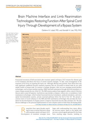
- 1. SYMPOSIUM ON REGENERATIVE MEDICINE Brain Machine Interface and Limb Reanimation Technologies: Restoring Function After Spinal Cord Injury Through Development of a Bypass System Darlene A. Lobel, MD, and Kendall H. Lee, MD, PhD CME Activity Target Audience: The target audience for Mayo Clinic Proceedings is primar-ily internal medicine physicians and other clinicians who wish to advance their current knowledge of clinical medicine and who wish to stay abreast of advances in medical research. Statement of Need: General internists and primary care physicians must maintain an extensive knowledge base on a wide variety of topics covering all body systems as well as common and uncommon disorders. Mayo Clinic Proceedings aims to leverage the expertise of its authors to help physicians understand best practices in diagnosis and management of conditions encountered in the clinical setting. Accreditation: Mayo Clinic College of Medicine is accredited by the Accred-itation Council for Continuing Medical Education to provide continuing med-ical education for physicians. Credit Statement: Mayo Clinic College of Medicine designates this journal-based CME activity for a maximum of 1.0 AMA PRA Category 1 Credit(s). Physicians should claim only the credit commensurate with the extent of their participation in the activity. Learning Objectives: On completion of this article, you should be able to (1) cite the advantages and disadvantages of cue based and self-paced brain machine interfaces (BMIs), (2) define the concept of central pattern genera-tors and explain its relevance to intraspinal microstimulation (ISMS) tech-niques, and (3) describe three challenges associated with developing a bypass system integrating BMI and ISMS technology. Disclosures: As a provider accredited by ACCME, Mayo Clinic College of Medicine (Mayo School of Continuous Professional Development) must ensure balance, independence, objectivity, and scientific rigor in its educa-tional activities. Course Director(s), Planning Committee members, Faculty, and all others who are in a position to control the content of this educational activity are required to disclose all relevant financial relationships with any commercial interest related to the subject matter of the educational activity. Safeguards against commercial bias have been put in place. Faculty also will disclose any off-label and/or investigational use of pharmaceuticals or instru-ments discussed in their presentation. Disclosure of this information will be published in course materials so that those participants in the activity may formulate their own judgments regarding the presentation. In their editorial and administrative roles, William L. Lanier, Jr, MD, Terry L. Jopke, Kimberly D. Sankey, and Nicki M. Smith, MPA, have control of the content of this program but have no relevant financial relationship(s) with industry. The authors report no competing interests. Method of Participation: In order to claim credit, participants must com-plete the following: 1. Read the activity. 2. Complete the online CME Test and Evaluation. Participants must achieve a score of 80% on the CME Test. One retake is allowed. Participants should locate the link to the activity desired at http://bit.ly/ Oq0RIB. On successful completion of the online test and evaluation, you can instantly download and print your certificate of credit. Estimated Time: The estimated time to complete each article is approxi-mately 1 hour. Hardware/Software: PC or MAC with Internet access. Date of Release: 05/01/2014 Expiration Date: 04/30/2016 (Credit can no longer be offered after it has passed the expiration date.) Privacy Policy: http://www.mayoclinic.org/global/privacy.html Questions? Contact dletcsupport@mayo.edu. Abstract Functional restoration of limb movement after traumatic spinal cord injury (SCI) remains the ultimate goal in SCI treatment and directs the focus of current research strategies. To date, most investigations in the treatment of SCI focus on repairing the injury site. Although offering some promise, these efforts have met with significant roadblocks because treatment measures that are successful in animal trials do not yield similar results in human trials. In contrast to biologic therapies, there are now emerging neural interface technologies, such as brain machine interface (BMI) and limb reanimation through electrical stimulators, to create a bypass around the site of the SCI. The BMI systems analyze brain signals to allow control of devices that are used to assist SCI patients. Such devices may include a computer, robotic arm, or exoskeleton. Limb reanimation technologies, which include functional electrical stimulation, epidural stimulation, and intra-spinal microstimulation systems, activate neuronal pathways below the level of the SCI. We present a concise review of recent advances in the BMI and limb reanimation technologies that provides the foun-dation for the development of a bypass system to improve functional outcome after traumatic SCI. We also discuss challenges to the practical implementation of such a bypass system in both these developing fields. ª 2014 Mayo Foundation for Medical Education and Research n Mayo Clin Proc. 2014;89(5):708-714 Researchers have spent decades search-ing for ways to restore function to those with traumatic spinal cord injury (SCI). Development of treatment strategies must begin with understanding how injury affects the nervous system. Injury to the spinal cord prevents cortical signals generated by the brain from reaching target muscles, resulting in From the Center for Neuro-logical Restoration, Depart-ment of Neurosurgery, Cleveland Clinic, Cleveland, OH (D.A.L.); and Depart-ment of Neurologic Surgery, Mayo Clinic, Rochester, MN (K.H.L.). 708 Mayo Clin Proc. n May 2014;89(5):708-714 n http://dx.doi.org/10.1016/j.mayocp.2014.02.003 www.mayoclinicproceedings.org n ª 2014 Mayo Foundation for Medical Education and Research
- 2. BRAIN MACHINE INTERFACE AND LIMB REANIMATION paralysis. Functional magnetic resonance imag-ing studies indicate that even after SCI, the brain continues to generate electrical signals in response to an individual’s intention to move.1 Additional studies indicate that electrophysio-logic stimuli applied to the muscles, peripheral nerves, or spinal cord, below the level of injury, can generate muscle activity.2 These discoveries offer a ray of hope in the treatment of SCI if we then conceive of paralysis as an information transfer lesion, where the information sent from the brain via the corticospinal tract does not reach the spinal cord. To restore limb function to individuals with SCI, this information transfer lesion must be either repaired or bypassed. To date, current research efforts have focused on ways to repair the damaged spinal cord or to prevent further injury after the initial insult to the spinal cord. Transplantation of stem cells at the site of the injury, introduction of tissue-bridging biomatrices and peripheral nerve transfers, and targeting of methods to increase expression of neurotrophins and cytokines via viral trans-duction are among the strategies being investi-gated. 3 Although offering promise in the preclinical setting, these investigations have met with limited success in clinical trials. The lack of an adequate animal model of SCI, along with safety concerns associated with some of these therapies,3 are cited as reasons for the poor translatability of these treatments in humans. Indeed, to date, there has been no report of restoration of limb movement using these biologic repair approaches. In part because of the limited success of techniques to directly repair lesions due to SCI, efforts have focused in recent years on rehabilitative strategies to restore functional in-dependence to individuals with SCI.2 Among these efforts are the development of brain ma-chine interface (BMI) systems. The BMI systems capture and analyze information from the brain and then deliver commands to an external de-vice that is then able to perform the function initially intended by the patient.4 Another strat-egy involves directly activating neuronal path-ways below the level of the SCI lesion. In this way, we can restore function to limbs that can no longer directly receive commands from the brain. This innovative concept, known as limb reanimation, includes functional electrical stimulation (FES) of peripheral nerves or target muscles and epidural stimulation or direct intraspinal microstimulation (ISMS) of the spi-nal cord itself.3 By combining the capabilities of the BMI and limb reanimation systems, a bypass of the information transfer lesion in SCI may be created, and the seemingly far-reaching goal of restoring limb function to SCI patients becomes possible (Figure 1). In this review, we discuss current advances in the BMI and limb reanimation systems and discuss how these technologies bring us closer to restoring function to paralyzed limbs in pa-tients with traumatic SCI. THE BMI SYSTEMS The BMI systems are designed to restore lost neurologic functions to individuals with SCI, stroke, or a neurodegenerative disorder, such as amyotrophic lateral sclerosis.4 A BMI first captures the electrical signals generated by the brain when the user intends to move. To oper-ate the BMI system, a user may simply imagine certain actions, such as squeezing the hand or moving the foot, or more complex movements, such as walking. This process, known as motor imagery, produces electrical activations in the regions of the motor, premotor, and supple-mentary motor cortices. These signals are captured by a variety of techniques, including electroencephalography, electrocorticography, direct recordings of action potentials (known as single-unit recordings), and near-infrared spectroscopy, to cite a few.4,5 The more invasive systems (single-unit recordings and electrocor-ticography) provide the best signal quality but do so at the highest risk to the patient. The least invasive systems ( electroencephalography and near-infrared spectroscopy) carry minimal risk to the user but yield the poorest signal quality. Signals from such noninvasive techniques may not provide sufficient quality to operate complex devices, such as prosthetic arms or exoskeletons, which require multiple degrees of freedom of control. Once the cortical signals are captured, they are analyzed using a computer-based algo-rithm to yield what is known as a signature. A signature is a specific pattern of electrical ac-tivity, composed of spatial-, temporal-, and frequency-based components, that is unique to a particular imagined movement. It is not necessary that the action imagined by the user correlate directly with the intended result; Mayo Clin Proc. n May 2014;89(5):708-714 n http://dx.doi.org/10.1016/j.mayocp.2014.02.003 www.mayoclinicproceedings.org 709
- 3. MAYO CLINIC PROCEEDINGS FIGURE 1. Brain machine interfaceedirected limb reanimation concept. A patient with spinal cord injury undergoes implantation with a cortical device to capture signals produced by the brain as he imagines movements. These signals are then processed and delivered wirelessly to an intraspinal microstimulation device, allowing him to walk. Image courtesy of Mayo Clinic in Rochester, Minnesota. what is important is that a unique signature be produced for each intended action.6 Once the signature is recognized, signal processing is per-formed by a software system, such as Open- Vibe7 or BCI 2000,8 and then a command is delivered to a device, known as an effector. Such commands can vary from the simplistic, such as controlling a computer mouse on a screen,9 to highly complex, controlling a 7-df prosthetic arm10 or, theoretically, even a full exoskeleton. One of the major challenges in the BMI sys-tem design is developing systems that can be safely and effectively used at home. An ideal system may be activated at any time and will safely and seamlessly function in whatever ca-pacity is needed. To meet this challenge, asyn-chronous (or self-paced) BMI systems have been developed. Such systems are available to use at any time. These differ from synchronous or cue-based systems that will work only at specific times determined by the BMI. Although asynchronous systems offer a significant degree of user autonomy, currently these systems yield a high number of unintentional (false-positive) activations by the system, thus introducing safety concerns.11 Cue-based systems, although significantly reducing false-positive activations, come at the cost of offering less user control.12 Optimizing the technology of asynchronous systems is the next challenge in the BMI design. To date, signal processing algorithms have been designed for asynchronous BMI control13; however, no complete BMI systems have pro-duced a sufficient online efficiency rate with a low enough false-positive rate to provide a reasonable clinical safety profile. Along the same lines, specific movements by an effector (such as a robotic arm) may be controlled primarily by the user or the BMI sys-tem itself. Systems that allow the user to have precise control of the effector require the 710 Mayo Clin Proc. n May 2014;89(5):708-714 n http://dx.doi.org/10.1016/j.mayocp.2014.02.003 www.mayoclinicproceedings.org
- 4. BRAIN MACHINE INTERFACE AND LIMB REANIMATION exertion of continuous control of brain signals from the moment of task initiation to its conclusion, which can be tiring after prolonged system use.14 In contrast to such process control systems, goal selection systems only require the user to control the system for a brief period, long enough for the system to ascertain the user’s intent.14 In doing so, these systems provide a high degree of accuracy, faster speed of activation, and less user fatigue because the system takes over once the intent of the user is known. However, such systems require move-ments to be preprogrammed, thus limiting the adaptability of such systems to user needs. In a recent clinical trial, a 52-year-old quad-riplegic patient who underwent implantation with dual 96-contact intracortical electrode ar-rays learned to control a 7-df prosthetic arm in a 4-month training period after electrode im-plantation. 10 Although the system still required a cue for activation, the patient was able to con-trol the movements of the prosthetic arm, inde-pendent of computer assistance, after 10 weeks of training. This is the first example of an efficient process control system that allowed a patient to consistently perform natural and complexmovements, without significant effects of fatigue. LIMB REANIMATION Once the intended movement has been identi-fied by analyzing cortical signals, the next step in developing a bypass system is delivering commands to the intended muscles. Just as the brain continues to generate electrical signals after SCI, studies have found that the muscles below the level of an SCI continue to respond to an applied electrical stimulus. Electrical stimulation may be applied directly to the mus-cles via surface or intramuscular techniques or to the motor neurons in FES procedures. Furthermore, stimulation of the spinal cord below the level of injury, using either epidural or ISMS techniques, produces a contraction in one or more muscles.15 During a recent clin-ical trial, a paraplegic patient was able to stand for a period during stimulation with electrodes implanted in the epidural space.16 As has been noted with many FES systems, the patient experienced muscle fatigue after prolonged epidural stimulation. In contrast, the force and durability of muscle contraction are greater with ISMS systems, which may allow smaller current requirement and greater fidelity of muscle control. Furthermore, intraspinal sys-tems avoid problems such as muscle fatigue, stimulation spillover, and reverse motor unit recruitment seen with more superficial stimula-tion systems.17-19 Thus, despite the risks associ-ated with placing an invasive spinal stimulation system, the ISMS system may provide the best long-term solution to achieve limb reanimation (Figure 2). Limb reanimation studies with the ISMS sys-tems are in the early research stages. Currently, no consensus exists regarding electrode design, optimization of electrode implantation location and stimulation parameters, or delivery system strategies. State-of-the-art systems use fine microwire electrodes, measuring on the order of tens to hundreds of micrometers in diameter. These microwires are inserted into the motor neuron pools of the lumbar enlargement in the spinal cord of small animal models.20 Currently, variability exists in selecting target areas for elec-trode insertion, which may improve as spinal cord mapping in animal models becomes further refined. To date, mapping studies have been conducted in the rat, frog, and cat. A recent study reported limb movement in rodents who had undergone T4 lesioning fol-lowed by implantation of a thin microwire in the lumbar enlargement of the spinal cord. Hind limb movements indicated a graded response to increasing levels of stimulation amplitude using an intraspinal microstimula-tion device (Peter A. Grahn, BA, unpublished data, 2013). Although these results are en-couraging, questions remain about whether successful trials in small animals will translate to large animal models and humans. Information has come to light in the past few years regarding the concept of central pattern generators, which are neuronal net-works located in the spinal cord that are thought to be responsible for locomotion. Much of the work with the ISMS system for limb reanimation was initiated by Vivian Mush-ahwar, who recently found almost full-strength stepping ability in anesthetized cats with ISMS electrodes implanted in the lumbar enlarge-ment of the spinal cord, targeting these central pattern generators.21 The effect of SCI on cen-tral pattern generators is currently unknown and must be further investigated to determine how electrode targeting needs to be adjusted Mayo Clin Proc. n May 2014;89(5):708-714 n http://dx.doi.org/10.1016/j.mayocp.2014.02.003 www.mayoclinicproceedings.org 711
- 5. FIGURE 2. Intraspinal microstimulation (ISMS) implantation concept. A, Spinal cord injury located at T12, producing a complete lesion. Laminectomy (B) and pedicle screw placement (C) performed as standard treatment for spinal fractures. D, Spinal cord cross-section showing electrode array implantation location in the ventral horn region below the level of the injury. E, Patient with implanted intraspinal microelec-trode array now able to walk via wirelessly controlled ISMS system. Image courtesy of Mayo Clinic in after injury. The ISMS studies in large animal models of SCI combined with advancements in magnetic resonance imaging of the spinal cord may provide more insight into a functional map of the spinal cord in both animal models and humans. Beyond optimizing electrode design and targeting, another significant challenge is development of a delivery system for electrode implantation. A stereotactic spinal delivery system is necessary to achieve precise implan-tation of intraspinal microelectrodes. Use of stereotactic frames or frameless localization systems in spine surgery has been hindered by problems with inaccuracy because of vari-ability of surface landmarks that are used as fixation points. The target accuracy for these systems must be at the submillimeter level, significantly more precise than current spinal stereotactic targeting systems.22 DEVELOPMENT OF A BYPASS SYSTEM FOR FUNCTIONAL RESTORATION OF PARALYZED LIMBS The concept of SCI as an information transfer lesion creates the possibility of developing a bypass system around the lesion to deliver intended commands to target muscles. The BMI systems allow us to ascertain information regarding a patient’s intent to move a limb, whereas the FES and ISMS systems permit us to apply specific patterns of electrical stimuli Rochester, Minnesota. MAYO CLINIC PROCEEDINGS 712 Mayo Clin Proc. n May 2014;89(5):708-714 n http://dx.doi.org/10.1016/j.mayocp.2014.02.003 www.mayoclinicproceedings.org
- 6. to target muscles or to motor neurons them-selves. By forming a wireless link between these 2 systems, we can conceive of an integrated bypass system that will restore motor function to a paralyzed patient. The complexities of forming a link between these 2 systems are sig-nificant. On a basic level, this type of bypass sys-tem will not account for activity in subcortical pathways because current BMI systems are not able to capture such signals. Therefore, details regarding how the signals are delivered tomotor neurons to effect simple limb movements, such as flexion and extension of specific muscles, as well as more complex movements such as gait, will have to be surmised from experimental ISMS studies. Presenting a further challenge, the BMI soft-ware, which processes cortical signals, must be integrated with the software systems that control the FES and ISMS devices. Alternatively, a new software system that is capable of delivering cortical signals obtained from the BMI directly to the limb reanimation electrodes could be developed. A benefit in developing a new direct software interface is the possibility of implement-ing a 2-way information transfer system, which would integrate sensory feedback fromthe spinal stimulation device to the BMI, to allow dynamic system control. This is an important adjunct to such a system because muscle response to a descending signal is thought to depend in part on sensory feedback fromspinal interneurons.15 Furthermore, advancements must be made in both the BMI and FES or ISMS systems to sup-port wireless control of the systems, which is essential to allow fully implantable devices that can be used at will in the patient’s home. To date, only 1 wireless BMI implant23 and 2 wire-less ISMS systems exist.24,25 Finally, safety pro-files of the individual system components and the overall bypass system must be carefully eval-uated before proceeding with implementation of these technologies. CONCLUSION Combination of the innovative technologies of the BMI and limb reanimation systems intro-duces hope to restore limb function to the para-lyzed. Control of a robotic arm and other effectors has already been produced using BMI technology. In addition, epidural spinal stimu-lation has permitted a paraplegic patient to stand. This achievement suggests the feasibility of integrating the capabilities of both systems to create a bypass system for patients with SCI and thus restore a degree of autonomy to these individuals. Because both the BMI and ISMS systems are in relatively early stages of develop-ment, we are afforded an opportunity to tailor the design of combined bypass systems to include functions such as providing sensory feedback that will maximize the benefit for pa-tients with SCI. ACKNOWLEDGMENTS We gratefully acknowledge Veneliza Salcedo of Mayo Clinic for her artistry in the creation of illustrations for this article. Abbreviations and Acronyms: BMI = brain machine interface; FES = functional electrical stimulation; ISMS = intraspinal microstimulation; SCI = spinal cord injury Grant Support: This work was supported by The Grainger Foundation Grant awarded to Dr Lee. Potential Competing Interests: Dr Lobel reports a minor consultant relationship with St. Jude Medical. Correspondence: Address to Darlene A. Lobel, MD, Cen-ter for Neurological Restoration, Department of Neurosur-gery, Cleveland Clinic, 9500 Euclid Ave, S31, Cleveland, OH (lobeld@ccf.org). Individual reprints of this article and a bound reprint of the entire Symposium on Regenerative Medicine will be available for purchase from our website www.mayoclinic.proceedings.org The Symposium on Regenerative Medicine will continue in an upcoming issue. REFERENCES 1. Freund P, Weiskopf N, Ward NS, et al. Disability, atrophy and cortical reorganization following spinal cord injury. Brain. 2011; 134(pt 6):1610-1622. 2. Jackson A, Zimmermann JB. Neural interfaces for the brain and spinal corddrestoring motor function. Nat Rev Neurol. 2012; 8(12):690-699. 3. Filli L, Schwab ME. The rocky road to translation in spinal cord repair. Ann Neurol. 2012;72(4):491-501. 4. Shih JJ, Krusienski DJ, Wolpaw JR. Brain-computer interfaces in medicine. Mayo Clin Proc. 2012;87(3):268-279. 5. Birbaumer N, Weber C, Neuper C, Buch E, Haapen K, Cohen L. Physiological regulation of thinking: brain-computer interface (BCI) research. Prog Brain Res. 2006;159:369-391. 6. Leuthardt E, Schalk G, Wolpaw JR, Ojemann JG, Moran DW. A brain-computer interface using electrocorticographic signals in humans. J Neural Eng. 2004;1(2):63-71. 7. Renard Y, Lotte F, Gibert G, et al. OpenViBE: an open-source software platform to design, test, and use brain-computer inter-faces in real and virtual environments. Presence Teleoperators Virtual Environ. 2010;19(1):35-53. 8. Schalk G, McFarland DJ, Hinterberger T, Birbaumer N, Wolpaw JR. BCI2000: a general-purpose brain-computer inter-face (BCI) system. IEEE Trans Biomed Eng. 2004;51(6):1034-1043. BRAIN MACHINE INTERFACE AND LIMB REANIMATION Mayo Clin Proc. n May 2014;89(5):708-714 n http://dx.doi.org/10.1016/j.mayocp.2014.02.003 www.mayoclinicproceedings.org 713
- 7. 9. Wolpaw JR, McFarland DJ. Control of a two-dimensional move-ment signal by a non-invasive brain-computer interface in humans. Proc Natl Acad Sci U S A. 2004;101(51):17849-17854. 10. Collinger JL, Wodlinger B, Downey JE, et al. High-performance neuroprosthetic control by an individual with tetraplegia. Lancet. 2013;381(9866):557-564. 11. Fatourechi M, Ward RK, Birch GE. A self-paced brainecom-puter interface system with a low false positive rate. J Neural Eng. 2008;5(1):9-23. 12. Müller-Putz GR, Scherer R, Pfurtscheller G, Rupp R. Brain-com-puter interfaces for control of neuroprostheses: from synchro-nous to asynchronous mode of operation. Biomed Tech (Berl). 2006;51(2):57-63. 13. Eliseyev A, Moro C, Costecalde T, et al. Iterative N-way partial least squares for a binary self-paced brainecomputer interface in freely moving animals. J Neural Eng. 2011;8(4):046012. 14. Royer AS, Rose ML, He B. Goal selection versus process con-trol while learning to use a brain-computer interface. J Neural Eng. 2011;8(3):036012. 15. Loeb GE, Davoodi R. The functional reanimation of paralyzed limbs. IEEE Eng Med Biol Mag. 2005;24(5):45-51. 16. Harkema S, Gerasimenko Y, Hodes J, et al. Effect of epidural stimulation of the lumbosacral spinal cord on voluntary move-ment, standing, and assisted stepping after motor complete paraplegia: a case study. Lancet. 2011;377(9781):1938-1947. 17. Crago PE, Peckham PH, Thrope GB. Modulation of muscle force by recruitment during intramuscular stimulation. IEEE Trans Biomed Eng. 1980;27(12):679-684. MAYO CLINIC PROCEEDINGS 18. Peckham PH, Knutson JS. Functional electrical stimulation for neuromuscular applications. Annu Rev Biomed Eng. 2005;7: 327-360. 19. Troyk PR, Donaldson Nde N. Implantable FES stimulation sys-tems: what is needed? Neuromodulation. 2001;4(4):196-204. 20. Bamford JA, Mushahwar VK. Intraspinal microstimulation for the recovery of function following spinal cord injury. Prog Brain Res. 2011;194:227-239. 21. Holinski BJ, Mazurek KA, Everaert DG, Stein RB, Mushahwar VK. Restoring stepping after spinal cord injury using intraspinal micro-stimulation and novel control strategies. Conf Proc IEEE Eng Med Biol Soc. 2011;2011:5798-5801. 22. Rath SA, Moszko S, Schäffner PM, et al. Accuracy of pedicle screw insertion in the cervical spine for internal fixation using frameless stereotactic guidance. J Neurosurg Spine. 2008;8(3): 237-245. 23. Charvet G, Foerster M, Chatalic G, et al. A wireless 64-channel ECoG recording electronic for implantable monitoring and BCI applications: WIMAGINE. Conf Proc IEEE Eng Med Biol Soc. 2012;2012:783-786. 24. Troyk PR, Mushahwar VK, Stein RB, et al. An implantable neural stimulator for intraspinal microstimulation. Conf Proc IEEE Eng Med Biol Soc. 2012;2012:900-903. 25. Chang SY, Kimble CJ, Kim I, et al. Development of the Mayo Investigational Neuromodulation Control System (MINCS): toward a closed-loop electrochemical feedback system for Deep Brain Stimulation. J Neurosurg. 2013;119(6): 1556-1565. CORRECTION In the article “Validation of a Novel Protocol for Calculating Estimated Energy Require-ments and Average Daily Physical Activity Ratio for the US Population: 2005-2006,” published in the December 2013 issue of Mayo Clinic Proceedings (2013;88(12):1398- 1407), the P values in the last sentence in the results section of the abstract were incor-rect. The sentence should read: “Obese men and women had lower APAR values than normal weight individuals (P¼.023 and P¼.015, respectively),.”. http://dx.doi.org/10.1016/j.mayocp.2014.04.001 714 Mayo Clin Proc. n May 2014;89(5):708-714 n http://dx.doi.org/10.1016/j.mayocp.2014.02.003 www.mayoclinicproceedings.org