Uvea anatomy
•Transferir como PPTX, PDF•
7 gostaram•2,593 visualizações

This document provides an overview of the anatomy of the uveal tract, which includes the iris, ciliary body, and choroid. It begins with an introduction and overview of the embryology and development of the uveal tract. It then discusses the anatomy and microstructure of each part of the uveal tract in detail, including their nerve and blood supply. It also briefly discusses some congenital anomalies that can affect the uveal tract.

Recomendados
Mais conteúdo relacionado
Mais procurados
Mais procurados (20)
Semelhante a Uvea anatomy
Semelhante a Uvea anatomy (20)
Mais de Mero Eye
Mais de Mero Eye (20)
Último
Último (20)
Uvea anatomy
- 1. UVEA: ANATOMY, NERVE & VASCULAR SUPPLY
- 2. 1. Introduction 2. Embryology of the uveal tract 3. Congenital anomalies of uveal tract 4. Iris: anatomy and nerve supply 5. Ciliary body: anatomy and nerve supply 6. Choroid: anatomy 7. Blood supply of uveal tract 8. Clinical Correlation Presentation layout
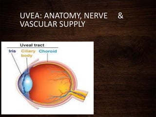
- 3. •Coined from latin word Uva- grape •Middle vascular coat of eyeball •From anterior to posterior: - iris - ciliary body - choroid INTRODUCTION
- 4. Uveal tract firmly attached to sclera only at 3 sites: • the scleral spur • the exit points of vortex veins • the optic nerve
- 5. c 1)Epithelium- Neuroectodermal 2)Sphincter & Dilator pupillae- Neuroectodermal 3)Stroma&blood vessels- Mesenchyme Epithelium- Neuroectodermal Stroma,ciliary muscles & blood vessels - Mesenchyme Mainly derived from inner vascular layer of mesenchyme that surrounds the optic cup. Melanocytes – originate from neural crest Embryology
- 6. Milestones 9th week of gestation Ciliary body begins to appear 12th week of gestation Sphincter pupillae appears 4th month Ciliary processes fully formed 5th month Iris and choroid are formed 6th month Dilator muscles begin to form Sphincter muscle fully differentiated Postnatal period dilator muscles reach adult proportion by 5 years
- 7. Towards the end of gestation , the central iris stroma disappers forming pupil. At birth dilator pupillae is poorly developed & pupil response poorly to mydriatics.
- 8. Congenital anomalies of uveal tract Aniridia (Iridremia) -congenital absence of Iris -true aniridia i.e complete absence of iris rare -a peripheral rim of iris present & this is called clinical Aniridia
- 9. Heterochromia of iris Heterochromia iridium Heterochromia iridis Color of one iris differs from the other One sector of iris differs from the remainder of iris
- 10. Uveal coloboma Typical coloboma: Located inferonasally in the region of closure of embryonic fissure a.Complete coloboma: Extends from pupil to optic nerve Includes retina, choroid, ciliary body, iris b. Incomplete coloboma: Involve the iris alone, or iris and ciliary body, or iris, ciliary body & part of choroid
- 11. Atypical coloboma - Occasionally found in other positions i.e. not related to fissure closure - It is usually incomplete Persistent pupillary membrane - Represents the remnants of vascular sheath of lens - Strands arise from and insert into the iris collarette.
- 12. IRIS • Greek Word Iris - color haloes/rainbows • Anterior most part of the uvea • Diameter -12 mm • Thickness -0.5 to 0.6 mm • 3 to 4 mm aperture slightly nasally- pupil • Attached to the middle of anterior surface of ciliary body • Thinnest at root and tears easily- Iridodialysis
- 13. POINT TO BE NOTED Pupillary margin rests lightly on anterior surface of lens so when lens is removed the iris is flat and often tremendous
- 14. Macroscopic structure Anterior surface 1. ciliary zone: - radial steaks - crypts: peripheral & central - contraction furrows 2. pupillary zone: - about 1.6mm wide, lies between collarette and pigmented pupillary frill Collarette: represents the attachment of pupillary m/m lies 2mm from the pupil margin thickest region of iris
- 16. Posterior surface - dark brown or black - looks smooth under magnification: -schwalbe’s contraction folds: radial furrows, commence 1mm from pupillary border -schwalbe’s structural furrows: start 1.5mm from pupillary border, narrow and deep to start with but becomes wide and shallow as they approach the ciliary margin -circular furrows: finer than radial furrows cross the structural furrows at regular interval more marked near the pupil
- 17. Contact between the posterior surface of iris and anterior capsule of the lens is also altered by pupillary size It is greatest on mid dilatation Precipitate angle closure glaucoma in some predisposed eye with narrow angle
- 18. Microscopic structure 1. Anterior limiting layer 2. Iris stroma 3. Anterior epithelial layer 4. Posterior pigmented epithelial layer
- 19. 1.Anterior limiting membrane •Condensed part of the stroma •Consists of melanocytes and fibroblasts •Deficient in areas of crypts, very thin at contraction furrows •Determines the color of iris 3 types of intercellular junctions present - gap junctions - intermediate junctions - discontinuous tight junctions
- 20. 2. Stroma • Main bulk of iris tissue • Consists of loosely arranged collagenous network with mucopolysaccharide ground substance contains -the sphincter pupillae muscles - dilator pupillae muscles -the vessels and nerves of iris -cellular elements: fibroblast, melanocytes, clump cells and mast cells
- 21. Sphincter pupillae muscles - 0.7mm wide , 0.1-0.17mm thick - Encircles pupillary margin - lies in stroma deep to the surface - - Even after broad iridectomy , which removes a sector of iris sphincter,the remaining sphincter can still constrict the remaining pupil margin Origin is from anterior epithelium,but actually separated from this layer by a thin sheet of collagen & dilator fibre processes, to which it is firmly bound. -Innervated by parasympathetic via short ciliary nerve
- 22. Dilator pupillae muscle - 60um long &7um wide - Filled with myofilaments - Extend from iris root towards pupil - When the muscle contract,it pulls the pupillary margin towards the ciliary body, dilating the pupil - Innervated by sympathetics via long ciliary nerve At birth dilator is poorly developed &pupil response poorly to mydriatics
- 23. Parasympathetic Control of pupillary Size Sphincter Pupillae Short ciliary Nerve Ciliary Ganglion Inferior Oblique Muscle Oculomoter Nerve Edinger Westpal Nucleus
- 24. Dilator muscle Long ciliary nerve Ciliary ganglion Ophthalmic division of Vth nerve Cervical ganglion Ciliospinal centre of Budge Posterior Hypothalamus Sympathetic Control of Pupillary Size:
- 26. Blood vessels - form bulk of iris stroma - they arise mainly from circulus arteriosis major - some also arise directly from anterior ciliary arteries - responsible for radial streaks seen on anterior surface of iris Peculiarities of iris : Absence of internal elastic lamina non fenestrated capillary endothelium
- 27. Cellular elements of stroma 1) Fibroblast • most common stromal cell • found around blood vessels, nerves, muscle tissue and throughout the iris substance 2) Melanocytes • branching elements with processes • contain melanin granules
- 28. 3) Clump cells • large round pigment cells without processes • filled with inclusion granules 4) Mast cells • They are round • Have villous processes Extracellular matrix of stroma -Contain type VI collagen -Laminin and fibronectin
- 29. 3. Anterior epithelial layer • anterior continuation of pigment epithelium of retina and ciliary body • lacking in melanocytes • basal processes of the cells of this layer give rise to dilator pupillae muscle
- 30. 4. Posterior pigmented epithelial layer • anterior continuation of non pigmented epithelium of ciliary body which in turn is the continuation of sensory retina • this layer derived from the internal layer of optic cup • contain abundant columnar type pigment cells • It curves around the pupillary margin and extends for a short distance onto anterior border layer of iris stroma as the pigment ruff.
- 31. - In Rubeosis iridis, this pigmented layer extends farther onto the anterior surface of the iris, a condition called Ectropion uveae Clinical significance
- 32. Forward bowing of iris Angle closure glaucoma Contact betweet the posterior surface of iris and lens creates a relative pupillary block to the flow of aqueous humor through the pupil,which is more marked in mid dilatation
- 33. Applied anatomy Iris nodules - Accumulated deposits of epithelioid cells and lymphocytes deposited onto the iris without tissue destruction. Two types: Koeppes nodule at pupillary border Busaccas nodules near collarette
- 34. Iris atrophy - areas of degeneration of iris - Commonly seen in iridocyclitis
- 36. • Forward continuation of the choroid at ora serrata. • In cut section, triangular in shape CILIARY BODY
- 37. • Anterior side of triangle- part of anterior chamber angle • in middle- attached to the iris • Outer side of triangle lies against the sclera with a suprachoroidal space in between • Inner side of triangle divided into 2 parts: 1) pars plicata - anterior 2) pars plana - posterior
- 38. 1) Pars plicata / corona ciliaris • anterior part • about 2 to 2.5 mm long • contain ciliary muscles • have finger like ciliary processes 2) Pars plana / orbicularis ciliaris • posterior smooth part • 5mm wide temporally • 3 mm wide nasally • Viterous base gain attachment to the epithelium of pars plana
- 40. Microscopic structure From without inwards, consists of five layers: 1) Supraciliary lamina 2) Stroma of the ciliary body 3) Layer of pigmented epithelium 4) Layer of non-pigmented epithelium 5) Internal limiting membrane
- 41. 1) Supraciliary lamina • outermost condensed part of stroma • consists of pigmented collagen fibres • Posteriorly,continuation of suprachoroidal lamina • Anteriorly, continues with the anterior limiting membrane of iris 2) Stroma of the ciliary body • consists of collagenous connective tissue and fibroblast • embedded in it: a. ciliary muscle b. blood vessels c. nerves
- 42. Ciliary muscle • non striated, smooth muscle • occupies most of the outer part of the ciliary body three main groups: 1) the longitudinal or meridional fibres 2) the oblique or radial fibres 3) the circular fibres
- 43. 1) Longitudinal or meridional fibres - most external and closest to the sclera - pass posteriorly into the stroma of ciliary body 2) Oblique or radial fibres - occupy the midportion of the ciliary body - radiate out from the scleral spur 3) Circular fibres - occupy anterior and inner portion of the ciliary body - nearest to the lens - runs parallel to the limbus - directly act as sphincter
- 44. Main action of all parts of ciliary muscles is to slacken the suspensory ligament of lens & thus helps in accommodation. -Longitudinal muscle fibres form tendinous attachment to the scleral spur: contraction increases aqueous flow by opening up the spaces of trabecular meshwork
- 45. Contraction of the ciliary muscle, especially longitudinal and circular fibres pulls the ciliary body forward in accommodation.
- 46. Vascular stroma -contain major arterial circle just in front of circular fibres of the muscle - Arterial circle is formed by the anastomosis between the long posterior ciliary arteries and anterior short ciliary arteries and send branches to iris and ciliary body
- 47. 3) Layer of pigmented epithelium • forward continuation of RPE • anteriorly, continuous with anterior epithelium of iris 4) Layer of non-pigmented epithelium • consists mainly of low columnar or cuboidal cells • forward continuation of sensory retina which stops at ora serrata. • continues anteriorly with posterior pigmented epithelium of the iris
- 48. 5) Internal limiting membrane • lines the non-pigmented epithelium • forward continuation of internal limiting membrane of the retina.
- 49. Nerve supply of the ciliary body • these run from the nasociliary branch of the ophthalmic division of the trigeminal nerve, running in the long ciliary nerve • these fibres enter the ciliary body and terminate in iris, cornea and ciliary muscle. Sensory fibres:
- 50. Sympathetic fibres: Cervical sympathetic trunk Synapse in superior cervical ganglion Long ciliary nerve Ciliary muscles Edinger Westpal nucleus Oculomoter nerve Ciliary ganglion Short ciliary nerve Ciliary muscle Parasympathetic fibres
- 51. Ciliary processes • Whitish finger-like projections from pars plicata part of the ciliary body • 70 to 80 in number • Each process is about 2mm long and 0.5mm in diameter • Form the site of aqueous production
- 52. Ultrastructure of ciliary processes Consists of: a. The network of capillaries b. Stroma of ciliary processes c. Two layers of epithelium
- 53. a. The network of capillaries • occupies the centre of each process • each capillary consists of a very thin endothelium with fenestration • lined by basement m/m • mural cells or pericytes present within basement membrane
- 54. b) Stroma of the ciliary process • very thin • separates capillary network from epithelial layers • consists of ground substance: mucopolysaccharide, proteins & solute of plasma • few collagen connective tissue fibres • wandering macrophages
- 55. c) Two layers of epithelium • their apical surfaces in apposition to each other outer pigmented epithelium: • contains numerous melanin granules inner non-pigmented epithelium: • contain mitochondria, zonula occludentes & lateral and surface interdigitations. • the tight junction between cells of this layer form blood aqueous barrier
- 57. • Posterior portion of the middle vascular coat • Extremely vascular • Extends from optic disc to ora serrata • The inner surface: smooth, brown and lies in contact with RPE • The outer surface: rough and attached to sclera • Posteriorly-0.22 mm thick • anteriorly-0.1 mm CHOROID
- 58. • Firmly attached to the margin of the optic disc • Loosely at points where vessels and nerves enter it • Attachment to sclera is strongest behind the sclera
- 59. Microscopic structure From without inwards, consists of four layers: 1) Suprachoroidal lamina (lamina fusca) 2) Stroma of the choroid 3) Choriocapillaries 4) bruch’s membrane (basal lamina or lamina vitrae)
- 60. 1) Suprachoroidal lamina • thin membrane 10 to 34 μm • made of condensed collagen fibres, melanocytes and fibroblasts • continues anteriorly with supraciliary lamina • space between this m/m and sclera: suprachoroidal space (contain long & short posterior ciliary arteries and nerves)
- 61. -contains vessels, nerves, cells & connective tissue -stromal cells include: a. melanocytes b. fibrocytes c. macrophages d. mast cells e. plasma cells 2) Stroma of the choroid Main bulk is formed by vessels, arranged in two layers: a. Haller’s layer: outer layer of large vessels b. Sattler’s layer: inner layer of medium vessels
- 62. • consists of a rich bed of wide bore fenestrated capillaries (18 to 50μm) • receives most of its blood from medium & large vessels of stroma • nourishes RPE & outer layers of sensory retina • density greatest at macula 3) Choriocapillaries • choriocapillaries are divided into non overlapping lobules or hexagonal patches
- 63. • innermost layer of choroid • thin non cellular lamina • lies between choriocapillaries and pigment epithelium of the retina • 2 to 4 μm thickness 4) Bruch’s membrane
- 64. Comprises of five layers a. Basal lamina of RPE b. Inner collagen layer c. Middle elastic layer d. Outer collagen layer e. Basal lamina of choriocapillaries
- 65. • Choroidal ischaemia often occurs as a pale hexagonal patches (mosaic pattern) • during choroidal phase of FFA, these lobules fill independently from one another, giving a transiently patched or blotched appearance Clinical significance
- 66. Bruchs membrane become thickened with increasing age and produces hyaline excrescence known as drusens
- 67. Uveal tract supplied by 3 sets of arteries: 1) Short posterior ciliary arteries 2) Long posterior ciliary arteries 3) Anterior ciliary arteries BLOOD SUPPLY OF THE UVEAL TRACT
- 68. 1) Short posterior ciliary arteries Arise as two trunks from the ophthalmic artery Each trunk divides into 10 t0 20 branches Pierce the sclera around the optic nerve Supply the choroid in segmental manner
- 70. 2) Long posterior ciliary arteries Arise as nasal and temporal branch from the ophthalmic artery Pierce the sclera obliquely on medial & lateral side of optic nerve Run forward in suprachoroidal space to reach ciliary muscle without giving any branch Anastomose with each other & with the anterior ciliary arteries to form major arterial circle Also give branches which supply the ciliary body
- 71. 3) Anterior ciliary arteries Derived from muscular branch of ophthalmic artery 7 in number: 2 each from arteries of SR, IR & MR, 1 from that of LR These pass anteriorly in episclera Give branches to sclera, limbus & conjunctiva Ultimately pierce the sclera near the limbus to enter ciliary muscle
- 72. Anastomose with two long posterior ciliary arteries to form major arterial circle Branches arise from major arterial circle to supply ciliary process (one branch for each process) Many branches from major arterial circle run radially through iris towards pupillary margin Anastomose with each other to form minor arterial circle
- 74. • four in number (superior temporal, inferior temporal, superior nasal and inferior nasal) • pierce sclera obliquely on each side of SR and IR muscles about 6 mm behind the equator -drain blood from: • whole of choroid • receive small veins from ONH • small veins from retina • from iris, ciliary process, ciliary muscle, anterior part of choroid 3) Venae vorticosae (vortex veins or posterior ciliary veins)
- 75. • two superior vortex veins open into superior ophthalmic vein • two inferior vortex veins open into inferior ophthalmic vein
- 76. Uveitis: Inflammation of uveal tissue only Classification: 1.Anterior uveitis: Some clinically applied aspects Inflammation of uveal tissue from iris upto pars plicata of ciliary body -Iritis: inflammation predominantly affect iris -Iridocyclitis: iris and pars plica part of ciliary body are involved -Anterior cyclitis: pars plicata part of ciliary body is predominantly affected
- 77. 2.Intemediate uveitis: Inflammation of pars plana and peripheral part of retina and underlying choroid 3.Posterior uveitis: Inflammation of choroid and retina Hence the term choroiditis, chorioretinitis, retinochoroiditis or neurouveitis is used 4.Panuveitis: Inflammation of whole uvea
- 78. Pain Redness Photophobia Salient features of uveitis
- 79. Posterior synechiae Cataract Glaucoma due to PAS Band keratopathy Complications of uveitis
- 80. Aqueous cells: -presence of inflammatory cells in the anterior chamber -the inflammatory response causes white blood cells such as neutrophils, monocytes and lymphocytes to leave the inflamed iris vessels to reach aqueous humour. Grading +1: 1-5cells +2: 6-15cells +3: 26-50cells +4: over 50
- 81. Aqueous flare - Turbidity of the aqueous humour caused by increased protein level - Blood aqueous barrier breakdown results in protein (albumin) exudation in the anterior chamber giving the normally clear, colorless aqueous humour a milky appearance k/a flare Grading +1:faint(just detectable) +2:moderate flare with clear iris and lens +3: marked flare(iris and lens details hazy) +4: intense flare(Fibrin or plastic aqueous
Notas do Editor
- Anterior balloons figure
- Both layer of epithelium r derived frm marginal region of optic cup i.e neuroectodermal. Sphi n dil r derived frm anterior epithelium.stroma n bv dev frm vascular layer of mesenchyme +nt anterior to optic cp.CM-epi frm anterior part of 2 layer of optic cp.ciliary epi undergo convuating or folding mvmnt to form 70-75 ciliary process.stroma.. Frm vascular lyer of mesenchyme
- Sometimes few strands of this tissue r left as persistant pupillary membrane
- 1st-Waardenburg syndrome(genetic condn asso wid hearing loss,neurofibromatosis(disorder of melanin)2nd- Malignent melanoma,trauma,blood in ac for long duration lead to iron deposition frm breakdown of blood particles
- Coloboma : a condition where a portion of the structure is missing
- As pupil dilates the pupillary edge approach near collarette so that d pupillary remnant when present may seem to arise from pupil margin.
- Nasal 3-4 mm aperture k/a pupil
- Anterior surface of iris can be divided into a ciliary zne n pupillary zn by a zigzag line ka collarette.Ciiary zn presents series of radial streaks due to underlying radial blood vessels n crypts are d depression where superficial layer of iris is missing.They are arranged in 2 rows-periphera +nt near iris root n central +nt near collarette.
- However when seen under magnification it presents radial n circular furrows n folds Circular furrow r formed due to difference in thickness of pigmented epithelium.
- Contact between d posterior surface of iris n anterior capsule of lens is also altered by pupil size n is said to be greatest with pupil in mid dilatation so that the relative pupillary block is created which bulges the iris forward slightly .In some predisposed eye wid narrow angle may precipitate angle closure
- Iris consist of 4 layers which from anterior to posterior r
- Anterormost condensed part of stroma.The definitive colour of iris depend upon this layer.In blue iris this layer is thin n contain few pigment cell.While in brown iris it is thick n densly pigmented.
- In which are embeeded the sphincter pup n dilater pupille, vessels n nerves and other cells which include fibroblast….
- Measures about ..
- Preganglionic fibre for both ciliary muscle n pupi arises from EWN which move along oculomoter nerve. At the anterior part of cavernous sinus the fibre moves along IO muscle n synapse at ciliary ganglion.From here 6-8 post ganglionic short ciliary nerve arises which then supplies to sphincter pupillae.
- 1st order neuron starts from posterior hypothalamus which runs through pons n medulla n finally synapse at C8-T2. 2nd order preganglionic axon travel along ventral root of c8-T2 n synapse at superior cervical ganglion at carotid bifurcation.3rd order neuron send their post ganglionic fibre through oph div of V nerve which enter into ciliary ganglion n reach dilator muscle as long ciliary nerve.
- Sphincter muscle encircles d pupil n innervated by parasym nerve endings n its contraction constrict d pupil.The dilator muscle runs radially n innervation is chiefly by sympathetic n contraction dilates d pupil
- Radial vessels are straight when pupil constrict and become wavy when pupil dilates.
- Mel r branching element wid process which contain melanin granu
- More marked in mid dilatation.This may encourage forward bowing of iris which in predisposed narrow angle may precipitate angle closure glaucoma
- Condn in which area of iris is degenerated
- Ciliary processes become longer n thinner wid age.
- Posterior it is d continuation of suprachoroidal lamina and anteriorly it become continuous wid anterior limiting membrane of iris. Structures embeeded in it are,,,,
- It occupies mst of the outer part of CB. It is non striated muscle having 3 parts. Main action of all parts of ciliary muscle is to slacken the suspensory ligament of lens n thus helps in accomodatn.
- 1st order neuron starts from posterior hypothal 2nd order preganglionic axon travel along c8-t2& synapse at superior cervical ganglion.3rd order neuron send their post ganglionic fibre through oph div of V CN which enter into ciliary gangl n reach ciliary muscle as long ciliary nerve..
- 2 layer of epi r arranged wid apical surface in apposition to each other
- Patchy filling in ffa figure
- But clinically there is always some associated inflammation of adjacent structures such as retina, vitreous,sclera n cornea
- Pain –ciliary spasm since ciliary body is innervated by trigeimina nerve.Redness due to dilated episcleral vessels.photophobia coz of inflammation of iris and cornea results irritation of corneal nerve n 2dary to ciliary spasm.
- Now we r at the end of this pptn. Common clinical signs of ueitis are
- If the flare is