1. Sudheerkumar kamarapu
Assistant Professor
Sri Shivani college of pharmacy
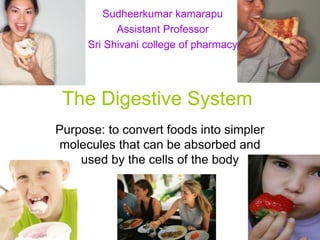
The Digestive System
Purpose: to convert foods into simpler
molecules that can be absorbed and
used by the cells of the body
2. Why is Digestion Important?
• When we eat such things as bread, meat,
and vegetables, they are not in a form that
the body can use as nourishment.
• Our food and drink must be changed into
smaller molecules of nutrients before they
can be absorbed into the blood and carried
to cells throughout the body.
• Digestion is the process by which food and
drink are broken down into their smallest
parts so that the body can use them to build
and nourish cells and to provide energy.
3. Let’s Break It Down…
• The digestive system it is a tube running from
mouth to anus.
• This tube is like an assembly line, or more
properly, a disassembly line.
• Its chief goal is to break down huge
macromolecules (proteins, fats and starch),
which cannot be absorbed intact, into smaller
molecules (amino acids, fatty acids and glucose)
that can be absorbed across the wall of the tube,
and into the circulatory system for dissemination
around your body.
4. Components of the Digestive
System
The Major Players The Accessory Structures
• Mouth • Salivary Glands
• Pharynx • Pancreas
• Esophagus • Liver
• Stomach • Teeth
• Small Intestine • Tongue
• Large Intestine
All of these organs work together to carry out…
5.
6.
7. 3 Fundamental Processes:
• Secretion: Delivery of enzymes, mucus,
ions and the like into the lumen, and
hormones into blood.
• Absorption: Transport of water, ions and
nutrients from the lumen, across the
epithelium and into blood.
• Motility: Contractions of smooth muscle in
the wall of the tube that crush, mix and
propel its contents.
8. The Path of Digestion
• Digestion begins in the mouth (oral cavity)
– Food is broken down by chewing
(mechanical) and enzymes in the saliva
(chemical)
– Teeth coated with enamel cut and tear food
– Saliva contains the enzyme salivary amylase
(breaks down starch)
– Food is turned into a soft ball called a bolus
– Bolus travels from the mouth to the pharynx
(throat)
9.
10. The Path of Digestion
• Swallowing is a result of the combined
effort of the tongue and throat muscles
• Epiglottis (small flap of connective tissue),
prevents food from going into the trachea
(airway)
• Next, food travels Into the esophagus
• Peristalsis squeezes
food along into the
stomach
12. The Path of Digestion
• A ring of muscles called the cardiac sphincter
closes the esophagus after food passes into the
stomach
• Stomach continues chemical and mechanical
digestion
– Chemical: Glands release mucous, HCl,
pepsin (enzyme that breaks down protein)
– Mechanical: Muscles contact and churn
contents into chyme (a thin watery liquid)
13. Anatomy and function of
digestive organs
• GIT-PERITONEUM
– peritoneum is a serous membrane which lines
the abdomen cavity and covers the abdominal
organs.
– It consists of two layers
1) Parietal peritoneum-lines the walls of abdominal
cavity.
2) Visceral peritoneum-which covers the
abdominal organs.
The space between these two layers is called as
peritoneal space.
14. • Organs completely covered by peritoneum
are – stomach, liver and intestines.
• Organs partly covered by peritoneum –
kidneys.
Folds of peritoneum
omenta : the folds of peritoneum connected to
the stomach are called omenta.
They divided into
Greater omentum – which hangs from the lower
border of stomach to the front surface of small
intestine.
Lesser omentum – which extends from the lower
border of liver to the lesser curvature of stomach.
15. Mesentry : it is the fold of peritoneum which attaches the
different parts of small intestine to the posterior
abdominal wall.
Blood vessels, nerves and lymphatics enter the intestine
only through mesentry.
Peritoneal ligaments : they are folds of peritoneum which
connect organs like liver and uterus to the posterior
abdominal wall.
Pelvic peritoneum : it is the part of peritoneum present in
the pelvic region.
The pelvic peritoneum is actually the continuation of
peritoneum in the abdominal cavity.
The arrangement is different in males and females due
to the presence of uterus and fallopian tubes in females.
16. Functions of peritoneum
• It forms a complete or partial covering for abdominal
organs.
• It forms the smooth lining which enables the abdominal
organs to move over each other without friction.
• The ligaments and mesentries of peritoneum hold the
abdominal organs in position.
• Omentum and mesentry serve as store house for fat.
• The fats of peritoneum prevents infections being carried
to abdominal organs.
• The peritoneum itself can absorb large quantities of
fluids.
17. WALL STRUCTURE
• SEROSA:- THE OUTERMOST LAYER MADE
OF A SEROUS MEMBRANE & CONNECTIVE
TISSUES. ALSO CALLED THE PERITONIUM.
• MUSCULAR LAYER:- MADE OF TWO
CIRCULAR & LONGITUDINAL SMOOTH
MUSCLE LAYERS.
• SUBMUCOSA:- MADE OF LOOSE
CONNECTIVE TISSUE CONTAINING NERVES,
GLANDS, BLOOD AND LYMPHATIC VESSELS.
• MUCOSA:- MADE OF EPITHELIUM,
CONNECTIVE TISSUE AND SOME SMOOTH
MUSCLE FIBERS.
22. THE MOUTH
• FORM THE ORAL CAVITY WHICH IS
INVOLVED IN THE INTAKE OF FOOD,
BREAKING IT, MIXING IT WITH SALIVA
AND SWALLOWING IT.
• CONTAIN THE TEETH & IS
SURROUNDED BY THE CHEEKS,
PALATE & TONGUE.
• MECHANICAL DIGESTION BY
CHEWING.
• CHEMICAL DIGESTION BY SALIVARY
AMYLASE ENZYME.
25. THE SALIVARY GLANDS
• THEY SECRETE SALIVA THAT CLEAN &
LUBRICATE THE MOUTH, DISSOLVE,
BIND AND DIGEST FOOD PARTICLES.
• PAROTID GLANDS:- IN FRONT OF EAR.
SECRETE AMYLASE ENZYME.
• SUBMANDIBULAR GLANDS:- IN FLOOR
OF THE MOUTH.
• SUBLINGUAL GLANDS:- UNDER
TONGUE.
27. PHARYNX & ESOPHAGUS
• THE PHARYNX TRANSPORT FOOD
FROM THE MOUTH TO THE
ESOPHAGUS.
• THE ESOPHAGUS IS A MUSCULAR
TUBE THAT CARRY FOOD FROM THE
PHARYNX INTO THE STOMACH.
• HAS AN UPPER ESOPHAGIAL
SPHINCTER AND A LOWER
ESOPHAGIAL SPHINCTER.
29. THE STOMACH
• A POUCHLIKE PART OF THE
DIGESTIVE SYSTEM THAT FUNCTION
FOR STORAGE, MIXING AND
DIGESTION OF FOOD.
MADE OF FOUR REGIONS:-
• CARDIAC – FUNDIC – BODY – PYLORIC
• ENTERANCE IS GUARDED BY THE
LOWER ESOPHAGIAL SPHINCTER
WHILE EXIT IS GUARDED BY THE
PYLORIC SPHINCTER.
31. THE STOMACH
• A THICK FOLDED MEMBRANE COATED
WITH MUCUS LINE THE STOMACH.
• GASTRIC GLANDS SECRETE
DIGESTIVE ENZYMES &
HYDOCHLORIC ACID.
• PEPSIN ENZYME START THE
DIGESTION OF PROTEINS TO FORM
SMALL PEPTIDES
• HYDROCHLORIC ACID ACTIVATE
PEPSIN & KILL ANY
MICROORGANISMS IN FOOD.
34. Pancreas
• Pancreas is a long, slender gland which lines
transversely across the posterior abdominal
wall.
• It lies behind the stomach at the level of Ist and
2nd lumbar vertebrae.
• Parts: pancreas consists of head, body and tail.
• Head lies in the C-shaped curve of duodenum
• Body lies in front of the bodies of lumbar
vertebrae.
• Tail lies in contact with the hylum of spleen.
39. Structure
• The substance of pancreas contains a number
of lobules of secretory cells called “Acini”
• Inbetween the Acini, there are groups of
endocrine cells called “Islets of Langerhans”.
• Small ducts emerge from these lobules, these
ducts unite and reunite to form the “Pancreatic
duct”(duct of Wirsung)
• This duct begins at the tail and emerge from the
head of pancreas it enters the duodenum along
the common bile duct.
40. Secretions
• The secretions of pancreas can be classified into
– Exocrine secretion – digestive secretion
– Endocrine secretion – hormonal secretion
• THE EXOCRINE PANCREAS SECRETES
PANCREATIC JUICE INTO DUODENUM PART OF
THE SMALL INTESTINE.
• It is an alkaline fluid with a pH of around 8.
• Daily 1200-1500ml. Of pancreatic juice is
secreted.
• Pancreatic juice contains 97-98%wateralong
with the following digestive enzymes.
41. • PANCREATIC JUICE CONTAIN DIGESTIVE
ENZYMES & A BICARBONATE SOLUTION.
• PANCREATIC AMYLASE:- CARBOHYDRATES
DIGESTION - converts starch into maltose.
• PANCREATIC LIPASE:- FATS DIGESTION -
converts fats into fatty acids and glyserol.
• TRYPSIN:- PROTEINS DIGESTION - converts
proteins into aminoacids.
43. THE LIVER
• It IS THE LARGEST GLAND IN THE
BODY.
• It lies in the upper part of abdominal cavity
below the diaphragm and under the cover
of lower ribs.
• Externally, the liver contains two lobes (a
right lobe and a left lobe) and four
surfaces-superior surface, inferior surface,
anterior surface and posterior surface)
45. • Internally the liver consists of a large number of
liver cells called lobules.
• Each lobules has a central vein or intralobular
vein.
• The connective tissue lying in between the
lobules contains the branches of – portal vein,
hepatic artery and bile duct.
• Liver secretes the bile is carried through bile
ducts which are formed by the union of biliary
canaliculi.
• The biliary canaliculi are small biliary channels
present inbeween the lobules of liver.
46. • The bile ducts from right and left lobes of liver
unite to form common hepatic duct.
• The hepatic duct unites with cystic duct of gall
bladder to form common bile duct.
• Later, the common bile duct unites with the
pancreatic duct in the duodenum at a papilla
called ampulla of vater.
FUNCTIONS:-
• CLEANS HARMFUL CHEMICALS, DRUGS
AND DEAD CELLS FROM THE BLOOD.
• STORES FATS, GLYCOGEN, MINERALS &
VITAMINS.
• SECRETES BILE THAT BREAKS THE LARGE
FAT DROPS INTO SMALLER PARTICLES &
HELP THEIR ENZYMATIC DIGESTION.
47. THE GALLBLADDER
• Gall bladder is a pear shaped storage sac for
bile.
• It is situated in the under surface of the right lobe
of liver.
• It consists of a – fundus, body and neck.
• Coats of gall bladder: gall bladder consists of
three coats.
• Outer serous or peritoneal coat – which is continuous with
the peritoneum covering the liver.
• Middle muscular layer coat – made of unstriped muscles.
• Inner mucous coat – which is continuous with the lining of
bile ducts.
48. • Duct of gall bladder:
• The duct through which gall bladder opens
is called cystic duct – arises at neck of gall
bladder.
• The cystic duct unite with common hepatic
duct and forms common bile duct.
common bile duct joins with pancreatic
duct and opens into the duodenum.
• The sphincter present in the bile duct at
this termination in the duodenum is called
sphincter of Oddi.
49. Function of gall bladder
• BILE IS STORED IN THE
GALLBLADDER.
• WHEN FATTY FOOD ENTER
DUODENUM, CHOLECYTOKININ {CCK}
HORMONE IS SECRETED BY THE
INTESTINAL CELLS.
• {CCK} STIMULATES THE
GALLBLADDER WALLS CONTRACTION
TO EMPTY BILE INTO THE DUODENUM.
51. THE SMALL INTESTINE
• A LONG TUBE EXTENDING BETWEEN
THE STOMACH AND THE LARGE
INTESTINE.
• ENTRANCE IS GUARDED BY THE
PYLORIC SPHINCTER WHILE EXIT IS
GUARDED BY THE ILEOCECAL
SPHINCTER.
• CONSIST OF THREE PARTS:-
• DUODENUM – JEJUNUM – ILEUM
• MOST OF DIGESTION & ABSORPTION
OF FOOD OCCUR IN THE SMALL
54. SMALL INTESTINE ENZYMES
• MALTASE DIGEST MALTOSE TO
GLUCOSE
• SUCRASE DIGEST SUCROSE TO
GLUCOSE AND FRUCTOSE.
• LACTASE DIGEST LACTOSE TO
GLUCOSE AND GALACTOSE.
• LIPASE DIGEST FATS TO FATTY
ACIDS.
• PEPTIDASE DIGEST SMALL PEPTIDES
TO SINGLE AMINO ACIDS.
56. THE LARGE INTESTINE
• EXTEND FROM THE ILEUM TO THE
ANUS AND IS MADE OF THREE
SEGMENTS:-
• CECUM – COLON – RECTUM.
• FUNCTION FOR ABSORPTION OF
WATER, MINERALS AND VITAMINS.
• ENTRANCE GUARDED BY THE
ILEOCECAL SPHINCTER WHILE THE
ANAL EXIST IS GUARDED BY
INTERNAL AND EXTERNAL ANAL
SPHINCTERS.