Monitoring neural activities by optical imaging
•Transferir como PPTX, PDF•
1 gostou•775 visualizações

Monitoring neural activities by optical imaging along with the use of genetic modification provides better spatio-temporal resolution to study single neural firing and hence very useful in understanding the neural process and dynamics. This is just a glimpse of few articles reported their outcome of such imaging.

Recomendados
Recomendados
Mais conteúdo relacionado
Mais procurados
Mais procurados (20)
Destaque
Destaque (8)
Semelhante a Monitoring neural activities by optical imaging
Semelhante a Monitoring neural activities by optical imaging (20)
Mais de Md Kafiul Islam
Mais de Md Kafiul Islam (20)
Último
Último (20)
Monitoring neural activities by optical imaging
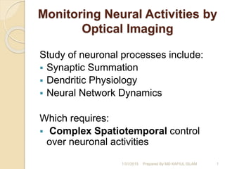
- 1. Monitoring Neural Activities by Optical Imaging Study of neuronal processes include: Synaptic Summation Dendritic Physiology Neural Network Dynamics Which requires: Complex Spatiotemporal control over neuronal activities 1/31/2015 1Prepared By MD KAFIUL ISLAM
- 2. Novel Approaches to Monitor and Manipulate Single Neurons In Vivo (Brecht et al. 2004, JNS) The combined application of the optical, electrophysiological, and genetic techniques outlined here will allow us to refine our focus to the structure and function of identified neurons in the intact brain Monitoring neurons with improved cellular resolution. A, Long-term in vivo imaging of GFP-expressing axons in the adult mouse barrel cortex. Note gained (e.g., green arrowhead), lost (blue arrowhead), and stable (yellow arrowheads) recognized synaptic terminals B, Ca2+ transients evoked by whisker deflection. Left, A high magnification image of layer 2/3 neurons in vivo (depth, 130m) in the barrel cortex of a 13-d-old mouse. Right, Line-scan recordings of Ca2+ transients evoked in two neurons by a deflection of the majority of whiskers on the contra lateral side of the mouse’s snout. The position of the scanned line and the cells analyzed are indicated (left) 1/31/2015 2 Prepared By MD KAFIUL ISLAM
- 3. Optical monitoring of brain function in vivo: from neurons to networks (Garaschuk et. al. 2006, EJP) Here is the review of the multi-cell bolus loading (MCBL) method that involves the use of membrane permanent fluorescent indicator dyes. It is shown that this approach is useful for the functional analysis of clusters of neurons and glial cells in vivo. And also MCBL and two-photon imaging can be easily combined with other labeling techniques, particularly with those involving the use of genetically encoded, green- fluorescent-protein-based indicators. Somatic Ca2+ signals in MCBL-stained cells require action potentials. a Left panel: a merged image of layer 2/3 cells in the rat somatosensory cortex stained in vivo with OG-1 AM (green) and sulforhodamine 101 (red). Neurons are green and astrocytes are yellow. Right panel: a simultaneous recording of action potentials in the cell-attached configuration (upper trace) and associated Ca2+ transients (lower trace) obtained from the neuron in the middle. The position of pipette is shown by an arrow. Sensory-driven Ca2+ transients in individual cortical neurons. a Individual neurons in the mouse barrel cortex (left) and the corresponding Ca2+ transients (right) evoked by the deflection of the majority of whiskers at the contralateral side of the snout. The transients were recorded using the line-scan mode (5 ms/line). The position of the scanned line is indicated 1/31/2015 3Prepared By MD KAFIUL ISLAM
- 4. In Vivo Calcium Imaging of Neural Network Function (Werner et. al. 2007, @APS ) Network activity can be measured in vivo using two-photon imaging of cell populations that are labeled with fluorescent calcium indicators. 1/31/2015 4Prepared By MD KAFIUL ISLAM
- 5. An Optical Neural Interface: in vivo control of rodent motor cortex with integrated fiber-optic & opto- genetic technology (A M Aravanis et al, JNE, 2007) Here they report the first in vivo behavioral demonstration of a functional optical neural interface (ONI) in intact animals, involving integrated fiberoptic & Optogenetic technology. Channelrhodopsin-2 (ChR2), an algal light-activated ion channel has been developed for use In mammals, can give rise to safe, light-driven stimulation of CNS neurons on a timescale of milliseconds. 1/31/2015 Prepared By MD KAFIUL ISLAM 5
- 6. Multi-site optical excitation using ChR2 and micro-LED array (Grossman et. al. 2010, JNE) The recent development of neural photosensitization tools as ChR2 (Channelrhodopsin-2 ), offers new opportunities for non-invasive, flexible and cell- specific “Neuronal Stimulation” Dendritic excitation. (A) Illumination (1:1) of three proximal dendrites and cell soma of ChR2- expressing hippocampal neuron. (B) Currents evoked by 10 ms of 1 mW mm−2 illumination. (C) ChR2-evoked input currents were synchronized to maximize spiking 1/31/2015 6Prepared By MD KAFIUL ISLAM
- 7. Optical Recording of Neuronal Activity With A Genetically Encoded Calcium Indicator in Anesthetized & Freely Moving Mice (Lütcke et al.2010, FNC) Bulk recording of spontaneous & sensory- evoked YC3.60 Ca2+ signals in barrel cortex.(B) Large-area Ca2+ imaging using single- photon excitation and a camera and simultaneous local field potential (LFP) recording in barrel cortex of an anesthetized mouse and The mean YC3.60 fluorescence signal (ΔF/F in YFP- channel; red traces) correlated well with the LFP for both spontaneous activity (top) and upon air-puff whisker stimulation (bottom; dashed vertical lines). (C) Fiber-optic bulk recording of YC3.60 signals in barrel cortex in an anesthetized mouse (left schematic). Fluorescence excitation and detection were both accomplished through the optical fiber, the tip of which was placed on the cortical 1/31/2015 7Prepared By MD KAFIUL ISLAM