SlideShare uma empresa Scribd logo
1 de 40
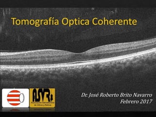
Tomografía Optica Coherente
Dr. José Roberto Brito Navarro
Febrero 2017
DRUSEN PEQUEÑOS DRUSEN GRANDES DEPOSITOS
SUBRRETINIANOS
DUROS CUTICULARES BLANDOS PSEUDODRUSEN
RETICULARES
MENOS DE 63 MICRAS NUMEROSOS MÁS DE 50
ENTRE 25 Y 75 MICRAS
GRANDES ARRIBA DE 125
MICRAS
DIAMETRO VARIABLE.
PEQUEÑOS DEPOSITOS
SUB-EPR
HIPERREFECTIVOS.
A VECES MENOS
DISCERNIBES QUE EN A
FOTOGRAFIA A COLOR
SUB-EPR
FORMA ALARGADA.
MODERADA
HIPERREFLECTIVIDAD
CONFIGURACIÓN
DENTADA O SIERRA
DRUSEN MAS GRANDES
ERODAN EN LA MONOCAPA
DE EPR.
DEPOSITOS MÁS GRANDES
SUB-EPR
HIPERREFECTIVOS
DEPOSITOS ENTRE EL EPR
Y ZONA ELIPSOIDES.
PUEDEN ROMPER LA ZONA
ELIPSOIDE.
GRACIAS

Mais conteúdo relacionado

Mais procurados

Degeneración macular relacionada con la edad, - Age related macular degenera...
Degeneración macular relacionada con la edad,  - Age related macular degenera...Degeneración macular relacionada con la edad,  - Age related macular degenera...
Degeneración macular relacionada con la edad, - Age related macular degenera...Diego Alejandro Valera-Cornejo
 
Miopia degenerativa
Miopia degenerativaMiopia degenerativa
Miopia degenerativaalejandra
 
Interpretacion del campo visual computadorizado
Interpretacion  del campo visual computadorizadoInterpretacion  del campo visual computadorizado
Interpretacion del campo visual computadorizadoEdwin Wilder Casaverde
 
EXPLORACIÓN FUNCIONAL DEL NERVIO ÓPTICO MEDIANTE CAMPIMETRÍA
EXPLORACIÓN FUNCIONAL DEL NERVIO ÓPTICO MEDIANTE CAMPIMETRÍAEXPLORACIÓN FUNCIONAL DEL NERVIO ÓPTICO MEDIANTE CAMPIMETRÍA
EXPLORACIÓN FUNCIONAL DEL NERVIO ÓPTICO MEDIANTE CAMPIMETRÍAElviraLopezArroquia
 
Taller de lámpara de hendidura
Taller de lámpara de hendiduraTaller de lámpara de hendidura
Taller de lámpara de hendiduraMónica Márquez
 
Estrategia SITA Faster y Programa 24-2C
Estrategia SITA Faster y Programa 24-2CEstrategia SITA Faster y Programa 24-2C
Estrategia SITA Faster y Programa 24-2CNataly Osses
 
Degeneración macular asociada a la edad
Degeneración macular asociada a la edadDegeneración macular asociada a la edad
Degeneración macular asociada a la edadDrMAZapata
 
Degeneracion macular relacionada con la edad seca - Dry Age-Related Macular D...
Degeneracion macular relacionada con la edad seca - Dry Age-Related Macular D...Degeneracion macular relacionada con la edad seca - Dry Age-Related Macular D...
Degeneracion macular relacionada con la edad seca - Dry Age-Related Macular D...Diego Alejandro Valera-Cornejo
 
Síndrome de dispersión pigmentaria y glaucoma pigmentario
Síndrome de dispersión pigmentaria y glaucoma pigmentarioSíndrome de dispersión pigmentaria y glaucoma pigmentario
Síndrome de dispersión pigmentaria y glaucoma pigmentarioEder Díaz Dorado
 

Mais procurados (20)

Degeneración macular relacionada con la edad, - Age related macular degenera...
Degeneración macular relacionada con la edad,  - Age related macular degenera...Degeneración macular relacionada con la edad,  - Age related macular degenera...
Degeneración macular relacionada con la edad, - Age related macular degenera...
 
Desprendimiento de retina
Desprendimiento de retinaDesprendimiento de retina
Desprendimiento de retina
 
CAMPO VISUAL
CAMPO VISUALCAMPO VISUAL
CAMPO VISUAL
 
Miopia degenerativa
Miopia degenerativaMiopia degenerativa
Miopia degenerativa
 
Retinoscopía
RetinoscopíaRetinoscopía
Retinoscopía
 
Interpretacion del campo visual computadorizado
Interpretacion  del campo visual computadorizadoInterpretacion  del campo visual computadorizado
Interpretacion del campo visual computadorizado
 
Retinoscopia
RetinoscopiaRetinoscopia
Retinoscopia
 
EXPLORACIÓN FUNCIONAL DEL NERVIO ÓPTICO MEDIANTE CAMPIMETRÍA
EXPLORACIÓN FUNCIONAL DEL NERVIO ÓPTICO MEDIANTE CAMPIMETRÍAEXPLORACIÓN FUNCIONAL DEL NERVIO ÓPTICO MEDIANTE CAMPIMETRÍA
EXPLORACIÓN FUNCIONAL DEL NERVIO ÓPTICO MEDIANTE CAMPIMETRÍA
 
Angiofluoresceinografía
AngiofluoresceinografíaAngiofluoresceinografía
Angiofluoresceinografía
 
Taller de lámpara de hendidura
Taller de lámpara de hendiduraTaller de lámpara de hendidura
Taller de lámpara de hendidura
 
Biomicroscopia con lámpara de hendidura
Biomicroscopia con lámpara de hendiduraBiomicroscopia con lámpara de hendidura
Biomicroscopia con lámpara de hendidura
 
Entendiendo angiografía con fluoresceína
Entendiendo angiografía con fluoresceínaEntendiendo angiografía con fluoresceína
Entendiendo angiografía con fluoresceína
 
Cristalino
CristalinoCristalino
Cristalino
 
Estrategia SITA Faster y Programa 24-2C
Estrategia SITA Faster y Programa 24-2CEstrategia SITA Faster y Programa 24-2C
Estrategia SITA Faster y Programa 24-2C
 
Degeneración macular asociada a la edad
Degeneración macular asociada a la edadDegeneración macular asociada a la edad
Degeneración macular asociada a la edad
 
Degeneracion macular relacionada con la edad seca - Dry Age-Related Macular D...
Degeneracion macular relacionada con la edad seca - Dry Age-Related Macular D...Degeneracion macular relacionada con la edad seca - Dry Age-Related Macular D...
Degeneracion macular relacionada con la edad seca - Dry Age-Related Macular D...
 
Síndrome de dispersión pigmentaria y glaucoma pigmentario
Síndrome de dispersión pigmentaria y glaucoma pigmentarioSíndrome de dispersión pigmentaria y glaucoma pigmentario
Síndrome de dispersión pigmentaria y glaucoma pigmentario
 
Ultrasonografia ocular clase uno
Ultrasonografia ocular clase unoUltrasonografia ocular clase uno
Ultrasonografia ocular clase uno
 
Anatomia retina
Anatomia  retinaAnatomia  retina
Anatomia retina
 
Sindrome de duane
Sindrome de duaneSindrome de duane
Sindrome de duane
 

Destaque (11)

Estrella Macular
Estrella MacularEstrella Macular
Estrella Macular
 
OCT: Interpretacion clínica
OCT: Interpretacion clínica OCT: Interpretacion clínica
OCT: Interpretacion clínica
 
Ultrasonido y Doppler Ocular
Ultrasonido y Doppler OcularUltrasonido y Doppler Ocular
Ultrasonido y Doppler Ocular
 
Caso Necrosis Retiniana Aguda
Caso Necrosis Retiniana AgudaCaso Necrosis Retiniana Aguda
Caso Necrosis Retiniana Aguda
 
Baja vision
Baja visionBaja vision
Baja vision
 
Maculopatia Miopica
Maculopatia MiopicaMaculopatia Miopica
Maculopatia Miopica
 
Exploración Ocular
Exploración OcularExploración Ocular
Exploración Ocular
 
Sics 1 construccion de herida sics español
Sics 1   construccion de herida sics españolSics 1   construccion de herida sics español
Sics 1 construccion de herida sics español
 
Exploracion ocular 2
Exploracion ocular 2Exploracion ocular 2
Exploracion ocular 2
 
Ovcr
OvcrOvcr
Ovcr
 
Generalidades de Uveitis
Generalidades de UveitisGeneralidades de Uveitis
Generalidades de Uveitis
 

Último

Sistema Nervioso Periférico (1).pdf
Sistema Nervioso Periférico      (1).pdfSistema Nervioso Periférico      (1).pdf
Sistema Nervioso Periférico (1).pdfNjeraMatas
 
(2024-04-30). ACTUALIZACIÓN EN PREP FRENTE A VIH (PPT)
(2024-04-30). ACTUALIZACIÓN EN PREP FRENTE A VIH (PPT)(2024-04-30). ACTUALIZACIÓN EN PREP FRENTE A VIH (PPT)
(2024-04-30). ACTUALIZACIÓN EN PREP FRENTE A VIH (PPT)UDMAFyC SECTOR ZARAGOZA II
 
infografía seminario.pdf.................
infografía seminario.pdf.................infografía seminario.pdf.................
infografía seminario.pdf.................ScarletMedina4
 
TEXTO PRN 8VA ESPAÑOL.pdf reanimacion neonatal
TEXTO PRN 8VA ESPAÑOL.pdf reanimacion neonatalTEXTO PRN 8VA ESPAÑOL.pdf reanimacion neonatal
TEXTO PRN 8VA ESPAÑOL.pdf reanimacion neonatalJanKarlaCanaviriDelg1
 
DETERMINISMO DEL TRABAJO DE PARTO-1.pptx
DETERMINISMO DEL TRABAJO DE PARTO-1.pptxDETERMINISMO DEL TRABAJO DE PARTO-1.pptx
DETERMINISMO DEL TRABAJO DE PARTO-1.pptxfiorellaanayaserrano
 
Historia Clínica y Consentimiento Informado en Odontología
Historia Clínica y Consentimiento Informado en OdontologíaHistoria Clínica y Consentimiento Informado en Odontología
Historia Clínica y Consentimiento Informado en OdontologíaJorge Enrique Manrique-Chávez
 
CLASE 2 de mecanismo de agresión y defensa I
CLASE 2 de mecanismo de agresión y defensa ICLASE 2 de mecanismo de agresión y defensa I
CLASE 2 de mecanismo de agresión y defensa IAnaB593936
 
1. PRESENTACION DE MANEJO DE CLAVE ROJA
1. PRESENTACION DE  MANEJO DE CLAVE ROJA1. PRESENTACION DE  MANEJO DE CLAVE ROJA
1. PRESENTACION DE MANEJO DE CLAVE ROJAanamamani2023
 
Celulas del sistema nervioso clase medicina
Celulas del sistema nervioso clase medicinaCelulas del sistema nervioso clase medicina
Celulas del sistema nervioso clase medicinaSalomeLoor1
 
SISTEMA OBLIGATORIO GARANTIA DE LA CALIDAD EN SALUD SOGCS.pdf
SISTEMA OBLIGATORIO GARANTIA DE LA CALIDAD EN SALUD SOGCS.pdfSISTEMA OBLIGATORIO GARANTIA DE LA CALIDAD EN SALUD SOGCS.pdf
SISTEMA OBLIGATORIO GARANTIA DE LA CALIDAD EN SALUD SOGCS.pdfTruGaCshirley
 
TRANSMISION DE LA INFORMACIÓN GENETICA - Clase 1.pptx
TRANSMISION DE LA INFORMACIÓN GENETICA - Clase 1.pptxTRANSMISION DE LA INFORMACIÓN GENETICA - Clase 1.pptx
TRANSMISION DE LA INFORMACIÓN GENETICA - Clase 1.pptxJoshueXavierE
 
ANÁLISIS ORGANOLÉPTICOS EXPOSICION (2).pptx
ANÁLISIS ORGANOLÉPTICOS EXPOSICION (2).pptxANÁLISIS ORGANOLÉPTICOS EXPOSICION (2).pptx
ANÁLISIS ORGANOLÉPTICOS EXPOSICION (2).pptxRazorzen
 
6.METODOLOGIA ATENEA MICHAEL. ZAPATA.pdf
6.METODOLOGIA ATENEA MICHAEL. ZAPATA.pdf6.METODOLOGIA ATENEA MICHAEL. ZAPATA.pdf
6.METODOLOGIA ATENEA MICHAEL. ZAPATA.pdfbibianavillazoo
 
Cuidados de enfermeria en RN con bajo peso y prematuro.pdf
Cuidados de enfermeria en RN con bajo peso y prematuro.pdfCuidados de enfermeria en RN con bajo peso y prematuro.pdf
Cuidados de enfermeria en RN con bajo peso y prematuro.pdfHelenReyes29
 
GENERALIDADES DEL SISTEMA HEMATOPOYETICO.pptx
GENERALIDADES DEL SISTEMA HEMATOPOYETICO.pptxGENERALIDADES DEL SISTEMA HEMATOPOYETICO.pptx
GENERALIDADES DEL SISTEMA HEMATOPOYETICO.pptxRuthHudtwalcker1
 
PRIMEROS AUXILIOS BOMBEROS 2024 actualizado
PRIMEROS AUXILIOS BOMBEROS 2024 actualizadoPRIMEROS AUXILIOS BOMBEROS 2024 actualizado
PRIMEROS AUXILIOS BOMBEROS 2024 actualizadoNestorCardona13
 
Limpieza y Acondicionamiento del instrumental quirurgico
Limpieza y Acondicionamiento del instrumental quirurgicoLimpieza y Acondicionamiento del instrumental quirurgico
Limpieza y Acondicionamiento del instrumental quirurgicobritezleyla26
 
OXIGENO TERAPIA: AEROSOLTERAPIA EN PACIENTES
OXIGENO TERAPIA: AEROSOLTERAPIA  EN PACIENTESOXIGENO TERAPIA: AEROSOLTERAPIA  EN PACIENTES
OXIGENO TERAPIA: AEROSOLTERAPIA EN PACIENTESandrescacha
 
Generalidades del sistema endocrino-Anatomía.pptx
Generalidades del sistema endocrino-Anatomía.pptxGeneralidades del sistema endocrino-Anatomía.pptx
Generalidades del sistema endocrino-Anatomía.pptx Estefa RM9
 
SEGUNDA Y TERCERA SEMANA DEL DESARROLLO EMBRIONARIO.pptx
SEGUNDA  Y  TERCERA  SEMANA  DEL  DESARROLLO  EMBRIONARIO.pptxSEGUNDA  Y  TERCERA  SEMANA  DEL  DESARROLLO  EMBRIONARIO.pptx
SEGUNDA Y TERCERA SEMANA DEL DESARROLLO EMBRIONARIO.pptxArian753404
 

Último (20)

Sistema Nervioso Periférico (1).pdf
Sistema Nervioso Periférico      (1).pdfSistema Nervioso Periférico      (1).pdf
Sistema Nervioso Periférico (1).pdf
 
(2024-04-30). ACTUALIZACIÓN EN PREP FRENTE A VIH (PPT)
(2024-04-30). ACTUALIZACIÓN EN PREP FRENTE A VIH (PPT)(2024-04-30). ACTUALIZACIÓN EN PREP FRENTE A VIH (PPT)
(2024-04-30). ACTUALIZACIÓN EN PREP FRENTE A VIH (PPT)
 
infografía seminario.pdf.................
infografía seminario.pdf.................infografía seminario.pdf.................
infografía seminario.pdf.................
 
TEXTO PRN 8VA ESPAÑOL.pdf reanimacion neonatal
TEXTO PRN 8VA ESPAÑOL.pdf reanimacion neonatalTEXTO PRN 8VA ESPAÑOL.pdf reanimacion neonatal
TEXTO PRN 8VA ESPAÑOL.pdf reanimacion neonatal
 
DETERMINISMO DEL TRABAJO DE PARTO-1.pptx
DETERMINISMO DEL TRABAJO DE PARTO-1.pptxDETERMINISMO DEL TRABAJO DE PARTO-1.pptx
DETERMINISMO DEL TRABAJO DE PARTO-1.pptx
 
Historia Clínica y Consentimiento Informado en Odontología
Historia Clínica y Consentimiento Informado en OdontologíaHistoria Clínica y Consentimiento Informado en Odontología
Historia Clínica y Consentimiento Informado en Odontología
 
CLASE 2 de mecanismo de agresión y defensa I
CLASE 2 de mecanismo de agresión y defensa ICLASE 2 de mecanismo de agresión y defensa I
CLASE 2 de mecanismo de agresión y defensa I
 
1. PRESENTACION DE MANEJO DE CLAVE ROJA
1. PRESENTACION DE  MANEJO DE CLAVE ROJA1. PRESENTACION DE  MANEJO DE CLAVE ROJA
1. PRESENTACION DE MANEJO DE CLAVE ROJA
 
Celulas del sistema nervioso clase medicina
Celulas del sistema nervioso clase medicinaCelulas del sistema nervioso clase medicina
Celulas del sistema nervioso clase medicina
 
SISTEMA OBLIGATORIO GARANTIA DE LA CALIDAD EN SALUD SOGCS.pdf
SISTEMA OBLIGATORIO GARANTIA DE LA CALIDAD EN SALUD SOGCS.pdfSISTEMA OBLIGATORIO GARANTIA DE LA CALIDAD EN SALUD SOGCS.pdf
SISTEMA OBLIGATORIO GARANTIA DE LA CALIDAD EN SALUD SOGCS.pdf
 
TRANSMISION DE LA INFORMACIÓN GENETICA - Clase 1.pptx
TRANSMISION DE LA INFORMACIÓN GENETICA - Clase 1.pptxTRANSMISION DE LA INFORMACIÓN GENETICA - Clase 1.pptx
TRANSMISION DE LA INFORMACIÓN GENETICA - Clase 1.pptx
 
ANÁLISIS ORGANOLÉPTICOS EXPOSICION (2).pptx
ANÁLISIS ORGANOLÉPTICOS EXPOSICION (2).pptxANÁLISIS ORGANOLÉPTICOS EXPOSICION (2).pptx
ANÁLISIS ORGANOLÉPTICOS EXPOSICION (2).pptx
 
6.METODOLOGIA ATENEA MICHAEL. ZAPATA.pdf
6.METODOLOGIA ATENEA MICHAEL. ZAPATA.pdf6.METODOLOGIA ATENEA MICHAEL. ZAPATA.pdf
6.METODOLOGIA ATENEA MICHAEL. ZAPATA.pdf
 
Cuidados de enfermeria en RN con bajo peso y prematuro.pdf
Cuidados de enfermeria en RN con bajo peso y prematuro.pdfCuidados de enfermeria en RN con bajo peso y prematuro.pdf
Cuidados de enfermeria en RN con bajo peso y prematuro.pdf
 
GENERALIDADES DEL SISTEMA HEMATOPOYETICO.pptx
GENERALIDADES DEL SISTEMA HEMATOPOYETICO.pptxGENERALIDADES DEL SISTEMA HEMATOPOYETICO.pptx
GENERALIDADES DEL SISTEMA HEMATOPOYETICO.pptx
 
PRIMEROS AUXILIOS BOMBEROS 2024 actualizado
PRIMEROS AUXILIOS BOMBEROS 2024 actualizadoPRIMEROS AUXILIOS BOMBEROS 2024 actualizado
PRIMEROS AUXILIOS BOMBEROS 2024 actualizado
 
Limpieza y Acondicionamiento del instrumental quirurgico
Limpieza y Acondicionamiento del instrumental quirurgicoLimpieza y Acondicionamiento del instrumental quirurgico
Limpieza y Acondicionamiento del instrumental quirurgico
 
OXIGENO TERAPIA: AEROSOLTERAPIA EN PACIENTES
OXIGENO TERAPIA: AEROSOLTERAPIA  EN PACIENTESOXIGENO TERAPIA: AEROSOLTERAPIA  EN PACIENTES
OXIGENO TERAPIA: AEROSOLTERAPIA EN PACIENTES
 
Generalidades del sistema endocrino-Anatomía.pptx
Generalidades del sistema endocrino-Anatomía.pptxGeneralidades del sistema endocrino-Anatomía.pptx
Generalidades del sistema endocrino-Anatomía.pptx
 
SEGUNDA Y TERCERA SEMANA DEL DESARROLLO EMBRIONARIO.pptx
SEGUNDA  Y  TERCERA  SEMANA  DEL  DESARROLLO  EMBRIONARIO.pptxSEGUNDA  Y  TERCERA  SEMANA  DEL  DESARROLLO  EMBRIONARIO.pptx
SEGUNDA Y TERCERA SEMANA DEL DESARROLLO EMBRIONARIO.pptx
 

Oct actualización

  • 1. Tomografía Optica Coherente Dr. José Roberto Brito Navarro Febrero 2017
  • 2.
  • 3.
  • 4.
  • 5.
  • 6.
  • 7.
  • 8.
  • 9.
  • 10. DRUSEN PEQUEÑOS DRUSEN GRANDES DEPOSITOS SUBRRETINIANOS DUROS CUTICULARES BLANDOS PSEUDODRUSEN RETICULARES MENOS DE 63 MICRAS NUMEROSOS MÁS DE 50 ENTRE 25 Y 75 MICRAS GRANDES ARRIBA DE 125 MICRAS DIAMETRO VARIABLE. PEQUEÑOS DEPOSITOS SUB-EPR HIPERREFECTIVOS. A VECES MENOS DISCERNIBES QUE EN A FOTOGRAFIA A COLOR SUB-EPR FORMA ALARGADA. MODERADA HIPERREFLECTIVIDAD CONFIGURACIÓN DENTADA O SIERRA DRUSEN MAS GRANDES ERODAN EN LA MONOCAPA DE EPR. DEPOSITOS MÁS GRANDES SUB-EPR HIPERREFECTIVOS DEPOSITOS ENTRE EL EPR Y ZONA ELIPSOIDES. PUEDEN ROMPER LA ZONA ELIPSOIDE.
  • 11.
  • 12.
  • 13.
  • 14.
  • 15.
  • 16.
  • 17.
  • 18.
  • 19.
  • 20.
  • 21.
  • 22.
  • 23.
  • 24.
  • 25.
  • 26.
  • 27.
  • 28.
  • 29.
  • 30.
  • 31.
  • 32.
  • 33.
  • 34.
  • 35.
  • 36.
  • 37.
  • 38.
  • 39.

Notas do Editor

  1. Al momento de analizar un OCT, se pueden obtener diferentes imágenes con información diferente, relevante en distintas enfermedades.
  2. Clasico corte de alta definición es el más útil. Ha sido comparado con un corte casi histológico de la retina.
  3. Clásicamente se han diferenciado las capas de la retina como líneas hiper o hiporeflectantes, sin embargo aún en situaciones normales, no existía consenso en algunas de estas capas, diferentes textos y artículos mostraban discordancias.
  4. MLE ES HIPERREFLECTIVA POR LAS BARRAS TERMINALES DE LAS CELULAS DE MULLER.
  5. Large soft drusen. A. Color photograph. The black arrows indicate the scan lines for the optical coherence tomographic (OCT) sections. B. The near-infrared reflectance scanning laser ophthalmoscopic (SLO) picture shows decreased brightness in the region of the soft drusen. C. There is a subtle increase in autofluorescence at the outer edges of several drusen. D. Representative OCT scans showing the deposition of material under the retinal pigment epithelium (RPE). Note that drusen color in A is not related to the thickness of sub-RPE material seen in D.
  6. Penetration of light through cuticular drusen revealed by OCT. A. Each druse shows thinning of the overlying RPE at its apex (red arrow) and RPE thickening at its base (open arrows). Penetration of light into the underlying choroid is blocked at sites adjacent to the drusen (bodies of the arrowheads) while transmission of light is increased in the center of each druse (at the points of the arrowhead). B, C. The transmission of light into the deeper layers thus varies not according to the thickness of the druse material, but by the thickness of the RPE.
  7. Categorias Serosos, vascuarizados o mixto
  8. This is an example of type 1 neovascularization.There is a vascularized PED with secondary elevation of the overlying retina and subretinal hemorrhage.Early in the fluorescein angiogram, there is an indistinct area of subpigment epithelial staining (top row, middle image). In the late stage of the angiogram, there is staining of the subpigment epithelial neovascular complex and leakage into the neurosensory detachment (top row, right image). The OCT shows that neovascularization is confined to the subpigment epithelial space with an overlying area of subretinal fluid.The vascularized PED may appear irregular in height and shape, as shown here.There may be varying degrees of sub-RPE hyper-reflective material comprised of neovascular tissue, exudation and hemorrhage.
  9. OCT visualizes the components of the neovascular membrane, a retinal pigment epithelial detachment (PED), detachment of the neuroepithelium (NED), intraretinal fluid, and subretinal hemorrhage. D: Drusenoid RPE detachments (∆), subrretinal hemorrhage (*), dense particles present in the subrretinal fluid (↓), intrarretinal migration of RPE cells (>).
  10. Spectral-domain optical coherence tomography at presentation demonstrated an intraretinal hyper-reflective focus and intra-retinal cysts (white arrow) at the area corresponding to intraretinal haemorrhage, subretinal fluid (green arrow) and a large pigment epithelial detachment (red arrow).
  11. The OCT shows a vascularized PED with heterogeneous sub-RPE signals and subretinal fluid.
  12. Subretinal fluid and dense subretinal hemorrhage adjacent to an area of "scrolled" retinal pigment epithelium (RPE) temporally to nasally; long white arrows point to the temporal edge of a well-demarcated area that lacks RPE, as a result of it curling nasally (white arrow heads) toward the optic nerve
  13. OCT, Right eye. Red arrow points to an area where the RPE ends abruptly. White arrow points to an area of "scrolled" RPE adjacent to moderate subretinal fluid.
  14. Tubulaciones: alteración de los fotorreceptores disrupción de la capa, invaginando células remanentes,
  15. La vasculopatía coroidea polipoidea (VCP) es una red de grandes vasos sanguíneos anormales, de paredes delgadas, que se originan en coroides. Se discute si la VCP es una entidad propia o es una variante de neovascularización coroidea(NVC) tipo 1, con una clara diferencia: los vasos terminan en lesiones polipoideas
  16. Fig. 1 Marked reduction of treatment-naïve polyp after 6 months of intravitreal aflibercept. a. EPIC study baseline ICG angiogram (left) with correlated OCT study (right). Note the hyperfluorescent polyp with hypofluorescent ring on ICG angiogram. On the OCT, an inverted U-shaped polyp (arrow) is seen with surrounding serous detachment. b. ICG angiogram (left) following 6 months of intravitreal aflibercept with corresponding OCT (right) reveals marked reduction in the polyp (arrow)
  17. las lesiones carac-terísticas de la VCP están por encima de la membrana deBruch. La VCP es una neovascularización tipo 1 con caracte-rísticas patognomónicas: 1) los DEP atípicos con morfologíaen M y complejo QRS; 2) los pólipos que se observan comocavidades ovaladas adheridas al EP, y 3) la NVC que se apreciacomo hiperreflectividad debajo del EP. Alshahrani et al.12, conSD-OCT, consideran que las lesiones en la VCP no están loca-lizadas en la coroides interna, sino entre el EP y su membranabasal. Khan et al.13, estudiaron 18 casos de VCP con OCT, loca-lizando sus lesiones en el espacio sub-EP. Otros estudios hanindicado una localización similar de las lesiones en la VCP
  18. La distrofia foveomacular viteliforme del adulto es un fenotipo macular pleomórfico de aparición tardía caracterizado por un depósito central amarillo entre la retina neurológica y el epitelio pigmentario retiniano (2). Este acúmulo da lugar funduscópicamente a una lesión subretiniana amarillenta, oval, única y levemente elevada de la zona central macular. Fue descrita por primera vez por Adams en 1883, en un paciente que demostraba unos cambios peculiares en la mácula. Posteriormente fue individualizada por Gass en 1974 (3). En un principio se incluyó dentro de las distrofias «en patrón» del epitelio pigmentado. Hoy se la considera como una entidad nosológica propia
  19. Fig. 3.20 Fibrovascular retinal pigmented epithelium detachment (PED). Cross-sectional B-scan of the right eye of an 87-year-old woman with a fibrovascular PED. The space below the retinal pigment epithelium is filled with solid layers of medium reflectivity separated by hyporeflective clefts. A small amount of subretinal fluid can be identified over the PED (arrow).