Aspergilles
•Transferir como PPTX, PDF•
23 gostaram•23,245 visualizações

life cycle of aspergilles

Recomendados
Mais conteúdo relacionado
Mais procurados
Mais procurados (20)
Destaque
Destaque (20)
Semelhante a Aspergilles
Semelhante a Aspergilles (20)
Último
Último (20)
Aspergilles
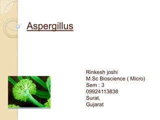
- 1. Aspergillus Rinkesh joshi M.Sc Bioscience ( Micro) Sem : 3 09924113838 Surat, Gujarat
- 2. Aspergillus Classification Division : Mycota Sub division : Eumycotina Class : Ascomycetes sub-class : Euascomycetidae Order : Aspergillales Family : Aspergillaceae Genus : Aspergillus
- 3. Occurrence Saprophytic fungus. There are 200 species of Aspergillus. Grows on decaying vegetable. On fatty media such as butter and ghee. On starchy media such as bread and rice. On preserved food such as jams and jellies. Also found on rotting oranges and other fruits.
- 4. Appearance Greenish and Smoky Patches along with Mucor, Rhizopus and Penicillium on moist bread. Other common shades are Yellow, Black, and Blue. Mostly appear in the conodial stage (imperfect stage). Very few produce cleistothecia (perfect stage).
- 5. Economic importance 33 species are reported in India. Aspergillus oryzae is utilized to make alcohol. Aspergillus niger is utilized in production of citric acid and other organic acid. Some species yield antibiotics. Culture of A.niger and A.oryzae yield a wide range of enzymes. Which are used for industrial fermentation.
- 6. Disease caused by Aspergillus Decay tobacco and cigar. Spoils nuts, bread and other food stuffs. In humid atmosphere it grows on leather and fabrics. Sometimes produce poisonous substance called micotoxins. Aspergillus Cause number of disease called aspergilloses. Eg. In humer ear cause otomycosis.
- 7. Mycelium Well developed structures. Made up of interwoven mass, branched and septate hyphae. Hyphae are branched and form mat on the substratum.
- 8. Some of the hyphae lie superficially upon the substratum and other penetrate deeply to absorb food and for mycelium.
- 9. Asexual reproduction 1. conidiopores Cells vigorously grow and mycelium become thick walled. Thick walled t shaped cells called foot cell. Each t cell produce erect branch called conidiopores.
- 10. Lenth of conidiopores is around 2.5mm. Swells at the tip and form globose called vesicle. Lumen of vesicle is continuous with upper part of conidiopores. From the surface of vesicle tubular cells grows outwords called strigmata or phialides. Phialides cover the whole surface of vesicle.
- 11. Development of phialides By dissolution of cell wall material thin tubular area formed. Cytoplasm, nucleus, mitochondria and other organelles migrate from vesicle to phialides. In maturity stage phialides cut off from vesicle from basal septum.
- 12. Abstrictions of conidia Phialides are uninucleate Nucleus divide by mitosis to form two daughter nuclei From two one migrates to tip of the phialides to form first conidium First conidium is cut off by basal septum at the phialide apex By fragmentation fungus produce asexual spores known as conidia
- 13. Later develop second conidium in same manner. This series of events repeated. Thus phialide continue to grow conidia one below to another. Consequently chain of conidia is formed at the tip of the phialides. The youngest is at the base and oldest is at the top.
- 14. Two advantages 1. Dispersel of mature conidia in the air 2. Proper nourishment of young conidia Conidia are black, green, brown, blue or yellow in color according to their species Conidial wall is thick consist of two layers outer epispore and inner endospore On falling of suitable substratum each conidium germinates First produce germ tube which grows into mycelium
- 15. Sexual reproduction Sexual reproduction is rare. Feamle sex organ is called ascogonium or archicarp. Male sex organ is called pollonidium or anthridium.
- 16. Ascogonium (female sex organ) Small, coiled septate branch. Terminal segment is longest and single celled called trigogyne contain 20 nuclei. Trigogyne function as a receptive part of female sex organ.
- 17. Anthredium (male sex organ) Male branch grows beside the ascogonium from the same hyphae. Anthredium is multi nucleate.
- 18. Plasmogamy Fusion of ascogonium and anthredium. Tip of anthredium fuse with trochogyne. Then intervening wall is dissolved. Content of anthredium pass into the trochogyne. Here haplophase ends. Male nuclei pair with female nuclei. Each pair is called dikaryon and phase is called dikaryophase.
- 19. Diplophase Diploid nucleus undergoes three successive division. 1st and 2nd division are meiosis. 3rd division is mitotic As a result 8 haploid daughter nucei form. Each haploid nucleus is surrounded by cytoplasm. Then formation of wall occur called ascopores. So 8 ascospores are formed.
- 20. The wall of asci is dissolved. Ascopores are released into cleistothesium. Then wall of cleistothesium decays to released ascopores into atmosphere. Each ascopores germinate to form mycelium.