SlideShare uma empresa Scribd logo
1 de 6
Baixar para ler offline
The Journal of Neuroscience, January 25, 2006 • 26(4):1269 –1274 • 1269


Brief Communications


A Critical Role of Erythropoietin Receptor in Neurogenesis
and Post-Stroke Recovery
Peter T. Tsai,1,4* John J. Ohab,2* Nathalie Kertesz,1 Matthias Groszer,1 Cheryl Matter,1,3 Jing Gao,1 Xin Liu,1,3,4
Hong Wu,1,4 and S. Thomas Carmichael2
Departments of 1Molecular and Medical Pharmacology, 2Neurology, and 3Pathology and Laboratory Medicine, and 4Molecular Biology Institute, University
of California at Los Angeles, Los Angeles, California 90095


Erythropoietin (EPO) is the principal growth factor regulating the production of red blood cells. Recent studies demonstrated that
exogenous EPO acts as a neuroprotectant and regulates neurogenesis. Using a genetic approach, we evaluate the roles of endogenous EPO
and its classical receptor (EPOR) in mammalian neurogenesis. We demonstrate severe and identical embryonic neurogenesis defects in
animals null for either the Epo or EpoR gene, suggesting that the classical EPOR is essential for EPO action during embryonic neurogen-
esis. Furthermore, by generating conditional EpoR knock-down animals, we demonstrate that brain-specific deletion of EpoR leads to
significantly reduced cell proliferation in the subventricular zone and impaired post-stroke neurogenesis. EpoR conditional knockdown
leads to a specific deficit in post-stroke neurogenesis through impaired migration of neuroblasts to the peri-infarct cortex. Our results
suggest that both EPO and EPOR are essential for early embryonic neural development and that the classical EPOR is important for adult
neurogenesis and for migration of regenerating neurons during post-injury recovery.
Key words: erythropoietin; erythropoietin receptor; neurogenesis; ischemia; post-injury recovery; subventricular zone


Introduction                                                                                                               (Brines et al., 2004). However, the endogenous role of EPO and
Erythropoietin (EPO) is a cytokine that plays critical roles in                                                            the role of the classical EPOR in neuroprotection and neurogen-
hematopoiesis. Both Epo and the classical EpoR are expressed in                                                            esis after stroke have not been defined. In this study, we evaluate
the nervous system during development and in adult (Digicayli-                                                             the roles for endogenous EPO and the classical EPOR in nervous
oglu et al., 1995), and their expression levels are upregulated after                                                      system development and repair using knockout of Epo and com-
hypoxic injury (Bernaudin et al., 1999). Pharmacological doses of                                                          plete and conditional knockout of the EpoR.
EPO promote neural stem cell proliferation, survival, and differ-
entiation in vitro (Studer et al., 2000; Shingo et al., 2001) and                                                          Materials and Methods
increased neuroblast migration to areas exposed to ischemic                                                                Generation of hGFAP-Cre;EpoRfloxp/floxp and hGFAP-Cre;EpoRfloxp/
damage in vivo (Wang et al., 2004). Exogenous EPO is neuropro-                                                             mice. Generation of Epo / and EpoR / mice have been described pre-
tective and neurotrophic in diverse CNS injury models, including                                                           viously (Wu et al., 1995). Timing of embryos at early gestational stages
hypoxic injury (Brines et al., 2000). Recently, an EPO derivative                                                          was determined by counting somite numbers (Lee et al., 2001). Condi-
has also been identified to have a protective role in neuronal                                                             tional knockout of EpoR receptor was done using the loxp/Cre system
tissue and to signal by binding to a heteroreceptor, consisting of                                                         (supplemental material, available at www.jneurosci.org). The specificity
                                                                                                                           of EpoR excision was evaluated by PCR (see Fig. 2), and EpoR knock-
the classical erythropoietin receptor (EPOR) (Leist et al., 2004)
                                                                                                                           down was evaluated by RT-PCR using EpoR and Actin primers described
and the common receptor ( cR) for granulocyte macrophage                                                                   previously (Digicaylioglu et al., 1995; Yamaji et al., 1996).
colony-stimulating factor and interleukin-3 and -5 receptors                                                                  In situ hybridization. Nonradioactive whole-mount mRNA in situ hy-
                                                                                                                           bridization for Epo and EpoR was performed as described previously (Lee
Received June 23, 2005; revised Dec. 9, 2005; accepted Dec. 14, 2005.
                                                                                                                           et al., 2001; Kertesz et al., 2004).
    This work was supported in part by the Larry L. Hillblom Foundation and the Nathan Shappell Fund (S.T.C.) and by          Stroke model. Stroke was produced in C57BL/6 mice and EpoR condi-
the Brain Tumor Society Award and the James S. McDonnell Foundation Award (X.L. and H.W.). P.T.T. and C.M. were            tional knock-down (hGFAP-Cre;EpoRfloxp/floxp) animals (Carmichael,
supported in part by the Medical Scientist Training Program training grant and the Engene V. Cota Robles Award by          2005) with distal branch of the middle cerebral artery cauterization fol-
the Graduate Division through the University of California Office of the President. H.W. is a V Foundation scholar. J.O.   lowed by 15 min bilateral common carotid occlusion.
was supported by the UCLA Training Program in Neural Repair. We thank Dr. Angus Sinclair (Amgen, Thousand Oaks,               Immunohistochemistry. Bromodeoxyuridine (BrdU; 50 mg/kg, i.p., in
CA) and members of our laboratories for helpful comments on this manuscript.                                               0.9% saline; Sigma, St. Louis, MO) was given twice daily for 2 d before the
    *P.T.T. and J.J.O. contributed equally to this work.                                                                   animals were killed, or pulse-labeled with a single injection (100 mg/kg,
    Correspondence should be addressed to either of the following: Dr. Hong Wu, Department of Molecular and
                                                                                                                           i.p.). At 3, 5, and 7 d after stroke and in control animals (n 4 – 6 for each
Medical Pharmacology, Geffen School of Medicine at University of California at Los Angeles, CHS 23-214, Los Ange-
les, CA 90095, E-mail: hwu@mednet.ucla.edu; or Dr. S. Thomas Carmichael, Department of Neurology, 635 Charles
                                                                                                                           group), tissue was processed for BrdU and doublecortin (DCX) immu-
Young Drive South, University of California at Los Angeles, Los Angeles, CA 90095, E-mail: scarmichael@                    nostaining (supplemental material, available at www.jneurosci.org). Sec-
mednet.ucla.edu.                                                                                                           tions were analyzed with bright-field and epifluorescence microscopy
    DOI:10.1523/JNEUROSCI.4480-05.2006                                                                                     (Leica DMLB) or with laser confocal microscopy (Leica TCS SP MP).
Copyright © 2006 Society for Neuroscience 0270-6474/06/261269-06$15.00/0                                                      Stereology. Stereological quantification was performed on serial sec-
1270 • J. Neurosci., January 25, 2006 • 26(4):1269 –1274                                                      Tsai et al. • Role of EPOR in Neurogenesis and Post-Stroke Recovery


tions through the anterior subventricular zone
(SVZ), olfactory bulb, and peri-infarct cortex
using unbiased counting with the optical frac-
tionator technique (supplemental material,
available at www.jneurosci.org).
   Infarct volume. Infarct volume was measured
7 d after stroke in hGFAP-Cre;EpoRfloxp/ and
control mice (n 5 and 4, respectively) (Kats-
man et al., 2003; Li et al., 2005).
   Statistics. Post-stroke SVZ counts were tested
with factorial ANOVA and Bonferroni’s post
hoc testing (Statview 5.0.1; SAS Institute, Cary,
NC). Infarct volume, SVZ, and olfactory bulb
counts were tested with two-sample t testing
assuming unequal variances (Excel; Microsoft,
Redmond, WA).

Results
Sequential expression of EpoR and Epo
in the developing brain and effects on
embryonic development
To understand the function of EPO/EPOR
in neurogenesis, Epo and EpoR expression
was first determined with in situ hybridiza-
tion. Whole-mount in situ hybridization
of EpoR and Epo in embryonic day 7.5 Figure 1. Epo and EpoR expression and neurogenesis defects in null animals. a–f, In situ hybridization of transverse sections
(E7.5) to E11 embryos shows expression with EpoR (a, c, e) or Epo (b, d ,f ) antisense probes on E8.5, E9.5, and E10.5 embryos. Inset photomicrographs are higher-
throughout the developing neural tube, magnification images from the neuroepithelium (a, c) and neural crest (e). g–l, Epo- (k, l ) and EpoR- (g–j) deficient embryos
with progressive expression in optic pla- show underdeveloped choroids plexus (i–l ) and forebrain (g, h) at E13.5. Con, Control; Mut, mutant.
code and forebrain and a delay of 0.5–1 d
between EpoR and Epo expression in the                                   and EpoR null animals results in systemic hypoxia (Wu et al.,
same region (supplemental Fig. 1a–f, available at www.jneurosci.         1995), and the roles for EPO/EPOR in adult tissues cannot be
org as supplemental material). At E8.5, EpoR expression is con-          evaluated in the null animals because of their early lethality
fined to the neuroepithelium, a zone of precursors with high             (Wu et al., 1995). To define the role of EPOR in these nonhe-
proliferation capacity (Fig. 1a, inset). Epo expression is absent        matopoietic tissues, we inserted loxp sites between the mini-
at E8.5 (Fig. 1b) but appears by E9.0 –E9.5 in the neuroepithe-          mal promoter and exons 1– 4 (supplemental Fig. 2, available at
lium (Fig. 1d). At E9.5–E10.5, EpoR expression rises in neural           www.jneurosci.org as supplemental material). In contrast to
crest- and mesenchyme-derived cells (Fig. 1c,e, insets), fol-            the early lethal phenotype observed in EpoR null embryos,
lowed by Epo expression in similar regions 1 d later (Fig. 1f ).         EpoRfloxp/floxp mice are viable and display no abnormal pheno-
The temporal and spatial regulation of Epo and EpoR suggests             types compared with wild-type (WT) littermates in gross de-
a role for this ligand–receptor pair in early neurogenesis.              velopment, fertility, and physiology (data not shown), sug-
    Both Epo and EpoR null embryos have incomplete neural tube
                                                                         gesting that introducing loxp sites into the endogenous EpoR
closure at E10.5 (supplemental Fig. 1h, available at www.jneurosci.
                                                                         locus does not disrupt its normal function.
org as supplemental material), especially in the hindbrain. By
E13.5, the neurogenesis defects become more evident, including
wavy neural tubes (supplemental Fig. 1j, arrow, available at             Brain-specific EpoR deletion
www.jneurosci.org as supplemental material), underdeveloped              EpoRfloxp/floxp mice were crossed with hGFAP-Cre transgenic mice
telencephalic regions (supplemental Fig. 1j,l, arrowhead, avail-         (Zhuo et al., 2001; Malatesta et al., 2003). The hGFAP promoter
able at www.jneurosci.org as supplemental material). Although            drives Cre expression in radial glial cells of the developing cortex,
no major structures are absent in Epo and EpoR null mice, the            hindbrain, and cerebellum (Zhuo et al., 2001; Yue et al., 2005),
mutant brains are consistently smaller and less developed than           resulting in efficient EpoR deletion in the brain and sparing of
their littermate controls, particularly in the SVZ of the develop-
                                                                         hematopoietic tissue (Fig. 2a,b, Mut). EpoRfloxp/floxp;hGFAP-Cre
ing forebrain (Fig. 1h). In addition, the choroid plexus is severely
                                                                         mutants are viable and fertile, yielding Mendelian offspring dis-
underdeveloped in both null embryos, with a flattened cubiodal
                                                                         tributions (data not shown), suggesting that neural-specific dele-
epithelium and underdeveloped capillary network (Fig, 1i,l ). The
                                                                         tion of EpoR after E14 is not essential for embryonic or adult
identical phenotypes of Epo and EpoR null animals suggest that
                                                                         survival. Although expression is undetectable in the brain tissues,
for early embryonic development, the classical EPOR is essential
                                                                         we cannot rule out the presence of remaining EpoR expression
for EPO action.
                                                                         beyond our levels of detection, so these animals are described as
Generation of EpoR conditional knock-down mice                           EpoR conditional knock-down animals. Hematocrit, brain
EPO and EPOR modulate cardiogenesis, vasculogenesis, and                 weight, and body weight of EpoR conditional knock-down mice
neurogenesis (Wu et al., 1999; Studer et al., 2000; Shingo et al.,       are indistinguishable from their WT littermates (Fig. 2c). Normal
2001; Kertesz et al., 2004; Wang et al., 2004), raising the ques-        brain architecture is preserved in EpoRfloxp/floxp;hGFAP-Cre an-
tion of whether the classical EPOR exerts these functions in a           imals in the cortex (Fig. 2d), olfactory bulbs, or cerebellum (data
cell autonomous manner. The erythrocyte deficiency in Epo                not shown).
Tsai et al. • Role of EPOR in Neurogenesis and Post-Stroke Recovery                                                               J. Neurosci., January 25, 2006 • 26(4):1269 –1274 • 1271


                                                                                                     proliferation, but not the normal pattern of neuroblast migration
                                                                                                     to the olfactory bulb.

                                                                                                     EPOR is not essential for protecting neurons from
                                                                                                     ischemic injury
                                                                                                     Previous studies that determined a neuroprotective effect of ex-
                                                                                                     ogenous EPO (Sakanaka et al., 1998; Bernaudin et al., 1999; Siren
                                                                                                     et al., 2001) either evaluated stroke size 24 h after stroke, when the
                                                                                                     area of infarction is not yet complete in most rodent stroke mod-
                                                                                                     els (Lipton, 1999), or used permanent focal ischemia, which pro-
                                                                                                     duces a different pattern of neuronal injury and death compared
                                                                                                     with stroke models that involve reperfusion (Lipton, 1999; Car-
                                                                                                     michael, 2005). We used a focal stroke model with the infarct and
                                                                                                     reperfusion injury restricted to the sensorimotor cortex (Kats-
                                                                                                     man et al., 2003; Carmichael et al., 2004). In this model, there is
                                                                                                     no statistically significant difference in infarct size between mu-
                                                                                                     tant (Fig. 4a) and controls (Fig. 4b) at day 3 (data not shown) and
                                                                                                     day 7 (Fig. 4c) after stroke ( p 0.31). This result suggests that,
                                                                                                     although higher doses of exogenous EPO may promote neuro-
                                                                                                     protection, endogenous levels of EPO and EPOR have limited
                                                                                                     roles in protecting neurons from ischemic reperfusion injury.

                                                                                                     EPOR is important for post-stroke neurogenesis
                                                                                                     Stroke induces migration of newly born neuroblasts from the
                                                                                                     SVZ into the area of injury in the first 7–14 d after the insult
                                                                                                     (Arvidsson et al., 2002; Parent et al., 2002; Jin et al., 2003), de-
                                                                                                     tected by the combination of pulse labeling with BrdU and stain-
                                                                                                     ing for DCX, a microtubule-associated protein present in imma-
                                                                                                     ture and migrating neurons (Feng and Walsh, 2001). As reported
                                                                                                     previously (Yang et al., 2004), there are no DCX cells in the
                                                                                                     cortex of either control or mutant mice before stroke (Fig. 4h).
                                                                                                     After stroke, DCX cells are found in subcortical white matter at
                                                                                                     day 3 (supplemental Fig. 3, available at www.jneurosci.org as
                                                                                                     supplemental material) and in the peri-infarct cortex at days 5
                                                                                                     and 7, resulting in 4000 DCX cells per animal in the peri-
                                                                                                     infarct cortex (Fig. 4h). In both control and mutant animals,
                                                                                                     DCX cells are labeled with BrdU given after the stroke (supple-
                                                                                                     mental Fig. 4, available at www.jneurosci.org as supplemental
                                                                                                     material), and these cells are also immunoreactive for other
                                                                                                     markers of immature neurons or neural progenitor cells, such as
Figure 2. Analysis of EpoR conditional knock-down animals. a, PCR. Floxed, WT, and excised           PSA-NCAM and musashi (Gleeson et al., 1999; Okano et al.,
alleles ( loxp) are indicated. b, RT-PCR analysis for EpoR expression levels. c, Hematocrit, brain   2005) (supplemental Fig. 5, available at www.jneurosci.org as
weight, and body weight are unchanged in the mutants. Error bars indicate SDs. d, Cresyl violet      supplemental material). The number of DCX cells between
sections from control (left) and mutant (right) animals. The bottom panels show higher-
                                                                                                     mutant and control animals remains the same on days 3 and 5
magnification views of the boxed areas. Con, Control; Mut, mutant.
                                                                                                     after stroke. However, EpoR conditional knock-down mice (Fig.
                                                                                                     4e,g) show a 2.6-fold reduction in the number of DCX neuro-
Neural-specific EpoR knockdown leads to reduced cell                                                 blasts present in the cortex at day 7 compared with controls (Fig.
proliferation in the SVZ                                                                             4d,f ).
Exogenous EPO promotes proliferation, survival, and neuronal                                             The reduction of DCX neuroblasts in the cortex of post-
differentiation of neural stem cells in vitro (Ling et al., 1998;                                    stroke EpoR conditional knock-down mice may be attributable to
Studer et al., 2000; Shingo et al., 2001;Yu et al., 2002). To examine                                reduced SVZ proliferation, reduced migration to the peri-infarct
neurogenesis in vivo, we compared the SVZ of EpoR mutant mice                                        cortex, or increased cell death in the peri-infarct cortex. To ex-
with littermate controls. The mutant SVZ is decreased in volume                                      amine proliferation, the number of BrdU cells in the SVZ was
(Fig. 3i) and has fewer cells compared with controls (Con,                                           quantified after stroke (Fig. 3h). Control mice show a significant
18,573 1523; Mut, 14,661 2131; p 0.03). After BrdU pulse,                                            decrease in BrdU incorporation at day 3 (F(5,21)         8.072; p
the mutant SVZ contains a significantly reduced number of                                            0.002) (supplemental Fig. 5d,h, available at www.jneurosci.org as
BrdU cells at postnatal day 15, which is further reduced at 6                                        supplemental material), during the period in which DCX cells
months (Fig. 3h). Despite reduced BrdU labeling in the SVZ,                                          migrate from the SVZ toward the peri-infarct cortex and then a
there is no difference in BrdU olfactory bulb cells between the                                      return to baseline at day 7 (Fig. 3f, h). In contrast, the mutant
two conditions (Con, 8431 1820 cells; Mut, 9068 1446 cells;                                          mice have a reduced number of BrdU cells and display no
p     0.16). These results provide the first evidence that EPOR                                      change at any time point after stroke (Fig. 3c,e,g,h). The SVZ
plays essential roles in adult neurogenesis in vivo and suggests                                     volume shows similar differences between control and mutant
that EPO/EPOR may regulate neural stem and progenitor cell                                           before and after stroke (Fig. 3i). Thus, the mutant SVZ not only
1272 • J. Neurosci., January 25, 2006 • 26(4):1269 –1274                                                                        Tsai et al. • Role of EPOR in Neurogenesis and Post-Stroke Recovery


has reduced BrdU labeling at baseline but
also lacks any significant response to
stroke. To determine cell migration and
death after stroke, a cohort of DCX cells
was labeled by administering BrdU on
days 3–5 after stroke and tracking these
cells in the peri-infarct cortex on days 5
and 7. In controls, 49 7% of DCX cells
are BrdU at day 5 with further increases
to 68 6% by day 7, indicating a contin-
ued migration of this cohort of DCX
cells into the peri-infarct cortex by day 7.
In mutants, the percentage of DCX /
BrdU cells on day 5 is similar to controls
( p 0.35) but fails to increase by day 7
(55 9%; p 0.53, MUT day 3 vs day 7).
This indicates that there is a failure of con-
tinued neuroblast migration into the peri-
infarct cortex in EpoR conditional knock-
down. To determine whether there is an
increase in cell death with EpoR con-
ditional knockdown, the number of ter-
minal deoxynucleotidyl transferase-                        Figure 3. Quantification of BrdU labeling in the SVZ. a, Cresyl violet section. The rectangle shows the location of b– g. b– g,
mediated biotinylated UTP nick end                         BrdU-labeled cells in control (b, d, f ) and EpoR conditional knock-down (c, e, g) animals in non-stroke (b, c), 3 d after stroke (d, e),
labeling (TUNEL)-positive cells was                        and 7 d after stroke (f, g) are shown. Scale bar, 50 m. h, i, The graphs show quantifications of BrdU-labeled cells (h) and SVZ
quantified in the peri-infarct cortex on                   volume before and after stroke (i). *p 0.004; #p 0.008; $p 0.016. Error bars indicate SDs. ctx, Cortex; str, stiatum; V,
days 5 and 7 after stroke. There is no sig-                ventricle; cc, corpus callosum; P15, postnatal day 15; mo, month; Con, control; Mut, mutant.
nificant difference in the number of
TUNEL cells in the per-infarct cortex at
these two time points (5 d Con, 45 19.6;
5 d Mut, 43.5 11.5; 7 d Con, 42.8 13.3;
7 d Mut, 37.8 14.2; p 0.63 for all com-
parisons). Coupled with the significant
loss of DCX cells in the mutant peri-
infarct cortex from days 5–7 (Fig. 4h),
these data show that neuroblasts migrate
to the peri-infarct cortex in which a subset
then undergo cell death. Continued mi-
gration of neuroblasts occurs in control
animals such that the overall number is
maintained, but there is no continued mi-
gration in mutant, and the overall number
of neuroblasts declines over time.

Discussion
In this study, we provide the first genetic
demonstration that EPO and EPOR play
essential roles in regulating embryonic
and adult neuronal development and
function. We demonstrate Epo and EpoR
expression during critical periods of neu-
ronal development as well as significantly
aberrant neurogenesis in null animals. Figure 4. EpoR conditional knock-down leads to reduced neuroblast migration. a, b, Cresyl violet sections through the frontal
Then, using a model whereby neuronal cortex of mutant (a) and control (b) 7 d after stroke. Scale bar, 500 m. c, Infarct volume 7 d after stroke was quantified. d– g,
EpoR expression is specifically reduced in DCX cells in control (d, f ) and mutant (e, g). The region within the box in d and e is enlarged in f and g. Scale bars: d, 100 m;
                                                                                                       #
the brain, we demonstrate a critical role f, 25 m. h, Stereological quantification of DCX cells. p 0.001 versus all other conditions; *p 0.008 versus Mut at
                                                                ˆ                          @                                              $
for EPOR in adult neurogenesis and in the non-stroke stage; p 0.02 versus Mut at 7 d; p 0.02 versus 3 d Mut/Con and non-stroke; p 0.002 versus 7 d control. Error
migration of newly born neuroblasts to ar-   bars indicate SDs.
eas of injury. Finally, EpoR conditional
knock-down mice display no significant differences in infarct size           EPO/EPOR are required for developmental neurogenesis
versus control mice when exposed to stroke, suggesting alterna-              The role of EPO in the nervous system extends beyond tissue
tive mechanisms underlying the neuroprotective functions of                  oxygenation. Epo and EpoR are expressed during critical periods
EPO.                                                                         of neuronal development, and Epo and EpoR null animals reveal
Tsai et al. • Role of EPOR in Neurogenesis and Post-Stroke Recovery                                    J. Neurosci., January 25, 2006 • 26(4):1269 –1274 • 1273


defects in neurogenesis (Yu et al., 2002; present study). Here,        roprotective effects of EPO in the conditional knock-down
we demonstrate that these defects are mediated through EPO             animals.
binding to the classical EPOR. Neuronal defects in Epo null
animals are identical to those seen in EpoR null animals, show-        References
ing that EPO is acting through the EPOR during neuronal                Arvidsson A, Collin T, Kirik D, Kokaia Z, Lindvall O (2002) Neuronal re-
development. Recent studies have suggested an alternative re-              placement from endogenous precursors in the adult brain after stroke.
ceptor for EPO neuronal effects (Leist et al., 2004) and that              Nat Med 8:963–970.
  cR is essential for EPO neuroprotective actions (Brines et al.,      Bernaudin M, Marti HH, Roussel S, Divoux D, Nouvelot A, MacKenzie ET,
                                                                           Petit E (1999) A potential role for erythropoietin in focal permanent
2004). The identical hematopoietic and neuronal develop-
                                                                           cerebral ischemia in mice. J Cereb Blood Flow Metab 19:643– 651.
mental phenotypes between Epo and EpoR null animals sug-               Brines M, Grasso G, Fiordaliso F, Sfacteria A, Ghezzi P, Fratelli M, Latini R,
gest that, for early embryonic development, the classical EPOR             Xie QW, Smart J, Su-Rick CJ, Pobre E, Diaz D, Gomez D, Hand C,
is essential for EPO action. Because cR null animals are viable            Coleman T, Cerami A (2004) Erythropoietin mediates tissue protection
without hematopoietic, cardiac, or neuronal defects, EPO                   through an erythropoietin and common beta-subunit heteroreceptor.
likely acts exclusively through EPOR homodimers for its roles              Proc Natl Acad Sci USA 101:14907–14912.
                                                                       Brines ML, Ghezzi P, Keenan S, Agnello D, de Lanerolle NC, Cerami C, Itri
in these tissues during development (Reed et al., 2000; Brines
                                                                           LM, Cerami A (2000) Erythropoietin crosses the blood-brain barrier to
et al., 2004).                                                             protect against experimental brain injury. Proc Natl Acad Sci USA
                                                                           97:10526 –10531.
EPOR is critical for adult and post-stroke neurogenesis                Carmichael ST (2005) Rodent stroke models: size, mechanism and purpose.
Exogenous EPO can promote neurogenesis in vitro (Ling et al.,              NeuroRx 2:396 – 409.
1998; Studer et al., 2000; Shingo et al., 2001). Our study, however,   Carmichael ST, Tatsukawa K, Katsman D, Tsuyuguchi N, Kornblum HI
provides the first evidence that EPOR plays essential roles in adult       (2004) Evolution of diaschisis in a focal stroke model. Stroke
                                                                           35:758 –763.
neurogenesis in vivo. EpoR conditional knock-down animals              Digicaylioglu M, Bichet S, Marti HH, Wenger RH, Rivas LA, Bauer C,
have significantly reduced levels of BrdU incorporation in the             Gassmann M (1995) Localization of specific erythropoietin binding
SVZ and diminished SVZ cell number and volume, suggesting                  sites in defined areas of the mouse brain. Proc Natl Acad Sci USA
that EPOR plays a critical function in maintaining dividing cell           92:3717–3720.
numbers in the adult SVZ.                                              Feng Y, Walsh CA (2001) Protein-protein interactions, cytoskeletal regula-
    Studies in large stroke models in the rat report increased la-         tion and neuronal migration. Nat Rev Neurosci 2:408 – 416.
                                                                       Gleeson JG, Lin PT, Flanagan LA, Walsh CA (1999) Doublecortin is a
beling of proliferating cells in the SVZ and migration of newly
                                                                           microtubule-associated protein and is expressed widely by migrating neu-
born neuroblasts from the SVZ to peri-infarct tissue (Arvidsson            rons. Neuron 23:257–271.
et al., 2002; Parent et al., 2002; Jin et al., 2003). In the present   Jin G, Omori N, Li F, Nagano I, Manabe Y, Shoji M, Abe K (2003) Protec-
study, focal stroke is restricted to the mouse somatosensory cor-          tion against ischemic brain damage by GDNF affecting cell survival and
tex, with 1 mm distance to the SVZ. There is a biphasic prolif-            death signals. Neurol Res 25:249 –253.
erative response in the SVZ in this model and a robust migration       Katsman D, Zheng J, Spinelli K, Carmichael ST (2003) Tissue microenvi-
                                                                           ronments within functional cortical subdivisions adjacent to focal stroke.
of neuroblasts to the peri-infarct cortex. Our study demonstrates
                                                                           J Cereb Blood Flow Metab 23:997–1009.
that EPOR also plays an essential role in this neurogenic response     Kertesz N, Wu J, Chen TH, Sucov HM, Wu H (2004) The role of erythro-
to ischemia. EpoR conditional knock-down animals do not dis-               poietin in regulating angiogenesis. Dev Biol 276:101–110.
play this biphasic SVZ response but instead maintain the reduced       Lee R, Kertesz N, Joseph SB, Jegalian A, Wu H (2001) Erythropoietin
control levels of BrdU labeling after stroke. Despite this lack of         (Epo) and EpoR expression and 2 waves of erythropoiesis. Blood
SVZ response, these animals appear to possess a normal initial             98:1408 –1415.
post-stroke neuroblast migration to the peri-infarct cortex. After     Leist M, Ghezzi P, Grasso G, Bianchi R, Villa P, Fratelli M, Savino C, Bianchi
                                                                           M, Nielsen J, Gerwien J, Kallunki P, Larsen AK, Helboe L, Christensen S,
this initial migration, significant numbers of neuroblasts are lost        Pedersen LO, Nielsen M, Torup L, Sager T, Sfacteria A, Erbayraktar S, et
in the peri-infarct cortex in the EpoR knock-down animals (Fig.            al. (2004) Derivatives of erythropoietin that are tissue protective but not
4h), and there is no continued migration of neuroblasts to replace         erythropoietic. Science 305:239 –242.
them.                                                                  Li S, Zheng J, Carmichel ST (2005) Differences in ischemic injury after
                                                                           stroke between agend and young adults. Neurobiol Dis 18:432– 440.
Alternative mechanisms for EPO-mediated neuroprotection                Ling ZD, Potter ED, Lipton JW, Carvey PM (1998) Differentiation of mes-
                                                                           encephalic progenitor cells into dopaminergic neurons by cytokines. Exp
Exogenous EPO has been shown to play a neuroprotective role
                                                                           Neurol 149:411– 423.
during the ischemic response of the brain by preventing neuronal       Lipton P (1999) Ischemic cell death in brain neurons. Physiol Rev
apoptosis (Sakanaka et al., 1998; Bernaudin et al., 1999; Siren et         79:1431–1568.
al., 2001). However, our data show no difference in infarct vol-       Malatesta P, Hack MA, Hartfuss E, Kettenmann H, Klinkert W, Kirchhoff F,
ume after conditional deletion of EpoR. This result suggests that          Gotz M (2003) Neuronal or glial progeny: regional differences in radial
although higher doses of exogenous EPO promotes neuroprotec-               glia fate. Neuron 37:751–764.
                                                                       Marti HH, Bernaudin M, Petit E, Bauer C (2000) Neuroprotection and an-
tion, there is a limited role of the endogenous EPO system in
                                                                           giogenesis: dual role of erythropoietin in brain ischemia. News Physiol Sci
neuroprotection. Alternatively, the neuroprotective actions of             15:225–229.
EPO may also be mediated via routes other than binding the             Okano H, Kawahara H, Toriya M, Nakao K, Shibata S, Imai T (2005) Func-
classical EPOR on neurons, such as through blood vessels. Exog-            tion of RNA-binding protein Musashi-1 in stem cells. Exp Cell Res
enous EPO has been demonstrated to increase cerebral vascula-              306:349 –356.
ture at infarct sites (Marti et al., 2000; Wang et al., 2004), with    Parent JM, Vexler ZS, Gong C, Derugin N, Ferriero DM (2002) Rat fore-
subsequent increased oxygen delivery perhaps resulting in neu-             brain neurogenesis and striatal neuron replacement after focal stroke.
                                                                           Ann Neurol 52:802– 813.
roprotection. EPO may also act as a neuroprotectant by binding a       Reed JA, Ikegami M, Robb L, Begley CG, Ross G, Whitsett JA (2000) Dis-
receptor that contains cR but lacks the classical EPOR (Brines et          tinct changes in pulmonary surfactant homeostasis in common beta-
al., 2004). Thus, upregulation of other receptor complexes may             chain- and GM-CSF-deficient mice. Am J Physiol Lung Cell Mol Physiol
compensate for loss of the classical EPOR and mediate the neu-             278:L1164 –L1171.
1274 • J. Neurosci., January 25, 2006 • 26(4):1269 –1274                                                Tsai et al. • Role of EPOR in Neurogenesis and Post-Stroke Recovery

Sakanaka M, Wen TC, Matsuda S, Masuda S, Morishita E, Nagao M, Sasaki R            throid BFU-E and CFU-E progenitors does not require erythropoietin or
    (1998) In vivo evidence that erythropoietin protects neurons from isch-        the erythropoietin receptor. Cell 83:59 – 67.
    emic damage. Proc Natl Acad Sci USA 95:4635– 4640.                          Wu H, Lee SH, Gao J, Liu X, Iruela-Arispe ML (1999) Inactivation of eryth-
Shingo T, Sorokan ST, Shimazaki T, Weiss S (2001) Erythropoietin regu-             ropoietin leads to defects in cardiac morphogenesis. Development
    lates the in vitro and in vivo production of neuronal progenitors by mam-      126:3597–3605.
    malian forebrain neural stem cells. J Neurosci 21:9733–9743.                Yamaji R, Okada T, Moriya M, Naito M, Tsuruo T, Miyatake K, Nakano Y
Siren AL, Fratelli M, Brines M, Goemans C, Casagrande S, Lewczuk P, Keenan         (1996) Brain capillary endothelial cells express two forms of erythropoi-
    S, Gleiter C, Pasquali C, Capobianco A, Mennini T, Heumann R, Cerami           etin receptor mRNA. Eur J Biochem 239:494 –500.
    A, Ehrenreich H, Ghezzi P (2001) Erythropoietin prevents neuronal ap-       Yu X, Shacka JJ, Eells JB, Suarez-Quian C, Przygodzki RM, Beleslin-Cokic B,
    optosis after cerebral ischemia and metabolic stress. Proc Natl Acad Sci       Lin CS, Nikodem VM, Hempstead B, Flanders KC, Costantini F, Noguchi
    USA 98:4044 – 4049.                                                            CT (2002) Erythropoietin receptor signaling is required for normal
Studer L, Csete M, Lee SH, Kabbani N, Walikonis J, Wold B, McKay R (2000)          brain development. Development 129:505–516.
    Enhanced proliferation, survival, and dopaminergic differentiation of       Yue Q, Groszer M, Gil JS, Berk AJ, Messing A, Wu H, Liu X (2005) PTEN
    CNS precursors in lowered oxygen. J Neurosci 20:7377–7383.                     deletion in Bergmann glia leads to premature differentiation and affects
Wang L, Zhang Z, Wang Y, Zhang R, Chopp M (2004) Treatment of stroke               laminar organization. Development 132:3281–3291.
    with erythropoietin enhances neurogenesis and angiogenesis and im-          Zhuo L, Theis M, Alvarez-Maya I, Brenner M, Willecke K, Messing A (2001)
    proves neurological function in rats. Stroke 35:1732–1737.                     hGFAP-cre transgenic mice for manipulation of glial and neuronal func-
Wu H, Liu X, Jaenisch R, Lodish HF (1995) Generation of committed ery-             tion in vivo. Genesis 31:85–94.

Mais conteúdo relacionado

Mais procurados

Genetic regulation of human brain aging
Genetic regulation of human brain agingGenetic regulation of human brain aging
Genetic regulation of human brain agingAlzforum
 
Neuropathological correlates imperial_college_4_sl_sh_2feb2018
Neuropathological correlates imperial_college_4_sl_sh_2feb2018Neuropathological correlates imperial_college_4_sl_sh_2feb2018
Neuropathological correlates imperial_college_4_sl_sh_2feb2018Klaus Schmierer
 
Development and implementation of a novel interactome platform in studies on ...
Development and implementation of a novel interactome platform in studies on ...Development and implementation of a novel interactome platform in studies on ...
Development and implementation of a novel interactome platform in studies on ...Ewelina Maliszewska-Cyna, PhD
 
Inestrosa idn 2011
Inestrosa idn 2011Inestrosa idn 2011
Inestrosa idn 2011Jorge Parodi
 
Protein-RNA Complexes, ALS and Aging
Protein-RNA Complexes, ALS and AgingProtein-RNA Complexes, ALS and Aging
Protein-RNA Complexes, ALS and AgingAlzforum
 
Alzforum print news
Alzforum   print newsAlzforum   print news
Alzforum print newsshiraknafo
 
Nogo receptor1confinesadisinhibitorymicrocircuittothe criticalperiodinvisualc...
Nogo receptor1confinesadisinhibitorymicrocircuittothe criticalperiodinvisualc...Nogo receptor1confinesadisinhibitorymicrocircuittothe criticalperiodinvisualc...
Nogo receptor1confinesadisinhibitorymicrocircuittothe criticalperiodinvisualc...Taruna Ikrar
 
KDM5 epigenetic modifiers as a focus for drug discovery
KDM5 epigenetic modifiers as a focus for drug discoveryKDM5 epigenetic modifiers as a focus for drug discovery
KDM5 epigenetic modifiers as a focus for drug discoveryChristopher Wynder
 
Proteus Syndrome Seminar
Proteus Syndrome SeminarProteus Syndrome Seminar
Proteus Syndrome SeminarSarisarestrepo
 
Effect Of Ethanol In Oligodendrocytes, research miami
Effect Of Ethanol In Oligodendrocytes, research miamiEffect Of Ethanol In Oligodendrocytes, research miami
Effect Of Ethanol In Oligodendrocytes, research miamiLydia Cortes
 
Strauss_et_al_2015_with_supp (1)
Strauss_et_al_2015_with_supp (1)Strauss_et_al_2015_with_supp (1)
Strauss_et_al_2015_with_supp (1)Abigail Benkert
 

Mais procurados (19)

ISX9 - Karthik Gopalakrishnan
ISX9 - Karthik GopalakrishnanISX9 - Karthik Gopalakrishnan
ISX9 - Karthik Gopalakrishnan
 
Genetic regulation of human brain aging
Genetic regulation of human brain agingGenetic regulation of human brain aging
Genetic regulation of human brain aging
 
ASN Neuro-2016-Nikolakopoulou-
ASN Neuro-2016-Nikolakopoulou-ASN Neuro-2016-Nikolakopoulou-
ASN Neuro-2016-Nikolakopoulou-
 
Neuropathological correlates imperial_college_4_sl_sh_2feb2018
Neuropathological correlates imperial_college_4_sl_sh_2feb2018Neuropathological correlates imperial_college_4_sl_sh_2feb2018
Neuropathological correlates imperial_college_4_sl_sh_2feb2018
 
Development and implementation of a novel interactome platform in studies on ...
Development and implementation of a novel interactome platform in studies on ...Development and implementation of a novel interactome platform in studies on ...
Development and implementation of a novel interactome platform in studies on ...
 
Inestrosa idn 2011
Inestrosa idn 2011Inestrosa idn 2011
Inestrosa idn 2011
 
Protein-RNA Complexes, ALS and Aging
Protein-RNA Complexes, ALS and AgingProtein-RNA Complexes, ALS and Aging
Protein-RNA Complexes, ALS and Aging
 
Alzforum print news
Alzforum   print newsAlzforum   print news
Alzforum print news
 
Nogo receptor1confinesadisinhibitorymicrocircuittothe criticalperiodinvisualc...
Nogo receptor1confinesadisinhibitorymicrocircuittothe criticalperiodinvisualc...Nogo receptor1confinesadisinhibitorymicrocircuittothe criticalperiodinvisualc...
Nogo receptor1confinesadisinhibitorymicrocircuittothe criticalperiodinvisualc...
 
Belayev, Ludmila
Belayev, LudmilaBelayev, Ludmila
Belayev, Ludmila
 
Herrmann STAT3 2008
Herrmann STAT3 2008Herrmann STAT3 2008
Herrmann STAT3 2008
 
KDM5 epigenetic modifiers as a focus for drug discovery
KDM5 epigenetic modifiers as a focus for drug discoveryKDM5 epigenetic modifiers as a focus for drug discovery
KDM5 epigenetic modifiers as a focus for drug discovery
 
Keane, Robert
Keane, RobertKeane, Robert
Keane, Robert
 
Proteus Syndrome Seminar
Proteus Syndrome SeminarProteus Syndrome Seminar
Proteus Syndrome Seminar
 
neu%2E2015%2E4008
neu%2E2015%2E4008neu%2E2015%2E4008
neu%2E2015%2E4008
 
nihms419319
nihms419319nihms419319
nihms419319
 
Effect Of Ethanol In Oligodendrocytes, research miami
Effect Of Ethanol In Oligodendrocytes, research miamiEffect Of Ethanol In Oligodendrocytes, research miami
Effect Of Ethanol In Oligodendrocytes, research miami
 
AN_A&D2016
AN_A&D2016AN_A&D2016
AN_A&D2016
 
Strauss_et_al_2015_with_supp (1)
Strauss_et_al_2015_with_supp (1)Strauss_et_al_2015_with_supp (1)
Strauss_et_al_2015_with_supp (1)
 

Semelhante a A Critical Role of Erythropoietin Receptor in Neurogenesis and Post-Stroke Recovery

Katoh_et_al-2015-Brain_and_Behavior
Katoh_et_al-2015-Brain_and_BehaviorKatoh_et_al-2015-Brain_and_Behavior
Katoh_et_al-2015-Brain_and_BehaviorRhea Kimpo
 
AdamsloftisASBposter-2
AdamsloftisASBposter-2AdamsloftisASBposter-2
AdamsloftisASBposter-2Kevin Loftiskg
 
Kimpo and Raymond_JNsci 2007
Kimpo and Raymond_JNsci 2007Kimpo and Raymond_JNsci 2007
Kimpo and Raymond_JNsci 2007Rhea Kimpo
 
Pten Deletion in Adult Neural Stem/Progenitor Cells Enhances Constitutive Neu...
Pten Deletion in Adult Neural Stem/Progenitor Cells Enhances Constitutive Neu...Pten Deletion in Adult Neural Stem/Progenitor Cells Enhances Constitutive Neu...
Pten Deletion in Adult Neural Stem/Progenitor Cells Enhances Constitutive Neu...johnohab
 
UTF-8''Final Assessing post-synaptic partners of Dentate Granule Cells in a M...
UTF-8''Final Assessing post-synaptic partners of Dentate Granule Cells in a M...UTF-8''Final Assessing post-synaptic partners of Dentate Granule Cells in a M...
UTF-8''Final Assessing post-synaptic partners of Dentate Granule Cells in a M...Grant Pizzo
 
Differentiation of neural_cells_in_human_embryonic_stem
Differentiation of neural_cells_in_human_embryonic_stemDifferentiation of neural_cells_in_human_embryonic_stem
Differentiation of neural_cells_in_human_embryonic_stemHoney Cheng
 
Tzekov_PLOSOne_2016 Publication Roskamp.PDF
Tzekov_PLOSOne_2016 Publication Roskamp.PDFTzekov_PLOSOne_2016 Publication Roskamp.PDF
Tzekov_PLOSOne_2016 Publication Roskamp.PDFMegan Orlando
 
Senior thesis(2015)_Ganesh
Senior thesis(2015)_GaneshSenior thesis(2015)_Ganesh
Senior thesis(2015)_GaneshArjun Ganesh
 
Inestrosa idn 2011
Inestrosa idn 2011Inestrosa idn 2011
Inestrosa idn 2011Jorge Parodi
 
mottin-2011-Jcbfm-nature-publishing
mottin-2011-Jcbfm-nature-publishingmottin-2011-Jcbfm-nature-publishing
mottin-2011-Jcbfm-nature-publishingStéphane MOTTIN
 
C-CEBH__ Current Trainees_2015
C-CEBH__ Current Trainees_2015C-CEBH__ Current Trainees_2015
C-CEBH__ Current Trainees_2015Aminah Sheikh
 
Heraud 2009 Neuro Image
Heraud 2009  Neuro ImageHeraud 2009  Neuro Image
Heraud 2009 Neuro Imageuvperson
 
Vascularización cerebral (parte ii)
Vascularización cerebral (parte ii)Vascularización cerebral (parte ii)
Vascularización cerebral (parte ii)Enrike G. Argandoña
 
Temporal-Spatial Expressions of Spy1 in Rat Sciatic Nerve After Crush
Temporal-Spatial Expressions of Spy1 in Rat Sciatic Nerve After CrushTemporal-Spatial Expressions of Spy1 in Rat Sciatic Nerve After Crush
Temporal-Spatial Expressions of Spy1 in Rat Sciatic Nerve After CrushJiao Yang
 
Stem Cell Transplantation for Stroke
Stem Cell Transplantation for StrokeStem Cell Transplantation for Stroke
Stem Cell Transplantation for StrokeAnkita-rastogi
 

Semelhante a A Critical Role of Erythropoietin Receptor in Neurogenesis and Post-Stroke Recovery (20)

Katoh_et_al-2015-Brain_and_Behavior
Katoh_et_al-2015-Brain_and_BehaviorKatoh_et_al-2015-Brain_and_Behavior
Katoh_et_al-2015-Brain_and_Behavior
 
AdamsloftisASBposter-2
AdamsloftisASBposter-2AdamsloftisASBposter-2
AdamsloftisASBposter-2
 
Kimpo and Raymond_JNsci 2007
Kimpo and Raymond_JNsci 2007Kimpo and Raymond_JNsci 2007
Kimpo and Raymond_JNsci 2007
 
Pten Deletion in Adult Neural Stem/Progenitor Cells Enhances Constitutive Neu...
Pten Deletion in Adult Neural Stem/Progenitor Cells Enhances Constitutive Neu...Pten Deletion in Adult Neural Stem/Progenitor Cells Enhances Constitutive Neu...
Pten Deletion in Adult Neural Stem/Progenitor Cells Enhances Constitutive Neu...
 
UTF-8''Final Assessing post-synaptic partners of Dentate Granule Cells in a M...
UTF-8''Final Assessing post-synaptic partners of Dentate Granule Cells in a M...UTF-8''Final Assessing post-synaptic partners of Dentate Granule Cells in a M...
UTF-8''Final Assessing post-synaptic partners of Dentate Granule Cells in a M...
 
Differentiation of neural_cells_in_human_embryonic_stem
Differentiation of neural_cells_in_human_embryonic_stemDifferentiation of neural_cells_in_human_embryonic_stem
Differentiation of neural_cells_in_human_embryonic_stem
 
Epha6 poster 2015 04 22
Epha6 poster 2015 04 22Epha6 poster 2015 04 22
Epha6 poster 2015 04 22
 
PTEN_Nogo-2
PTEN_Nogo-2PTEN_Nogo-2
PTEN_Nogo-2
 
Tzekov_PLOSOne_2016 Publication Roskamp.PDF
Tzekov_PLOSOne_2016 Publication Roskamp.PDFTzekov_PLOSOne_2016 Publication Roskamp.PDF
Tzekov_PLOSOne_2016 Publication Roskamp.PDF
 
Senior thesis(2015)_Ganesh
Senior thesis(2015)_GaneshSenior thesis(2015)_Ganesh
Senior thesis(2015)_Ganesh
 
Inestrosa idn 2011
Inestrosa idn 2011Inestrosa idn 2011
Inestrosa idn 2011
 
mottin-2011-Jcbfm-nature-publishing
mottin-2011-Jcbfm-nature-publishingmottin-2011-Jcbfm-nature-publishing
mottin-2011-Jcbfm-nature-publishing
 
6926.full
6926.full6926.full
6926.full
 
C-CEBH__ Current Trainees_2015
C-CEBH__ Current Trainees_2015C-CEBH__ Current Trainees_2015
C-CEBH__ Current Trainees_2015
 
ijrm-9-277
ijrm-9-277ijrm-9-277
ijrm-9-277
 
Heraud 2009 Neuro Image
Heraud 2009  Neuro ImageHeraud 2009  Neuro Image
Heraud 2009 Neuro Image
 
iirm 2011 nos
iirm 2011 nos iirm 2011 nos
iirm 2011 nos
 
Vascularización cerebral (parte ii)
Vascularización cerebral (parte ii)Vascularización cerebral (parte ii)
Vascularización cerebral (parte ii)
 
Temporal-Spatial Expressions of Spy1 in Rat Sciatic Nerve After Crush
Temporal-Spatial Expressions of Spy1 in Rat Sciatic Nerve After CrushTemporal-Spatial Expressions of Spy1 in Rat Sciatic Nerve After Crush
Temporal-Spatial Expressions of Spy1 in Rat Sciatic Nerve After Crush
 
Stem Cell Transplantation for Stroke
Stem Cell Transplantation for StrokeStem Cell Transplantation for Stroke
Stem Cell Transplantation for Stroke
 

Último

(Rocky) Jaipur Call Girl - 09521753030 Escorts Service 50% Off with Cash ON D...
(Rocky) Jaipur Call Girl - 09521753030 Escorts Service 50% Off with Cash ON D...(Rocky) Jaipur Call Girl - 09521753030 Escorts Service 50% Off with Cash ON D...
(Rocky) Jaipur Call Girl - 09521753030 Escorts Service 50% Off with Cash ON D...indiancallgirl4rent
 
Best Rate (Guwahati ) Call Girls Guwahati ⟟ 8617370543 ⟟ High Class Call Girl...
Best Rate (Guwahati ) Call Girls Guwahati ⟟ 8617370543 ⟟ High Class Call Girl...Best Rate (Guwahati ) Call Girls Guwahati ⟟ 8617370543 ⟟ High Class Call Girl...
Best Rate (Guwahati ) Call Girls Guwahati ⟟ 8617370543 ⟟ High Class Call Girl...Dipal Arora
 
Call Girls Ooty Just Call 9907093804 Top Class Call Girl Service Available
Call Girls Ooty Just Call 9907093804 Top Class Call Girl Service AvailableCall Girls Ooty Just Call 9907093804 Top Class Call Girl Service Available
Call Girls Ooty Just Call 9907093804 Top Class Call Girl Service AvailableDipal Arora
 
All Time Service Available Call Girls Marine Drive 📳 9820252231 For 18+ VIP C...
All Time Service Available Call Girls Marine Drive 📳 9820252231 For 18+ VIP C...All Time Service Available Call Girls Marine Drive 📳 9820252231 For 18+ VIP C...
All Time Service Available Call Girls Marine Drive 📳 9820252231 For 18+ VIP C...Arohi Goyal
 
Call Girls Siliguri Just Call 9907093804 Top Class Call Girl Service Available
Call Girls Siliguri Just Call 9907093804 Top Class Call Girl Service AvailableCall Girls Siliguri Just Call 9907093804 Top Class Call Girl Service Available
Call Girls Siliguri Just Call 9907093804 Top Class Call Girl Service AvailableDipal Arora
 
Call Girls Bareilly Just Call 9907093804 Top Class Call Girl Service Available
Call Girls Bareilly Just Call 9907093804 Top Class Call Girl Service AvailableCall Girls Bareilly Just Call 9907093804 Top Class Call Girl Service Available
Call Girls Bareilly Just Call 9907093804 Top Class Call Girl Service AvailableDipal Arora
 
Pondicherry Call Girls Book Now 9630942363 Top Class Pondicherry Escort Servi...
Pondicherry Call Girls Book Now 9630942363 Top Class Pondicherry Escort Servi...Pondicherry Call Girls Book Now 9630942363 Top Class Pondicherry Escort Servi...
Pondicherry Call Girls Book Now 9630942363 Top Class Pondicherry Escort Servi...Genuine Call Girls
 
VIP Hyderabad Call Girls Bahadurpally 7877925207 ₹5000 To 25K With AC Room 💚😋
VIP Hyderabad Call Girls Bahadurpally 7877925207 ₹5000 To 25K With AC Room 💚😋VIP Hyderabad Call Girls Bahadurpally 7877925207 ₹5000 To 25K With AC Room 💚😋
VIP Hyderabad Call Girls Bahadurpally 7877925207 ₹5000 To 25K With AC Room 💚😋TANUJA PANDEY
 
VIP Service Call Girls Sindhi Colony 📳 7877925207 For 18+ VIP Call Girl At Th...
VIP Service Call Girls Sindhi Colony 📳 7877925207 For 18+ VIP Call Girl At Th...VIP Service Call Girls Sindhi Colony 📳 7877925207 For 18+ VIP Call Girl At Th...
VIP Service Call Girls Sindhi Colony 📳 7877925207 For 18+ VIP Call Girl At Th...jageshsingh5554
 
College Call Girls in Haridwar 9667172968 Short 4000 Night 10000 Best call gi...
College Call Girls in Haridwar 9667172968 Short 4000 Night 10000 Best call gi...College Call Girls in Haridwar 9667172968 Short 4000 Night 10000 Best call gi...
College Call Girls in Haridwar 9667172968 Short 4000 Night 10000 Best call gi...perfect solution
 
Call Girls Cuttack Just Call 9907093804 Top Class Call Girl Service Available
Call Girls Cuttack Just Call 9907093804 Top Class Call Girl Service AvailableCall Girls Cuttack Just Call 9907093804 Top Class Call Girl Service Available
Call Girls Cuttack Just Call 9907093804 Top Class Call Girl Service AvailableDipal Arora
 
Top Rated Bangalore Call Girls Richmond Circle ⟟ 9332606886 ⟟ Call Me For Ge...
Top Rated Bangalore Call Girls Richmond Circle ⟟  9332606886 ⟟ Call Me For Ge...Top Rated Bangalore Call Girls Richmond Circle ⟟  9332606886 ⟟ Call Me For Ge...
Top Rated Bangalore Call Girls Richmond Circle ⟟ 9332606886 ⟟ Call Me For Ge...narwatsonia7
 
Top Rated Bangalore Call Girls Ramamurthy Nagar ⟟ 9332606886 ⟟ Call Me For G...
Top Rated Bangalore Call Girls Ramamurthy Nagar ⟟  9332606886 ⟟ Call Me For G...Top Rated Bangalore Call Girls Ramamurthy Nagar ⟟  9332606886 ⟟ Call Me For G...
Top Rated Bangalore Call Girls Ramamurthy Nagar ⟟ 9332606886 ⟟ Call Me For G...narwatsonia7
 
Call Girls Gwalior Just Call 8617370543 Top Class Call Girl Service Available
Call Girls Gwalior Just Call 8617370543 Top Class Call Girl Service AvailableCall Girls Gwalior Just Call 8617370543 Top Class Call Girl Service Available
Call Girls Gwalior Just Call 8617370543 Top Class Call Girl Service AvailableDipal Arora
 
VIP Call Girls Indore Kirti 💚😋 9256729539 🚀 Indore Escorts
VIP Call Girls Indore Kirti 💚😋  9256729539 🚀 Indore EscortsVIP Call Girls Indore Kirti 💚😋  9256729539 🚀 Indore Escorts
VIP Call Girls Indore Kirti 💚😋 9256729539 🚀 Indore Escortsaditipandeya
 
Premium Bangalore Call Girls Jigani Dail 6378878445 Escort Service For Hot Ma...
Premium Bangalore Call Girls Jigani Dail 6378878445 Escort Service For Hot Ma...Premium Bangalore Call Girls Jigani Dail 6378878445 Escort Service For Hot Ma...
Premium Bangalore Call Girls Jigani Dail 6378878445 Escort Service For Hot Ma...tanya dube
 
The Most Attractive Hyderabad Call Girls Kothapet 𖠋 6297143586 𖠋 Will You Mis...
The Most Attractive Hyderabad Call Girls Kothapet 𖠋 6297143586 𖠋 Will You Mis...The Most Attractive Hyderabad Call Girls Kothapet 𖠋 6297143586 𖠋 Will You Mis...
The Most Attractive Hyderabad Call Girls Kothapet 𖠋 6297143586 𖠋 Will You Mis...chandars293
 
Best Rate (Patna ) Call Girls Patna ⟟ 8617370543 ⟟ High Class Call Girl In 5 ...
Best Rate (Patna ) Call Girls Patna ⟟ 8617370543 ⟟ High Class Call Girl In 5 ...Best Rate (Patna ) Call Girls Patna ⟟ 8617370543 ⟟ High Class Call Girl In 5 ...
Best Rate (Patna ) Call Girls Patna ⟟ 8617370543 ⟟ High Class Call Girl In 5 ...Dipal Arora
 
Lucknow Call girls - 8800925952 - 24x7 service with hotel room
Lucknow Call girls - 8800925952 - 24x7 service with hotel roomLucknow Call girls - 8800925952 - 24x7 service with hotel room
Lucknow Call girls - 8800925952 - 24x7 service with hotel roomdiscovermytutordmt
 
Call Girls Varanasi Just Call 9907093804 Top Class Call Girl Service Available
Call Girls Varanasi Just Call 9907093804 Top Class Call Girl Service AvailableCall Girls Varanasi Just Call 9907093804 Top Class Call Girl Service Available
Call Girls Varanasi Just Call 9907093804 Top Class Call Girl Service AvailableDipal Arora
 

Último (20)

(Rocky) Jaipur Call Girl - 09521753030 Escorts Service 50% Off with Cash ON D...
(Rocky) Jaipur Call Girl - 09521753030 Escorts Service 50% Off with Cash ON D...(Rocky) Jaipur Call Girl - 09521753030 Escorts Service 50% Off with Cash ON D...
(Rocky) Jaipur Call Girl - 09521753030 Escorts Service 50% Off with Cash ON D...
 
Best Rate (Guwahati ) Call Girls Guwahati ⟟ 8617370543 ⟟ High Class Call Girl...
Best Rate (Guwahati ) Call Girls Guwahati ⟟ 8617370543 ⟟ High Class Call Girl...Best Rate (Guwahati ) Call Girls Guwahati ⟟ 8617370543 ⟟ High Class Call Girl...
Best Rate (Guwahati ) Call Girls Guwahati ⟟ 8617370543 ⟟ High Class Call Girl...
 
Call Girls Ooty Just Call 9907093804 Top Class Call Girl Service Available
Call Girls Ooty Just Call 9907093804 Top Class Call Girl Service AvailableCall Girls Ooty Just Call 9907093804 Top Class Call Girl Service Available
Call Girls Ooty Just Call 9907093804 Top Class Call Girl Service Available
 
All Time Service Available Call Girls Marine Drive 📳 9820252231 For 18+ VIP C...
All Time Service Available Call Girls Marine Drive 📳 9820252231 For 18+ VIP C...All Time Service Available Call Girls Marine Drive 📳 9820252231 For 18+ VIP C...
All Time Service Available Call Girls Marine Drive 📳 9820252231 For 18+ VIP C...
 
Call Girls Siliguri Just Call 9907093804 Top Class Call Girl Service Available
Call Girls Siliguri Just Call 9907093804 Top Class Call Girl Service AvailableCall Girls Siliguri Just Call 9907093804 Top Class Call Girl Service Available
Call Girls Siliguri Just Call 9907093804 Top Class Call Girl Service Available
 
Call Girls Bareilly Just Call 9907093804 Top Class Call Girl Service Available
Call Girls Bareilly Just Call 9907093804 Top Class Call Girl Service AvailableCall Girls Bareilly Just Call 9907093804 Top Class Call Girl Service Available
Call Girls Bareilly Just Call 9907093804 Top Class Call Girl Service Available
 
Pondicherry Call Girls Book Now 9630942363 Top Class Pondicherry Escort Servi...
Pondicherry Call Girls Book Now 9630942363 Top Class Pondicherry Escort Servi...Pondicherry Call Girls Book Now 9630942363 Top Class Pondicherry Escort Servi...
Pondicherry Call Girls Book Now 9630942363 Top Class Pondicherry Escort Servi...
 
VIP Hyderabad Call Girls Bahadurpally 7877925207 ₹5000 To 25K With AC Room 💚😋
VIP Hyderabad Call Girls Bahadurpally 7877925207 ₹5000 To 25K With AC Room 💚😋VIP Hyderabad Call Girls Bahadurpally 7877925207 ₹5000 To 25K With AC Room 💚😋
VIP Hyderabad Call Girls Bahadurpally 7877925207 ₹5000 To 25K With AC Room 💚😋
 
VIP Service Call Girls Sindhi Colony 📳 7877925207 For 18+ VIP Call Girl At Th...
VIP Service Call Girls Sindhi Colony 📳 7877925207 For 18+ VIP Call Girl At Th...VIP Service Call Girls Sindhi Colony 📳 7877925207 For 18+ VIP Call Girl At Th...
VIP Service Call Girls Sindhi Colony 📳 7877925207 For 18+ VIP Call Girl At Th...
 
College Call Girls in Haridwar 9667172968 Short 4000 Night 10000 Best call gi...
College Call Girls in Haridwar 9667172968 Short 4000 Night 10000 Best call gi...College Call Girls in Haridwar 9667172968 Short 4000 Night 10000 Best call gi...
College Call Girls in Haridwar 9667172968 Short 4000 Night 10000 Best call gi...
 
Call Girls Cuttack Just Call 9907093804 Top Class Call Girl Service Available
Call Girls Cuttack Just Call 9907093804 Top Class Call Girl Service AvailableCall Girls Cuttack Just Call 9907093804 Top Class Call Girl Service Available
Call Girls Cuttack Just Call 9907093804 Top Class Call Girl Service Available
 
Top Rated Bangalore Call Girls Richmond Circle ⟟ 9332606886 ⟟ Call Me For Ge...
Top Rated Bangalore Call Girls Richmond Circle ⟟  9332606886 ⟟ Call Me For Ge...Top Rated Bangalore Call Girls Richmond Circle ⟟  9332606886 ⟟ Call Me For Ge...
Top Rated Bangalore Call Girls Richmond Circle ⟟ 9332606886 ⟟ Call Me For Ge...
 
Top Rated Bangalore Call Girls Ramamurthy Nagar ⟟ 9332606886 ⟟ Call Me For G...
Top Rated Bangalore Call Girls Ramamurthy Nagar ⟟  9332606886 ⟟ Call Me For G...Top Rated Bangalore Call Girls Ramamurthy Nagar ⟟  9332606886 ⟟ Call Me For G...
Top Rated Bangalore Call Girls Ramamurthy Nagar ⟟ 9332606886 ⟟ Call Me For G...
 
Call Girls Gwalior Just Call 8617370543 Top Class Call Girl Service Available
Call Girls Gwalior Just Call 8617370543 Top Class Call Girl Service AvailableCall Girls Gwalior Just Call 8617370543 Top Class Call Girl Service Available
Call Girls Gwalior Just Call 8617370543 Top Class Call Girl Service Available
 
VIP Call Girls Indore Kirti 💚😋 9256729539 🚀 Indore Escorts
VIP Call Girls Indore Kirti 💚😋  9256729539 🚀 Indore EscortsVIP Call Girls Indore Kirti 💚😋  9256729539 🚀 Indore Escorts
VIP Call Girls Indore Kirti 💚😋 9256729539 🚀 Indore Escorts
 
Premium Bangalore Call Girls Jigani Dail 6378878445 Escort Service For Hot Ma...
Premium Bangalore Call Girls Jigani Dail 6378878445 Escort Service For Hot Ma...Premium Bangalore Call Girls Jigani Dail 6378878445 Escort Service For Hot Ma...
Premium Bangalore Call Girls Jigani Dail 6378878445 Escort Service For Hot Ma...
 
The Most Attractive Hyderabad Call Girls Kothapet 𖠋 6297143586 𖠋 Will You Mis...
The Most Attractive Hyderabad Call Girls Kothapet 𖠋 6297143586 𖠋 Will You Mis...The Most Attractive Hyderabad Call Girls Kothapet 𖠋 6297143586 𖠋 Will You Mis...
The Most Attractive Hyderabad Call Girls Kothapet 𖠋 6297143586 𖠋 Will You Mis...
 
Best Rate (Patna ) Call Girls Patna ⟟ 8617370543 ⟟ High Class Call Girl In 5 ...
Best Rate (Patna ) Call Girls Patna ⟟ 8617370543 ⟟ High Class Call Girl In 5 ...Best Rate (Patna ) Call Girls Patna ⟟ 8617370543 ⟟ High Class Call Girl In 5 ...
Best Rate (Patna ) Call Girls Patna ⟟ 8617370543 ⟟ High Class Call Girl In 5 ...
 
Lucknow Call girls - 8800925952 - 24x7 service with hotel room
Lucknow Call girls - 8800925952 - 24x7 service with hotel roomLucknow Call girls - 8800925952 - 24x7 service with hotel room
Lucknow Call girls - 8800925952 - 24x7 service with hotel room
 
Call Girls Varanasi Just Call 9907093804 Top Class Call Girl Service Available
Call Girls Varanasi Just Call 9907093804 Top Class Call Girl Service AvailableCall Girls Varanasi Just Call 9907093804 Top Class Call Girl Service Available
Call Girls Varanasi Just Call 9907093804 Top Class Call Girl Service Available
 

A Critical Role of Erythropoietin Receptor in Neurogenesis and Post-Stroke Recovery

  • 1. The Journal of Neuroscience, January 25, 2006 • 26(4):1269 –1274 • 1269 Brief Communications A Critical Role of Erythropoietin Receptor in Neurogenesis and Post-Stroke Recovery Peter T. Tsai,1,4* John J. Ohab,2* Nathalie Kertesz,1 Matthias Groszer,1 Cheryl Matter,1,3 Jing Gao,1 Xin Liu,1,3,4 Hong Wu,1,4 and S. Thomas Carmichael2 Departments of 1Molecular and Medical Pharmacology, 2Neurology, and 3Pathology and Laboratory Medicine, and 4Molecular Biology Institute, University of California at Los Angeles, Los Angeles, California 90095 Erythropoietin (EPO) is the principal growth factor regulating the production of red blood cells. Recent studies demonstrated that exogenous EPO acts as a neuroprotectant and regulates neurogenesis. Using a genetic approach, we evaluate the roles of endogenous EPO and its classical receptor (EPOR) in mammalian neurogenesis. We demonstrate severe and identical embryonic neurogenesis defects in animals null for either the Epo or EpoR gene, suggesting that the classical EPOR is essential for EPO action during embryonic neurogen- esis. Furthermore, by generating conditional EpoR knock-down animals, we demonstrate that brain-specific deletion of EpoR leads to significantly reduced cell proliferation in the subventricular zone and impaired post-stroke neurogenesis. EpoR conditional knockdown leads to a specific deficit in post-stroke neurogenesis through impaired migration of neuroblasts to the peri-infarct cortex. Our results suggest that both EPO and EPOR are essential for early embryonic neural development and that the classical EPOR is important for adult neurogenesis and for migration of regenerating neurons during post-injury recovery. Key words: erythropoietin; erythropoietin receptor; neurogenesis; ischemia; post-injury recovery; subventricular zone Introduction (Brines et al., 2004). However, the endogenous role of EPO and Erythropoietin (EPO) is a cytokine that plays critical roles in the role of the classical EPOR in neuroprotection and neurogen- hematopoiesis. Both Epo and the classical EpoR are expressed in esis after stroke have not been defined. In this study, we evaluate the nervous system during development and in adult (Digicayli- the roles for endogenous EPO and the classical EPOR in nervous oglu et al., 1995), and their expression levels are upregulated after system development and repair using knockout of Epo and com- hypoxic injury (Bernaudin et al., 1999). Pharmacological doses of plete and conditional knockout of the EpoR. EPO promote neural stem cell proliferation, survival, and differ- entiation in vitro (Studer et al., 2000; Shingo et al., 2001) and Materials and Methods increased neuroblast migration to areas exposed to ischemic Generation of hGFAP-Cre;EpoRfloxp/floxp and hGFAP-Cre;EpoRfloxp/ damage in vivo (Wang et al., 2004). Exogenous EPO is neuropro- mice. Generation of Epo / and EpoR / mice have been described pre- tective and neurotrophic in diverse CNS injury models, including viously (Wu et al., 1995). Timing of embryos at early gestational stages hypoxic injury (Brines et al., 2000). Recently, an EPO derivative was determined by counting somite numbers (Lee et al., 2001). Condi- has also been identified to have a protective role in neuronal tional knockout of EpoR receptor was done using the loxp/Cre system tissue and to signal by binding to a heteroreceptor, consisting of (supplemental material, available at www.jneurosci.org). The specificity of EpoR excision was evaluated by PCR (see Fig. 2), and EpoR knock- the classical erythropoietin receptor (EPOR) (Leist et al., 2004) down was evaluated by RT-PCR using EpoR and Actin primers described and the common receptor ( cR) for granulocyte macrophage previously (Digicaylioglu et al., 1995; Yamaji et al., 1996). colony-stimulating factor and interleukin-3 and -5 receptors In situ hybridization. Nonradioactive whole-mount mRNA in situ hy- bridization for Epo and EpoR was performed as described previously (Lee Received June 23, 2005; revised Dec. 9, 2005; accepted Dec. 14, 2005. et al., 2001; Kertesz et al., 2004). This work was supported in part by the Larry L. Hillblom Foundation and the Nathan Shappell Fund (S.T.C.) and by Stroke model. Stroke was produced in C57BL/6 mice and EpoR condi- the Brain Tumor Society Award and the James S. McDonnell Foundation Award (X.L. and H.W.). P.T.T. and C.M. were tional knock-down (hGFAP-Cre;EpoRfloxp/floxp) animals (Carmichael, supported in part by the Medical Scientist Training Program training grant and the Engene V. Cota Robles Award by 2005) with distal branch of the middle cerebral artery cauterization fol- the Graduate Division through the University of California Office of the President. H.W. is a V Foundation scholar. J.O. lowed by 15 min bilateral common carotid occlusion. was supported by the UCLA Training Program in Neural Repair. We thank Dr. Angus Sinclair (Amgen, Thousand Oaks, Immunohistochemistry. Bromodeoxyuridine (BrdU; 50 mg/kg, i.p., in CA) and members of our laboratories for helpful comments on this manuscript. 0.9% saline; Sigma, St. Louis, MO) was given twice daily for 2 d before the *P.T.T. and J.J.O. contributed equally to this work. animals were killed, or pulse-labeled with a single injection (100 mg/kg, Correspondence should be addressed to either of the following: Dr. Hong Wu, Department of Molecular and i.p.). At 3, 5, and 7 d after stroke and in control animals (n 4 – 6 for each Medical Pharmacology, Geffen School of Medicine at University of California at Los Angeles, CHS 23-214, Los Ange- les, CA 90095, E-mail: hwu@mednet.ucla.edu; or Dr. S. Thomas Carmichael, Department of Neurology, 635 Charles group), tissue was processed for BrdU and doublecortin (DCX) immu- Young Drive South, University of California at Los Angeles, Los Angeles, CA 90095, E-mail: scarmichael@ nostaining (supplemental material, available at www.jneurosci.org). Sec- mednet.ucla.edu. tions were analyzed with bright-field and epifluorescence microscopy DOI:10.1523/JNEUROSCI.4480-05.2006 (Leica DMLB) or with laser confocal microscopy (Leica TCS SP MP). Copyright © 2006 Society for Neuroscience 0270-6474/06/261269-06$15.00/0 Stereology. Stereological quantification was performed on serial sec-
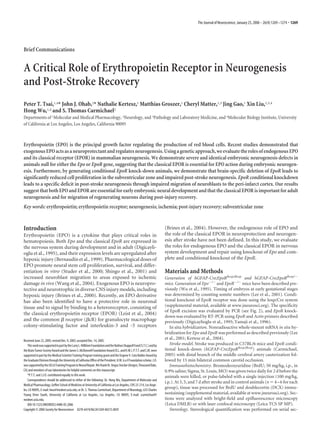
  • 2. 1270 • J. Neurosci., January 25, 2006 • 26(4):1269 –1274 Tsai et al. • Role of EPOR in Neurogenesis and Post-Stroke Recovery tions through the anterior subventricular zone (SVZ), olfactory bulb, and peri-infarct cortex using unbiased counting with the optical frac- tionator technique (supplemental material, available at www.jneurosci.org). Infarct volume. Infarct volume was measured 7 d after stroke in hGFAP-Cre;EpoRfloxp/ and control mice (n 5 and 4, respectively) (Kats- man et al., 2003; Li et al., 2005). Statistics. Post-stroke SVZ counts were tested with factorial ANOVA and Bonferroni’s post hoc testing (Statview 5.0.1; SAS Institute, Cary, NC). Infarct volume, SVZ, and olfactory bulb counts were tested with two-sample t testing assuming unequal variances (Excel; Microsoft, Redmond, WA). Results Sequential expression of EpoR and Epo in the developing brain and effects on embryonic development To understand the function of EPO/EPOR in neurogenesis, Epo and EpoR expression was first determined with in situ hybridiza- tion. Whole-mount in situ hybridization of EpoR and Epo in embryonic day 7.5 Figure 1. Epo and EpoR expression and neurogenesis defects in null animals. a–f, In situ hybridization of transverse sections (E7.5) to E11 embryos shows expression with EpoR (a, c, e) or Epo (b, d ,f ) antisense probes on E8.5, E9.5, and E10.5 embryos. Inset photomicrographs are higher- throughout the developing neural tube, magnification images from the neuroepithelium (a, c) and neural crest (e). g–l, Epo- (k, l ) and EpoR- (g–j) deficient embryos with progressive expression in optic pla- show underdeveloped choroids plexus (i–l ) and forebrain (g, h) at E13.5. Con, Control; Mut, mutant. code and forebrain and a delay of 0.5–1 d between EpoR and Epo expression in the and EpoR null animals results in systemic hypoxia (Wu et al., same region (supplemental Fig. 1a–f, available at www.jneurosci. 1995), and the roles for EPO/EPOR in adult tissues cannot be org as supplemental material). At E8.5, EpoR expression is con- evaluated in the null animals because of their early lethality fined to the neuroepithelium, a zone of precursors with high (Wu et al., 1995). To define the role of EPOR in these nonhe- proliferation capacity (Fig. 1a, inset). Epo expression is absent matopoietic tissues, we inserted loxp sites between the mini- at E8.5 (Fig. 1b) but appears by E9.0 –E9.5 in the neuroepithe- mal promoter and exons 1– 4 (supplemental Fig. 2, available at lium (Fig. 1d). At E9.5–E10.5, EpoR expression rises in neural www.jneurosci.org as supplemental material). In contrast to crest- and mesenchyme-derived cells (Fig. 1c,e, insets), fol- the early lethal phenotype observed in EpoR null embryos, lowed by Epo expression in similar regions 1 d later (Fig. 1f ). EpoRfloxp/floxp mice are viable and display no abnormal pheno- The temporal and spatial regulation of Epo and EpoR suggests types compared with wild-type (WT) littermates in gross de- a role for this ligand–receptor pair in early neurogenesis. velopment, fertility, and physiology (data not shown), sug- Both Epo and EpoR null embryos have incomplete neural tube gesting that introducing loxp sites into the endogenous EpoR closure at E10.5 (supplemental Fig. 1h, available at www.jneurosci. locus does not disrupt its normal function. org as supplemental material), especially in the hindbrain. By E13.5, the neurogenesis defects become more evident, including wavy neural tubes (supplemental Fig. 1j, arrow, available at Brain-specific EpoR deletion www.jneurosci.org as supplemental material), underdeveloped EpoRfloxp/floxp mice were crossed with hGFAP-Cre transgenic mice telencephalic regions (supplemental Fig. 1j,l, arrowhead, avail- (Zhuo et al., 2001; Malatesta et al., 2003). The hGFAP promoter able at www.jneurosci.org as supplemental material). Although drives Cre expression in radial glial cells of the developing cortex, no major structures are absent in Epo and EpoR null mice, the hindbrain, and cerebellum (Zhuo et al., 2001; Yue et al., 2005), mutant brains are consistently smaller and less developed than resulting in efficient EpoR deletion in the brain and sparing of their littermate controls, particularly in the SVZ of the develop- hematopoietic tissue (Fig. 2a,b, Mut). EpoRfloxp/floxp;hGFAP-Cre ing forebrain (Fig. 1h). In addition, the choroid plexus is severely mutants are viable and fertile, yielding Mendelian offspring dis- underdeveloped in both null embryos, with a flattened cubiodal tributions (data not shown), suggesting that neural-specific dele- epithelium and underdeveloped capillary network (Fig, 1i,l ). The tion of EpoR after E14 is not essential for embryonic or adult identical phenotypes of Epo and EpoR null animals suggest that survival. Although expression is undetectable in the brain tissues, for early embryonic development, the classical EPOR is essential we cannot rule out the presence of remaining EpoR expression for EPO action. beyond our levels of detection, so these animals are described as Generation of EpoR conditional knock-down mice EpoR conditional knock-down animals. Hematocrit, brain EPO and EPOR modulate cardiogenesis, vasculogenesis, and weight, and body weight of EpoR conditional knock-down mice neurogenesis (Wu et al., 1999; Studer et al., 2000; Shingo et al., are indistinguishable from their WT littermates (Fig. 2c). Normal 2001; Kertesz et al., 2004; Wang et al., 2004), raising the ques- brain architecture is preserved in EpoRfloxp/floxp;hGFAP-Cre an- tion of whether the classical EPOR exerts these functions in a imals in the cortex (Fig. 2d), olfactory bulbs, or cerebellum (data cell autonomous manner. The erythrocyte deficiency in Epo not shown).
  • 3. Tsai et al. • Role of EPOR in Neurogenesis and Post-Stroke Recovery J. Neurosci., January 25, 2006 • 26(4):1269 –1274 • 1271 proliferation, but not the normal pattern of neuroblast migration to the olfactory bulb. EPOR is not essential for protecting neurons from ischemic injury Previous studies that determined a neuroprotective effect of ex- ogenous EPO (Sakanaka et al., 1998; Bernaudin et al., 1999; Siren et al., 2001) either evaluated stroke size 24 h after stroke, when the area of infarction is not yet complete in most rodent stroke mod- els (Lipton, 1999), or used permanent focal ischemia, which pro- duces a different pattern of neuronal injury and death compared with stroke models that involve reperfusion (Lipton, 1999; Car- michael, 2005). We used a focal stroke model with the infarct and reperfusion injury restricted to the sensorimotor cortex (Kats- man et al., 2003; Carmichael et al., 2004). In this model, there is no statistically significant difference in infarct size between mu- tant (Fig. 4a) and controls (Fig. 4b) at day 3 (data not shown) and day 7 (Fig. 4c) after stroke ( p 0.31). This result suggests that, although higher doses of exogenous EPO may promote neuro- protection, endogenous levels of EPO and EPOR have limited roles in protecting neurons from ischemic reperfusion injury. EPOR is important for post-stroke neurogenesis Stroke induces migration of newly born neuroblasts from the SVZ into the area of injury in the first 7–14 d after the insult (Arvidsson et al., 2002; Parent et al., 2002; Jin et al., 2003), de- tected by the combination of pulse labeling with BrdU and stain- ing for DCX, a microtubule-associated protein present in imma- ture and migrating neurons (Feng and Walsh, 2001). As reported previously (Yang et al., 2004), there are no DCX cells in the cortex of either control or mutant mice before stroke (Fig. 4h). After stroke, DCX cells are found in subcortical white matter at day 3 (supplemental Fig. 3, available at www.jneurosci.org as supplemental material) and in the peri-infarct cortex at days 5 and 7, resulting in 4000 DCX cells per animal in the peri- infarct cortex (Fig. 4h). In both control and mutant animals, DCX cells are labeled with BrdU given after the stroke (supple- mental Fig. 4, available at www.jneurosci.org as supplemental material), and these cells are also immunoreactive for other markers of immature neurons or neural progenitor cells, such as Figure 2. Analysis of EpoR conditional knock-down animals. a, PCR. Floxed, WT, and excised PSA-NCAM and musashi (Gleeson et al., 1999; Okano et al., alleles ( loxp) are indicated. b, RT-PCR analysis for EpoR expression levels. c, Hematocrit, brain 2005) (supplemental Fig. 5, available at www.jneurosci.org as weight, and body weight are unchanged in the mutants. Error bars indicate SDs. d, Cresyl violet supplemental material). The number of DCX cells between sections from control (left) and mutant (right) animals. The bottom panels show higher- mutant and control animals remains the same on days 3 and 5 magnification views of the boxed areas. Con, Control; Mut, mutant. after stroke. However, EpoR conditional knock-down mice (Fig. 4e,g) show a 2.6-fold reduction in the number of DCX neuro- Neural-specific EpoR knockdown leads to reduced cell blasts present in the cortex at day 7 compared with controls (Fig. proliferation in the SVZ 4d,f ). Exogenous EPO promotes proliferation, survival, and neuronal The reduction of DCX neuroblasts in the cortex of post- differentiation of neural stem cells in vitro (Ling et al., 1998; stroke EpoR conditional knock-down mice may be attributable to Studer et al., 2000; Shingo et al., 2001;Yu et al., 2002). To examine reduced SVZ proliferation, reduced migration to the peri-infarct neurogenesis in vivo, we compared the SVZ of EpoR mutant mice cortex, or increased cell death in the peri-infarct cortex. To ex- with littermate controls. The mutant SVZ is decreased in volume amine proliferation, the number of BrdU cells in the SVZ was (Fig. 3i) and has fewer cells compared with controls (Con, quantified after stroke (Fig. 3h). Control mice show a significant 18,573 1523; Mut, 14,661 2131; p 0.03). After BrdU pulse, decrease in BrdU incorporation at day 3 (F(5,21) 8.072; p the mutant SVZ contains a significantly reduced number of 0.002) (supplemental Fig. 5d,h, available at www.jneurosci.org as BrdU cells at postnatal day 15, which is further reduced at 6 supplemental material), during the period in which DCX cells months (Fig. 3h). Despite reduced BrdU labeling in the SVZ, migrate from the SVZ toward the peri-infarct cortex and then a there is no difference in BrdU olfactory bulb cells between the return to baseline at day 7 (Fig. 3f, h). In contrast, the mutant two conditions (Con, 8431 1820 cells; Mut, 9068 1446 cells; mice have a reduced number of BrdU cells and display no p 0.16). These results provide the first evidence that EPOR change at any time point after stroke (Fig. 3c,e,g,h). The SVZ plays essential roles in adult neurogenesis in vivo and suggests volume shows similar differences between control and mutant that EPO/EPOR may regulate neural stem and progenitor cell before and after stroke (Fig. 3i). Thus, the mutant SVZ not only
  • 4. 1272 • J. Neurosci., January 25, 2006 • 26(4):1269 –1274 Tsai et al. • Role of EPOR in Neurogenesis and Post-Stroke Recovery has reduced BrdU labeling at baseline but also lacks any significant response to stroke. To determine cell migration and death after stroke, a cohort of DCX cells was labeled by administering BrdU on days 3–5 after stroke and tracking these cells in the peri-infarct cortex on days 5 and 7. In controls, 49 7% of DCX cells are BrdU at day 5 with further increases to 68 6% by day 7, indicating a contin- ued migration of this cohort of DCX cells into the peri-infarct cortex by day 7. In mutants, the percentage of DCX / BrdU cells on day 5 is similar to controls ( p 0.35) but fails to increase by day 7 (55 9%; p 0.53, MUT day 3 vs day 7). This indicates that there is a failure of con- tinued neuroblast migration into the peri- infarct cortex in EpoR conditional knock- down. To determine whether there is an increase in cell death with EpoR con- ditional knockdown, the number of ter- minal deoxynucleotidyl transferase- Figure 3. Quantification of BrdU labeling in the SVZ. a, Cresyl violet section. The rectangle shows the location of b– g. b– g, mediated biotinylated UTP nick end BrdU-labeled cells in control (b, d, f ) and EpoR conditional knock-down (c, e, g) animals in non-stroke (b, c), 3 d after stroke (d, e), labeling (TUNEL)-positive cells was and 7 d after stroke (f, g) are shown. Scale bar, 50 m. h, i, The graphs show quantifications of BrdU-labeled cells (h) and SVZ quantified in the peri-infarct cortex on volume before and after stroke (i). *p 0.004; #p 0.008; $p 0.016. Error bars indicate SDs. ctx, Cortex; str, stiatum; V, days 5 and 7 after stroke. There is no sig- ventricle; cc, corpus callosum; P15, postnatal day 15; mo, month; Con, control; Mut, mutant. nificant difference in the number of TUNEL cells in the per-infarct cortex at these two time points (5 d Con, 45 19.6; 5 d Mut, 43.5 11.5; 7 d Con, 42.8 13.3; 7 d Mut, 37.8 14.2; p 0.63 for all com- parisons). Coupled with the significant loss of DCX cells in the mutant peri- infarct cortex from days 5–7 (Fig. 4h), these data show that neuroblasts migrate to the peri-infarct cortex in which a subset then undergo cell death. Continued mi- gration of neuroblasts occurs in control animals such that the overall number is maintained, but there is no continued mi- gration in mutant, and the overall number of neuroblasts declines over time. Discussion In this study, we provide the first genetic demonstration that EPO and EPOR play essential roles in regulating embryonic and adult neuronal development and function. We demonstrate Epo and EpoR expression during critical periods of neu- ronal development as well as significantly aberrant neurogenesis in null animals. Figure 4. EpoR conditional knock-down leads to reduced neuroblast migration. a, b, Cresyl violet sections through the frontal Then, using a model whereby neuronal cortex of mutant (a) and control (b) 7 d after stroke. Scale bar, 500 m. c, Infarct volume 7 d after stroke was quantified. d– g, EpoR expression is specifically reduced in DCX cells in control (d, f ) and mutant (e, g). The region within the box in d and e is enlarged in f and g. Scale bars: d, 100 m; # the brain, we demonstrate a critical role f, 25 m. h, Stereological quantification of DCX cells. p 0.001 versus all other conditions; *p 0.008 versus Mut at ˆ @ $ for EPOR in adult neurogenesis and in the non-stroke stage; p 0.02 versus Mut at 7 d; p 0.02 versus 3 d Mut/Con and non-stroke; p 0.002 versus 7 d control. Error migration of newly born neuroblasts to ar- bars indicate SDs. eas of injury. Finally, EpoR conditional knock-down mice display no significant differences in infarct size EPO/EPOR are required for developmental neurogenesis versus control mice when exposed to stroke, suggesting alterna- The role of EPO in the nervous system extends beyond tissue tive mechanisms underlying the neuroprotective functions of oxygenation. Epo and EpoR are expressed during critical periods EPO. of neuronal development, and Epo and EpoR null animals reveal
  • 5. Tsai et al. • Role of EPOR in Neurogenesis and Post-Stroke Recovery J. Neurosci., January 25, 2006 • 26(4):1269 –1274 • 1273 defects in neurogenesis (Yu et al., 2002; present study). Here, roprotective effects of EPO in the conditional knock-down we demonstrate that these defects are mediated through EPO animals. binding to the classical EPOR. Neuronal defects in Epo null animals are identical to those seen in EpoR null animals, show- References ing that EPO is acting through the EPOR during neuronal Arvidsson A, Collin T, Kirik D, Kokaia Z, Lindvall O (2002) Neuronal re- development. Recent studies have suggested an alternative re- placement from endogenous precursors in the adult brain after stroke. ceptor for EPO neuronal effects (Leist et al., 2004) and that Nat Med 8:963–970. cR is essential for EPO neuroprotective actions (Brines et al., Bernaudin M, Marti HH, Roussel S, Divoux D, Nouvelot A, MacKenzie ET, Petit E (1999) A potential role for erythropoietin in focal permanent 2004). The identical hematopoietic and neuronal develop- cerebral ischemia in mice. J Cereb Blood Flow Metab 19:643– 651. mental phenotypes between Epo and EpoR null animals sug- Brines M, Grasso G, Fiordaliso F, Sfacteria A, Ghezzi P, Fratelli M, Latini R, gest that, for early embryonic development, the classical EPOR Xie QW, Smart J, Su-Rick CJ, Pobre E, Diaz D, Gomez D, Hand C, is essential for EPO action. Because cR null animals are viable Coleman T, Cerami A (2004) Erythropoietin mediates tissue protection without hematopoietic, cardiac, or neuronal defects, EPO through an erythropoietin and common beta-subunit heteroreceptor. likely acts exclusively through EPOR homodimers for its roles Proc Natl Acad Sci USA 101:14907–14912. Brines ML, Ghezzi P, Keenan S, Agnello D, de Lanerolle NC, Cerami C, Itri in these tissues during development (Reed et al., 2000; Brines LM, Cerami A (2000) Erythropoietin crosses the blood-brain barrier to et al., 2004). protect against experimental brain injury. Proc Natl Acad Sci USA 97:10526 –10531. EPOR is critical for adult and post-stroke neurogenesis Carmichael ST (2005) Rodent stroke models: size, mechanism and purpose. Exogenous EPO can promote neurogenesis in vitro (Ling et al., NeuroRx 2:396 – 409. 1998; Studer et al., 2000; Shingo et al., 2001). Our study, however, Carmichael ST, Tatsukawa K, Katsman D, Tsuyuguchi N, Kornblum HI provides the first evidence that EPOR plays essential roles in adult (2004) Evolution of diaschisis in a focal stroke model. Stroke 35:758 –763. neurogenesis in vivo. EpoR conditional knock-down animals Digicaylioglu M, Bichet S, Marti HH, Wenger RH, Rivas LA, Bauer C, have significantly reduced levels of BrdU incorporation in the Gassmann M (1995) Localization of specific erythropoietin binding SVZ and diminished SVZ cell number and volume, suggesting sites in defined areas of the mouse brain. Proc Natl Acad Sci USA that EPOR plays a critical function in maintaining dividing cell 92:3717–3720. numbers in the adult SVZ. Feng Y, Walsh CA (2001) Protein-protein interactions, cytoskeletal regula- Studies in large stroke models in the rat report increased la- tion and neuronal migration. Nat Rev Neurosci 2:408 – 416. Gleeson JG, Lin PT, Flanagan LA, Walsh CA (1999) Doublecortin is a beling of proliferating cells in the SVZ and migration of newly microtubule-associated protein and is expressed widely by migrating neu- born neuroblasts from the SVZ to peri-infarct tissue (Arvidsson rons. Neuron 23:257–271. et al., 2002; Parent et al., 2002; Jin et al., 2003). In the present Jin G, Omori N, Li F, Nagano I, Manabe Y, Shoji M, Abe K (2003) Protec- study, focal stroke is restricted to the mouse somatosensory cor- tion against ischemic brain damage by GDNF affecting cell survival and tex, with 1 mm distance to the SVZ. There is a biphasic prolif- death signals. Neurol Res 25:249 –253. erative response in the SVZ in this model and a robust migration Katsman D, Zheng J, Spinelli K, Carmichael ST (2003) Tissue microenvi- ronments within functional cortical subdivisions adjacent to focal stroke. of neuroblasts to the peri-infarct cortex. Our study demonstrates J Cereb Blood Flow Metab 23:997–1009. that EPOR also plays an essential role in this neurogenic response Kertesz N, Wu J, Chen TH, Sucov HM, Wu H (2004) The role of erythro- to ischemia. EpoR conditional knock-down animals do not dis- poietin in regulating angiogenesis. Dev Biol 276:101–110. play this biphasic SVZ response but instead maintain the reduced Lee R, Kertesz N, Joseph SB, Jegalian A, Wu H (2001) Erythropoietin control levels of BrdU labeling after stroke. Despite this lack of (Epo) and EpoR expression and 2 waves of erythropoiesis. Blood SVZ response, these animals appear to possess a normal initial 98:1408 –1415. post-stroke neuroblast migration to the peri-infarct cortex. After Leist M, Ghezzi P, Grasso G, Bianchi R, Villa P, Fratelli M, Savino C, Bianchi M, Nielsen J, Gerwien J, Kallunki P, Larsen AK, Helboe L, Christensen S, this initial migration, significant numbers of neuroblasts are lost Pedersen LO, Nielsen M, Torup L, Sager T, Sfacteria A, Erbayraktar S, et in the peri-infarct cortex in the EpoR knock-down animals (Fig. al. (2004) Derivatives of erythropoietin that are tissue protective but not 4h), and there is no continued migration of neuroblasts to replace erythropoietic. Science 305:239 –242. them. Li S, Zheng J, Carmichel ST (2005) Differences in ischemic injury after stroke between agend and young adults. Neurobiol Dis 18:432– 440. Alternative mechanisms for EPO-mediated neuroprotection Ling ZD, Potter ED, Lipton JW, Carvey PM (1998) Differentiation of mes- encephalic progenitor cells into dopaminergic neurons by cytokines. Exp Exogenous EPO has been shown to play a neuroprotective role Neurol 149:411– 423. during the ischemic response of the brain by preventing neuronal Lipton P (1999) Ischemic cell death in brain neurons. Physiol Rev apoptosis (Sakanaka et al., 1998; Bernaudin et al., 1999; Siren et 79:1431–1568. al., 2001). However, our data show no difference in infarct vol- Malatesta P, Hack MA, Hartfuss E, Kettenmann H, Klinkert W, Kirchhoff F, ume after conditional deletion of EpoR. This result suggests that Gotz M (2003) Neuronal or glial progeny: regional differences in radial although higher doses of exogenous EPO promotes neuroprotec- glia fate. Neuron 37:751–764. Marti HH, Bernaudin M, Petit E, Bauer C (2000) Neuroprotection and an- tion, there is a limited role of the endogenous EPO system in giogenesis: dual role of erythropoietin in brain ischemia. News Physiol Sci neuroprotection. Alternatively, the neuroprotective actions of 15:225–229. EPO may also be mediated via routes other than binding the Okano H, Kawahara H, Toriya M, Nakao K, Shibata S, Imai T (2005) Func- classical EPOR on neurons, such as through blood vessels. Exog- tion of RNA-binding protein Musashi-1 in stem cells. Exp Cell Res enous EPO has been demonstrated to increase cerebral vascula- 306:349 –356. ture at infarct sites (Marti et al., 2000; Wang et al., 2004), with Parent JM, Vexler ZS, Gong C, Derugin N, Ferriero DM (2002) Rat fore- subsequent increased oxygen delivery perhaps resulting in neu- brain neurogenesis and striatal neuron replacement after focal stroke. Ann Neurol 52:802– 813. roprotection. EPO may also act as a neuroprotectant by binding a Reed JA, Ikegami M, Robb L, Begley CG, Ross G, Whitsett JA (2000) Dis- receptor that contains cR but lacks the classical EPOR (Brines et tinct changes in pulmonary surfactant homeostasis in common beta- al., 2004). Thus, upregulation of other receptor complexes may chain- and GM-CSF-deficient mice. Am J Physiol Lung Cell Mol Physiol compensate for loss of the classical EPOR and mediate the neu- 278:L1164 –L1171.
  • 6. 1274 • J. Neurosci., January 25, 2006 • 26(4):1269 –1274 Tsai et al. • Role of EPOR in Neurogenesis and Post-Stroke Recovery Sakanaka M, Wen TC, Matsuda S, Masuda S, Morishita E, Nagao M, Sasaki R throid BFU-E and CFU-E progenitors does not require erythropoietin or (1998) In vivo evidence that erythropoietin protects neurons from isch- the erythropoietin receptor. Cell 83:59 – 67. emic damage. Proc Natl Acad Sci USA 95:4635– 4640. Wu H, Lee SH, Gao J, Liu X, Iruela-Arispe ML (1999) Inactivation of eryth- Shingo T, Sorokan ST, Shimazaki T, Weiss S (2001) Erythropoietin regu- ropoietin leads to defects in cardiac morphogenesis. Development lates the in vitro and in vivo production of neuronal progenitors by mam- 126:3597–3605. malian forebrain neural stem cells. J Neurosci 21:9733–9743. Yamaji R, Okada T, Moriya M, Naito M, Tsuruo T, Miyatake K, Nakano Y Siren AL, Fratelli M, Brines M, Goemans C, Casagrande S, Lewczuk P, Keenan (1996) Brain capillary endothelial cells express two forms of erythropoi- S, Gleiter C, Pasquali C, Capobianco A, Mennini T, Heumann R, Cerami etin receptor mRNA. Eur J Biochem 239:494 –500. A, Ehrenreich H, Ghezzi P (2001) Erythropoietin prevents neuronal ap- Yu X, Shacka JJ, Eells JB, Suarez-Quian C, Przygodzki RM, Beleslin-Cokic B, optosis after cerebral ischemia and metabolic stress. Proc Natl Acad Sci Lin CS, Nikodem VM, Hempstead B, Flanders KC, Costantini F, Noguchi USA 98:4044 – 4049. CT (2002) Erythropoietin receptor signaling is required for normal Studer L, Csete M, Lee SH, Kabbani N, Walikonis J, Wold B, McKay R (2000) brain development. Development 129:505–516. Enhanced proliferation, survival, and dopaminergic differentiation of Yue Q, Groszer M, Gil JS, Berk AJ, Messing A, Wu H, Liu X (2005) PTEN CNS precursors in lowered oxygen. J Neurosci 20:7377–7383. deletion in Bergmann glia leads to premature differentiation and affects Wang L, Zhang Z, Wang Y, Zhang R, Chopp M (2004) Treatment of stroke laminar organization. Development 132:3281–3291. with erythropoietin enhances neurogenesis and angiogenesis and im- Zhuo L, Theis M, Alvarez-Maya I, Brenner M, Willecke K, Messing A (2001) proves neurological function in rats. Stroke 35:1732–1737. hGFAP-cre transgenic mice for manipulation of glial and neuronal func- Wu H, Liu X, Jaenisch R, Lodish HF (1995) Generation of committed ery- tion in vivo. Genesis 31:85–94.