SlideShare uma empresa Scribd logo
1 de 41
ANATOMY AND PHYSIOLOGY OF
LUNG
JITHA.A.HARI
2ND YR MSc N
CNK
INTRODUCTION
The lungs are the primary organs of respiration in humans
and many other animals including a few fish and some
snails.
In mammals and most other vertebrates, two lungs are
located near the backbone on either side of the heart.
ANATOMY - LUNG
The lungs are located in the chest on either side of the heart in the rib cage. They are
conical in shape with a narrow rounded apex at the top and a broad base that rests on the
diaphragm
The apex of the lung extends into the root of the neck, reaching shortly above the level of
the sternal end of the first rib.
The front and outer sides of the lung face the ribs, which make light indendations on their
surfaces. The bottom of the lungs is smooth and rests on the diaphragm, matching its
concavity.
 The medial surface of the lungs faces towards the center of the chest, and lies against the
heart, great vessels, and the carina where the two main bronchi branch off from the base of
Gross anatomy
Both lungs have a central recession called the hilum at the root of the lung, where the blood
vessels and airways pass into the lungs. There are also bronchopulmonary lymph nodes on
the hilum.
The lungs are surrounded by the pulmonary pleurae. The pleurae are two serous
membranes; the outer parietal pleura lines the inner wall of the rib cage and the inner
visceral pleura directly lines the surface of the lungs. Between the pleurae is a potential
space called the pleural cavity containing pleural fluid.
Each lung is divided into lobes by the invaginations of the pleura as fissures. The fissures
are double folds of pleura that section the lungs and help in their expansion.
 The lobes of the lungs are further divided into broncho pulmonary segments based on
the locations of bronchioles.
Oblique fissure (Right & Left):
It starts at the 3rd thoracic spine while the arms are elevated, descends downwards,
laterally & anteriorly along the medial border of the scapula touching the inferior
angle of the scapula) cutting the midaxillary line in the 5th rib & ending at the 6th
costal cartilage 3 inches from the midline.
In cadaver it arise at the 2nd thoracic spine.
The transverse fissure (Right):
It arises at the 4th costal cartilage, runs horizontally to meet the oblique fissure in
the midaxillary line in the 5th rib.
LUNG FISSURES:
FISSURES & LOBES OF THE RIGHT LUNG
PLEURA
THE BORDERS OF THE PLEURA:
 The pleura runs close to the lung except at the lower border which
extends for 3-5 cm below it both anteriorly & posteriorly & 9-10 cm
below in the axilla.
 The lower border cuts the 8th rib in MCL, 10th rib in MAL and 12th rib in
Scapular line.
Parietal Pleura
Costal pleura:
Lines thoracic wall (ribs, intercostal spaces)
Mediastinal pleura:
Lines corresponding surface of mediastinum; reflected over root of lung & becomes continuous
with visceral pleura around hilum
Cervical pleura:
Extends into neck about 2 inches above 1st costal cartilage & one inch above medial 1/3 clavicle;
covers apex of lung
Diaphragmatic pleura:
Lines superior surface of diaphragm
Microanatomy
• The lungs contain the respiratory tract and its lining, which terminate in
alveoli, the tissue in between and veins, arteries, nerves and lymphatic vessels.
• The respiratory tract begins with the trachea and bronchi. These structures are lined
with columnar epithelial cells that possess cilia , small frond-like projections.
• Interspersed with the epithelial cells are goblet cells which produce mucous, and club
cells with actions similar to macrophages. Surrounding these in the trachea and
bronchi are cartilage rings, which help to maintain stability.
• The respiratory tract ends in lobules. These consist of a respiratory bronchiole,
which branches into alveolar ducts and alveolar sacs, which in turn divide into
alveoli.
Alveoli consist of two types of alveolar cell and an alveolar macrophage. The two types of
cell are known as type I and type II alveolar cells (also known as pneumocytes)
Types I and II make up the walls and septa of the alveoli.
Type I cells provide 95% of the surface area of each alveoli and are flat ("squamous"), and
Type II cells generally cluster in the corners of the alveoli and have a cuboidal shape.
Despite this, cells occur in a roughly equal ratio of 1:1 or 6:4
Type I are squamous epithelial cells that make up the alveolar wall structure. They have
extremely thin walls that enable an easy gas exchange.
These type I cells also make up the alveolar septa which separate each alveolus. The septa
consist of an epithelial lining and associated basement membranes. Type I cells are not able
to divide, and consequently rely on differentiation from Type II cells.
Type II are larger and they line the alveoli and produce and secrete ELF and
surfactant. Type II cells are able to divide and differentiate to Type 1 cells.
BLOOD SUPPLY
The human lung has a dual blood supply.
The tissue of the lungs receive oxygenated blood via the bronchial circulation, a series
of arteries that leave the aorta and are part of the systemic circulation. There are usually
three arteries, and they branch alongside the bronchi and bronchioles.
 The blood volume of the lungs is about 450 millilitres on average, about 9 per cent of the
total blood volume of the entire circulatory system. This quantity can easily fluctuate from
between one-half and twice the normal volume.
The lungs also receive deoxygenated blood from the heart and supply it with oxygen,
in a process known as respiration.
 In this process, venous blood in the body collects in the right atrium and is pumped
from the right ventricle through the pulmonary trunk and the pulmonary arteries into
the left and right lungs.
 Blood passes through small capillaries next to the alveoli in the lung, receives
oxygen, and travels back to the heart. This is called the pulmonary circulation. The
oxygenated blood is then pumped to the rest of the body.
NERVE SUPPLY
 The lungs are supplied by nerves of the autonomic nervous system. Input from the parasympathetic
nervous system occurs via the vagus nerve.
 When stimulated by acetylcholine, this causes constriction of the smooth muscle lining the bronchus and
bronchiole, and increases the secretions from glands.
 The lungs also have a sympathetic tone from norepinephrine acting on the beta 2 receptors in the
respiratory tract, which causes bronchodilation.
 The action of breathing takes place because of nerve signals sent by the respiratory centres in the
brainstem, along the phrenic nerve to the diaphragm.
PHYSIOLOGY OF LUNG
LUNG VOLUMES
Lung volumes and lung capacities refer to the volume of air associated with different
phases of the respiratory cycle. Lung volumes are directly measured; lung capacities are
inferred from lung volumes.
The average total lung capacity of an adult human male is about 6 litres of air.
Four types
1. Tidal volume
2. Inspiratory reserve volume
3. Expiratory persevere volume
4. Residual volume
TIDAL VOLUME
Normal volume of air inspired or expired during quiet breathing
TV = 500 ml
INSPIRATORY RESERVE VOLUME
 Extra volume of air inhaled after tidal volume by max inspiratory effort
 3000ml in adult male
(or)
 3300 / 1900 = M/F
EXPIRATORY RESERVE VOLUME
Extra volume of air that can be exhaled after tidal volume by max
expiratory efforts
1100 in a normal adult male
(or)
1200/700 = M/F
RESIDUAL VOLUME
Volume of the air left out in lungs after forceful expiration or
complete expiration
1200/1100 = M/F
These are combinations of two or more lung volumes
1. Inspiratory capacity
2. Expiratory capacity
3. Functional residual capacity
4. Vital capacity
5. Total lung capacity
INSPIRATORY CAPACITY
Max volume of air that can be inspired after normal tidal
expiration
IC = TV+IRV
= 500 +3000
= 3500 ml
EXPIRATORY CAPACITY
Max volume of air that can be expired after normal
tidal inspiration
EC=TV+ERV (500+1100=1600ml)
FUNCTIONAL RESIDUAL CAPACITY
Volume of air remaining in lungs after normal tidal
expiration
FRC= ERV + RV ( 1100 + 1200 = 2300ml)
VITAL CAPACITY
Max Amount of air expelled after deepest possible
inspiration
VC = TV+IRV+ERV
500+3000+1100= 4600ml
TOTAL LUNG CAPACITY
Volume of air present in lung after max inspiration
TLC = VC + RV ( 4600+1200 = 5800ml )
Anatomy and physiology of lung

Mais conteúdo relacionado

Mais procurados (20)

Bed sores / decubitis ulcer / pressure sores
Bed sores / decubitis ulcer / pressure soresBed sores / decubitis ulcer / pressure sores
Bed sores / decubitis ulcer / pressure sores
 
Pleural effusion
Pleural effusionPleural effusion
Pleural effusion
 
Cardiac assessment ppt
Cardiac assessment pptCardiac assessment ppt
Cardiac assessment ppt
 
Positions
PositionsPositions
Positions
 
Chronic obstructive pulmonary disorders COPD
Chronic obstructive pulmonary disorders COPDChronic obstructive pulmonary disorders COPD
Chronic obstructive pulmonary disorders COPD
 
Anatomy of kidneys
Anatomy of  kidneysAnatomy of  kidneys
Anatomy of kidneys
 
suctioning procedure ppt.pptx
suctioning procedure  ppt.pptxsuctioning procedure  ppt.pptx
suctioning procedure ppt.pptx
 
Deep breathing and coughing exercises
Deep breathing and coughing exercisesDeep breathing and coughing exercises
Deep breathing and coughing exercises
 
CHRONIC OBSTRUCTIVE PULMONARY DISEASE
CHRONIC OBSTRUCTIVE PULMONARY DISEASECHRONIC OBSTRUCTIVE PULMONARY DISEASE
CHRONIC OBSTRUCTIVE PULMONARY DISEASE
 
Care of an unconcious patient
Care of an unconcious patientCare of an unconcious patient
Care of an unconcious patient
 
Thoracentesis
Thoracentesis Thoracentesis
Thoracentesis
 
Cardiopulmonary resuscitation(cpr)
Cardiopulmonary resuscitation(cpr)Cardiopulmonary resuscitation(cpr)
Cardiopulmonary resuscitation(cpr)
 
Suctioning
SuctioningSuctioning
Suctioning
 
Bronchitis
BronchitisBronchitis
Bronchitis
 
O2 therapy procedure
O2 therapy procedureO2 therapy procedure
O2 therapy procedure
 
Empyema
EmpyemaEmpyema
Empyema
 
Respiratory System - Physiology
Respiratory System - PhysiologyRespiratory System - Physiology
Respiratory System - Physiology
 
Lungs abscess
Lungs abscessLungs abscess
Lungs abscess
 
Pneumonia
PneumoniaPneumonia
Pneumonia
 
Cpr ppt
Cpr ppt Cpr ppt
Cpr ppt
 

Semelhante a Anatomy and physiology of lung

Respiratory System - Human Anatomy and Physiology bPharm
Respiratory System - Human Anatomy and Physiology bPharmRespiratory System - Human Anatomy and Physiology bPharm
Respiratory System - Human Anatomy and Physiology bPharmPankaj Saha
 
Respiratory system and lungs.pptx
Respiratory system and lungs.pptxRespiratory system and lungs.pptx
Respiratory system and lungs.pptxShubham Shukla
 
Anatomy of lungs, pleura and diaphragm
Anatomy  of lungs, pleura and diaphragmAnatomy  of lungs, pleura and diaphragm
Anatomy of lungs, pleura and diaphragmDr. Ashish kumar
 
Lec.8 lungs pt&rc
Lec.8 lungs pt&rcLec.8 lungs pt&rc
Lec.8 lungs pt&rcDr Motawei
 
The lungs presentation
The lungs presentationThe lungs presentation
The lungs presentationglenda
 
Respiratory System by Roshni Kumari (1).pptx
Respiratory System by Roshni Kumari (1).pptxRespiratory System by Roshni Kumari (1).pptx
Respiratory System by Roshni Kumari (1).pptxAbhishek Kumar
 
Bronchial tree alveoli lungs review
Bronchial tree alveoli lungs  reviewBronchial tree alveoli lungs  review
Bronchial tree alveoli lungs reviewMichael Wrock
 
Respiratory System unit-I cop.pptx
Respiratory System unit-I cop.pptxRespiratory System unit-I cop.pptx
Respiratory System unit-I cop.pptxRaeesShahidBasharat
 
Gross anatomy and development of respiratory system
Gross anatomy and development of respiratory systemGross anatomy and development of respiratory system
Gross anatomy and development of respiratory systemDr Laxman Khanal
 

Semelhante a Anatomy and physiology of lung (20)

ASTHMA.docx
ASTHMA.docxASTHMA.docx
ASTHMA.docx
 
Surgery 5th year, 1st lecture (Dr. Ahmed Al-Azzawi)
Surgery 5th year, 1st lecture (Dr. Ahmed Al-Azzawi)Surgery 5th year, 1st lecture (Dr. Ahmed Al-Azzawi)
Surgery 5th year, 1st lecture (Dr. Ahmed Al-Azzawi)
 
Respiratory System - Human Anatomy and Physiology bPharm
Respiratory System - Human Anatomy and Physiology bPharmRespiratory System - Human Anatomy and Physiology bPharm
Respiratory System - Human Anatomy and Physiology bPharm
 
Respiratory system and lungs.pptx
Respiratory system and lungs.pptxRespiratory system and lungs.pptx
Respiratory system and lungs.pptx
 
Anatomy of lungs, pleura and diaphragm
Anatomy  of lungs, pleura and diaphragmAnatomy  of lungs, pleura and diaphragm
Anatomy of lungs, pleura and diaphragm
 
Lungs Grade 4
Lungs Grade 4Lungs Grade 4
Lungs Grade 4
 
Lec.8 lungs pt&rc
Lec.8 lungs pt&rcLec.8 lungs pt&rc
Lec.8 lungs pt&rc
 
LUNGS ANATOMY
LUNGS ANATOMYLUNGS ANATOMY
LUNGS ANATOMY
 
lungs.pptx
lungs.pptxlungs.pptx
lungs.pptx
 
The lungs presentation
The lungs presentationThe lungs presentation
The lungs presentation
 
Respiratory syst
Respiratory systRespiratory syst
Respiratory syst
 
Clinical Anatomy of lungs
Clinical Anatomy of lungsClinical Anatomy of lungs
Clinical Anatomy of lungs
 
Respiratory System by Roshni Kumari (1).pptx
Respiratory System by Roshni Kumari (1).pptxRespiratory System by Roshni Kumari (1).pptx
Respiratory System by Roshni Kumari (1).pptx
 
MJ ADENIYI, MSC
MJ ADENIYI, MSCMJ ADENIYI, MSC
MJ ADENIYI, MSC
 
Respiratory physiology
Respiratory physiologyRespiratory physiology
Respiratory physiology
 
Mj adeniyi msc
Mj adeniyi mscMj adeniyi msc
Mj adeniyi msc
 
Bronchial tree alveoli lungs review
Bronchial tree alveoli lungs  reviewBronchial tree alveoli lungs  review
Bronchial tree alveoli lungs review
 
Functional Anatomy of Respiratory system
Functional Anatomy of Respiratory systemFunctional Anatomy of Respiratory system
Functional Anatomy of Respiratory system
 
Respiratory System unit-I cop.pptx
Respiratory System unit-I cop.pptxRespiratory System unit-I cop.pptx
Respiratory System unit-I cop.pptx
 
Gross anatomy and development of respiratory system
Gross anatomy and development of respiratory systemGross anatomy and development of respiratory system
Gross anatomy and development of respiratory system
 

Último

(May 9, 2024) Enhanced Ultrafast Vector Flow Imaging (VFI) Using Multi-Angle ...
(May 9, 2024) Enhanced Ultrafast Vector Flow Imaging (VFI) Using Multi-Angle ...(May 9, 2024) Enhanced Ultrafast Vector Flow Imaging (VFI) Using Multi-Angle ...
(May 9, 2024) Enhanced Ultrafast Vector Flow Imaging (VFI) Using Multi-Angle ...Scintica Instrumentation
 
module for grade 9 for distance learning
module for grade 9 for distance learningmodule for grade 9 for distance learning
module for grade 9 for distance learninglevieagacer
 
Human genetics..........................pptx
Human genetics..........................pptxHuman genetics..........................pptx
Human genetics..........................pptxSilpa
 
biology HL practice questions IB BIOLOGY
biology HL practice questions IB BIOLOGYbiology HL practice questions IB BIOLOGY
biology HL practice questions IB BIOLOGY1301aanya
 
Grade 7 - Lesson 1 - Microscope and Its Functions
Grade 7 - Lesson 1 - Microscope and Its FunctionsGrade 7 - Lesson 1 - Microscope and Its Functions
Grade 7 - Lesson 1 - Microscope and Its FunctionsOrtegaSyrineMay
 
GBSN - Microbiology (Unit 2)
GBSN - Microbiology (Unit 2)GBSN - Microbiology (Unit 2)
GBSN - Microbiology (Unit 2)Areesha Ahmad
 
Locating and isolating a gene, FISH, GISH, Chromosome walking and jumping, te...
Locating and isolating a gene, FISH, GISH, Chromosome walking and jumping, te...Locating and isolating a gene, FISH, GISH, Chromosome walking and jumping, te...
Locating and isolating a gene, FISH, GISH, Chromosome walking and jumping, te...Silpa
 
GBSN - Microbiology (Unit 3)
GBSN - Microbiology (Unit 3)GBSN - Microbiology (Unit 3)
GBSN - Microbiology (Unit 3)Areesha Ahmad
 
Stages in the normal growth curve
Stages in the normal growth curveStages in the normal growth curve
Stages in the normal growth curveAreesha Ahmad
 
COMPUTING ANTI-DERIVATIVES (Integration by SUBSTITUTION)
COMPUTING ANTI-DERIVATIVES(Integration by SUBSTITUTION)COMPUTING ANTI-DERIVATIVES(Integration by SUBSTITUTION)
COMPUTING ANTI-DERIVATIVES (Integration by SUBSTITUTION)AkefAfaneh2
 
CURRENT SCENARIO OF POULTRY PRODUCTION IN INDIA
CURRENT SCENARIO OF POULTRY PRODUCTION IN INDIACURRENT SCENARIO OF POULTRY PRODUCTION IN INDIA
CURRENT SCENARIO OF POULTRY PRODUCTION IN INDIADr. TATHAGAT KHOBRAGADE
 
GBSN - Biochemistry (Unit 1)
GBSN - Biochemistry (Unit 1)GBSN - Biochemistry (Unit 1)
GBSN - Biochemistry (Unit 1)Areesha Ahmad
 
PSYCHOSOCIAL NEEDS. in nursing II sem pptx
PSYCHOSOCIAL NEEDS. in nursing II sem pptxPSYCHOSOCIAL NEEDS. in nursing II sem pptx
PSYCHOSOCIAL NEEDS. in nursing II sem pptxSuji236384
 
Porella : features, morphology, anatomy, reproduction etc.
Porella : features, morphology, anatomy, reproduction etc.Porella : features, morphology, anatomy, reproduction etc.
Porella : features, morphology, anatomy, reproduction etc.Silpa
 
Zoology 5th semester notes( Sumit_yadav).pdf
Zoology 5th semester notes( Sumit_yadav).pdfZoology 5th semester notes( Sumit_yadav).pdf
Zoology 5th semester notes( Sumit_yadav).pdfSumit Kumar yadav
 
Introduction of DNA analysis in Forensic's .pptx
Introduction of DNA analysis in Forensic's .pptxIntroduction of DNA analysis in Forensic's .pptx
Introduction of DNA analysis in Forensic's .pptxrohankumarsinghrore1
 

Último (20)

(May 9, 2024) Enhanced Ultrafast Vector Flow Imaging (VFI) Using Multi-Angle ...
(May 9, 2024) Enhanced Ultrafast Vector Flow Imaging (VFI) Using Multi-Angle ...(May 9, 2024) Enhanced Ultrafast Vector Flow Imaging (VFI) Using Multi-Angle ...
(May 9, 2024) Enhanced Ultrafast Vector Flow Imaging (VFI) Using Multi-Angle ...
 
module for grade 9 for distance learning
module for grade 9 for distance learningmodule for grade 9 for distance learning
module for grade 9 for distance learning
 
Human genetics..........................pptx
Human genetics..........................pptxHuman genetics..........................pptx
Human genetics..........................pptx
 
PATNA CALL GIRLS 8617370543 LOW PRICE ESCORT SERVICE
PATNA CALL GIRLS 8617370543 LOW PRICE ESCORT SERVICEPATNA CALL GIRLS 8617370543 LOW PRICE ESCORT SERVICE
PATNA CALL GIRLS 8617370543 LOW PRICE ESCORT SERVICE
 
biology HL practice questions IB BIOLOGY
biology HL practice questions IB BIOLOGYbiology HL practice questions IB BIOLOGY
biology HL practice questions IB BIOLOGY
 
Grade 7 - Lesson 1 - Microscope and Its Functions
Grade 7 - Lesson 1 - Microscope and Its FunctionsGrade 7 - Lesson 1 - Microscope and Its Functions
Grade 7 - Lesson 1 - Microscope and Its Functions
 
GBSN - Microbiology (Unit 2)
GBSN - Microbiology (Unit 2)GBSN - Microbiology (Unit 2)
GBSN - Microbiology (Unit 2)
 
Locating and isolating a gene, FISH, GISH, Chromosome walking and jumping, te...
Locating and isolating a gene, FISH, GISH, Chromosome walking and jumping, te...Locating and isolating a gene, FISH, GISH, Chromosome walking and jumping, te...
Locating and isolating a gene, FISH, GISH, Chromosome walking and jumping, te...
 
GBSN - Microbiology (Unit 3)
GBSN - Microbiology (Unit 3)GBSN - Microbiology (Unit 3)
GBSN - Microbiology (Unit 3)
 
Stages in the normal growth curve
Stages in the normal growth curveStages in the normal growth curve
Stages in the normal growth curve
 
COMPUTING ANTI-DERIVATIVES (Integration by SUBSTITUTION)
COMPUTING ANTI-DERIVATIVES(Integration by SUBSTITUTION)COMPUTING ANTI-DERIVATIVES(Integration by SUBSTITUTION)
COMPUTING ANTI-DERIVATIVES (Integration by SUBSTITUTION)
 
CURRENT SCENARIO OF POULTRY PRODUCTION IN INDIA
CURRENT SCENARIO OF POULTRY PRODUCTION IN INDIACURRENT SCENARIO OF POULTRY PRODUCTION IN INDIA
CURRENT SCENARIO OF POULTRY PRODUCTION IN INDIA
 
GBSN - Biochemistry (Unit 1)
GBSN - Biochemistry (Unit 1)GBSN - Biochemistry (Unit 1)
GBSN - Biochemistry (Unit 1)
 
PSYCHOSOCIAL NEEDS. in nursing II sem pptx
PSYCHOSOCIAL NEEDS. in nursing II sem pptxPSYCHOSOCIAL NEEDS. in nursing II sem pptx
PSYCHOSOCIAL NEEDS. in nursing II sem pptx
 
Porella : features, morphology, anatomy, reproduction etc.
Porella : features, morphology, anatomy, reproduction etc.Porella : features, morphology, anatomy, reproduction etc.
Porella : features, morphology, anatomy, reproduction etc.
 
Site Acceptance Test .
Site Acceptance Test                    .Site Acceptance Test                    .
Site Acceptance Test .
 
Clean In Place(CIP).pptx .
Clean In Place(CIP).pptx                 .Clean In Place(CIP).pptx                 .
Clean In Place(CIP).pptx .
 
Zoology 5th semester notes( Sumit_yadav).pdf
Zoology 5th semester notes( Sumit_yadav).pdfZoology 5th semester notes( Sumit_yadav).pdf
Zoology 5th semester notes( Sumit_yadav).pdf
 
Introduction of DNA analysis in Forensic's .pptx
Introduction of DNA analysis in Forensic's .pptxIntroduction of DNA analysis in Forensic's .pptx
Introduction of DNA analysis in Forensic's .pptx
 
+971581248768>> SAFE AND ORIGINAL ABORTION PILLS FOR SALE IN DUBAI AND ABUDHA...
+971581248768>> SAFE AND ORIGINAL ABORTION PILLS FOR SALE IN DUBAI AND ABUDHA...+971581248768>> SAFE AND ORIGINAL ABORTION PILLS FOR SALE IN DUBAI AND ABUDHA...
+971581248768>> SAFE AND ORIGINAL ABORTION PILLS FOR SALE IN DUBAI AND ABUDHA...
 

Anatomy and physiology of lung

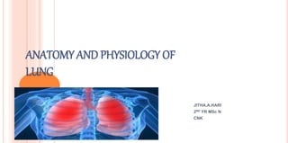
  • 1. ANATOMY AND PHYSIOLOGY OF LUNG JITHA.A.HARI 2ND YR MSc N CNK
  • 2. INTRODUCTION The lungs are the primary organs of respiration in humans and many other animals including a few fish and some snails. In mammals and most other vertebrates, two lungs are located near the backbone on either side of the heart.
  • 4. The lungs are located in the chest on either side of the heart in the rib cage. They are conical in shape with a narrow rounded apex at the top and a broad base that rests on the diaphragm The apex of the lung extends into the root of the neck, reaching shortly above the level of the sternal end of the first rib. The front and outer sides of the lung face the ribs, which make light indendations on their surfaces. The bottom of the lungs is smooth and rests on the diaphragm, matching its concavity.  The medial surface of the lungs faces towards the center of the chest, and lies against the heart, great vessels, and the carina where the two main bronchi branch off from the base of Gross anatomy
  • 5. Both lungs have a central recession called the hilum at the root of the lung, where the blood vessels and airways pass into the lungs. There are also bronchopulmonary lymph nodes on the hilum. The lungs are surrounded by the pulmonary pleurae. The pleurae are two serous membranes; the outer parietal pleura lines the inner wall of the rib cage and the inner visceral pleura directly lines the surface of the lungs. Between the pleurae is a potential space called the pleural cavity containing pleural fluid. Each lung is divided into lobes by the invaginations of the pleura as fissures. The fissures are double folds of pleura that section the lungs and help in their expansion.
  • 6.  The lobes of the lungs are further divided into broncho pulmonary segments based on the locations of bronchioles.
  • 7.
  • 8. Oblique fissure (Right & Left): It starts at the 3rd thoracic spine while the arms are elevated, descends downwards, laterally & anteriorly along the medial border of the scapula touching the inferior angle of the scapula) cutting the midaxillary line in the 5th rib & ending at the 6th costal cartilage 3 inches from the midline. In cadaver it arise at the 2nd thoracic spine. The transverse fissure (Right): It arises at the 4th costal cartilage, runs horizontally to meet the oblique fissure in the midaxillary line in the 5th rib. LUNG FISSURES:
  • 9. FISSURES & LOBES OF THE RIGHT LUNG
  • 11. THE BORDERS OF THE PLEURA:  The pleura runs close to the lung except at the lower border which extends for 3-5 cm below it both anteriorly & posteriorly & 9-10 cm below in the axilla.  The lower border cuts the 8th rib in MCL, 10th rib in MAL and 12th rib in Scapular line.
  • 12.
  • 13. Parietal Pleura Costal pleura: Lines thoracic wall (ribs, intercostal spaces) Mediastinal pleura: Lines corresponding surface of mediastinum; reflected over root of lung & becomes continuous with visceral pleura around hilum Cervical pleura: Extends into neck about 2 inches above 1st costal cartilage & one inch above medial 1/3 clavicle; covers apex of lung Diaphragmatic pleura: Lines superior surface of diaphragm
  • 14. Microanatomy • The lungs contain the respiratory tract and its lining, which terminate in alveoli, the tissue in between and veins, arteries, nerves and lymphatic vessels. • The respiratory tract begins with the trachea and bronchi. These structures are lined with columnar epithelial cells that possess cilia , small frond-like projections. • Interspersed with the epithelial cells are goblet cells which produce mucous, and club cells with actions similar to macrophages. Surrounding these in the trachea and bronchi are cartilage rings, which help to maintain stability.
  • 15. • The respiratory tract ends in lobules. These consist of a respiratory bronchiole, which branches into alveolar ducts and alveolar sacs, which in turn divide into alveoli.
  • 16.
  • 17. Alveoli consist of two types of alveolar cell and an alveolar macrophage. The two types of cell are known as type I and type II alveolar cells (also known as pneumocytes) Types I and II make up the walls and septa of the alveoli. Type I cells provide 95% of the surface area of each alveoli and are flat ("squamous"), and Type II cells generally cluster in the corners of the alveoli and have a cuboidal shape. Despite this, cells occur in a roughly equal ratio of 1:1 or 6:4 Type I are squamous epithelial cells that make up the alveolar wall structure. They have extremely thin walls that enable an easy gas exchange.
  • 18. These type I cells also make up the alveolar septa which separate each alveolus. The septa consist of an epithelial lining and associated basement membranes. Type I cells are not able to divide, and consequently rely on differentiation from Type II cells. Type II are larger and they line the alveoli and produce and secrete ELF and surfactant. Type II cells are able to divide and differentiate to Type 1 cells.
  • 19.
  • 20. BLOOD SUPPLY The human lung has a dual blood supply. The tissue of the lungs receive oxygenated blood via the bronchial circulation, a series of arteries that leave the aorta and are part of the systemic circulation. There are usually three arteries, and they branch alongside the bronchi and bronchioles.  The blood volume of the lungs is about 450 millilitres on average, about 9 per cent of the total blood volume of the entire circulatory system. This quantity can easily fluctuate from between one-half and twice the normal volume.
  • 21. The lungs also receive deoxygenated blood from the heart and supply it with oxygen, in a process known as respiration.  In this process, venous blood in the body collects in the right atrium and is pumped from the right ventricle through the pulmonary trunk and the pulmonary arteries into the left and right lungs.  Blood passes through small capillaries next to the alveoli in the lung, receives oxygen, and travels back to the heart. This is called the pulmonary circulation. The oxygenated blood is then pumped to the rest of the body.
  • 22.
  • 23. NERVE SUPPLY  The lungs are supplied by nerves of the autonomic nervous system. Input from the parasympathetic nervous system occurs via the vagus nerve.  When stimulated by acetylcholine, this causes constriction of the smooth muscle lining the bronchus and bronchiole, and increases the secretions from glands.  The lungs also have a sympathetic tone from norepinephrine acting on the beta 2 receptors in the respiratory tract, which causes bronchodilation.  The action of breathing takes place because of nerve signals sent by the respiratory centres in the brainstem, along the phrenic nerve to the diaphragm.
  • 25.
  • 26. LUNG VOLUMES Lung volumes and lung capacities refer to the volume of air associated with different phases of the respiratory cycle. Lung volumes are directly measured; lung capacities are inferred from lung volumes. The average total lung capacity of an adult human male is about 6 litres of air. Four types 1. Tidal volume 2. Inspiratory reserve volume 3. Expiratory persevere volume 4. Residual volume
  • 27. TIDAL VOLUME Normal volume of air inspired or expired during quiet breathing TV = 500 ml
  • 28.
  • 29. INSPIRATORY RESERVE VOLUME  Extra volume of air inhaled after tidal volume by max inspiratory effort  3000ml in adult male (or)  3300 / 1900 = M/F
  • 30. EXPIRATORY RESERVE VOLUME Extra volume of air that can be exhaled after tidal volume by max expiratory efforts 1100 in a normal adult male (or) 1200/700 = M/F
  • 31. RESIDUAL VOLUME Volume of the air left out in lungs after forceful expiration or complete expiration 1200/1100 = M/F
  • 32. These are combinations of two or more lung volumes 1. Inspiratory capacity 2. Expiratory capacity 3. Functional residual capacity 4. Vital capacity 5. Total lung capacity
  • 33. INSPIRATORY CAPACITY Max volume of air that can be inspired after normal tidal expiration IC = TV+IRV = 500 +3000 = 3500 ml
  • 34. EXPIRATORY CAPACITY Max volume of air that can be expired after normal tidal inspiration EC=TV+ERV (500+1100=1600ml)
  • 35. FUNCTIONAL RESIDUAL CAPACITY Volume of air remaining in lungs after normal tidal expiration FRC= ERV + RV ( 1100 + 1200 = 2300ml)
  • 36. VITAL CAPACITY Max Amount of air expelled after deepest possible inspiration VC = TV+IRV+ERV 500+3000+1100= 4600ml
  • 37. TOTAL LUNG CAPACITY Volume of air present in lung after max inspiration TLC = VC + RV ( 4600+1200 = 5800ml )