Cerebellum ibms
•Transferir como PPTX, PDF•
15 gostaram•2,011 visualizações

anatomy of the cerebellum

Recomendados
Mais conteúdo relacionado
Mais procurados
Mais procurados (20)
Destaque
Destaque (20)
Semelhante a Cerebellum ibms
Semelhante a Cerebellum ibms (20)
Mais de Jamil Anwar
Mais de Jamil Anwar (10)
Último
Último (20)
Cerebellum ibms
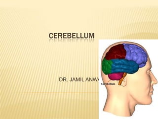
- 1. CEREBELLUM DR. JAMIL ANWAR
- 2. The cerebellum is situated in the posterior cranial fossa. It is the largest part of the hindbrain and lies posterior to the fourth ventricle, the pons, and the medulla oblongata.
- 3. CEREBELLUM The cerebellum is located behind the dorsal aspect of the pons and the medulla. A midline portion, the vermis, separates two lateral lobes, or cerebellar hemispheres. The cerebellum consists of the cerebellar cortex and the underlying cerebellar white matter Four paired deep cerebellar nuclei are located within the white matter of the cerebellum
- 4. The cerebellum is divided into three main lobes: the anterior lobe the middle(posterior) lobe the flocculonodular lobe. A deep horizontal fissure that is found along the margin of the cerebellum separates the superior from the inferior surfaces; it is of no morphologic or functional significance
- 7. Structure of the Cerebellum The cerebellum is composed of an outer covering of gray matter called the cortex and inner white matter.
- 8. A section made through the cerebellum parallel with the median plane divides the folia at right angles, and the cut surface has a branched appearance, called the arbor vitae.
- 9. The gray matter of the cortex throughout its extent may be divided into three layers: (1) an external layer, the molecular layer (2) a middle layer, the Purkinje cell layer (3) an internal layer, the granular layer .
- 11. Functional Areas of the Cerebellar Cortex Clinically it is possible to divide up the cerebellar cortex into three functional areas. 1.The cortex of the vermis influences the movements of the long axis of the body, namely, the neck, the shoulders, the thorax, the abdomen, and the hips. 2.Immediately lateral to the vermis is a so-called intermediate zone of the cerebellar hemisphere. This area has been shown to control the muscles of the distal parts of the limbs, especially the hands and feet. 3.The lateral zone of each cerebellar hemisphere appears to be concerned with the planning of sequential movements of the entire body and is involved with the conscious assessment of movement errors.
- 12. Intracerebellar Nuclei Four masses of gray matter are embedded in the white matter of the cerebellum on each side of the midline. From lateral to medial, these nuclei are the dentate, the emboliform, the globose, and the fastigial.
- 13. White Matter There is a small amount of white matter in the vermis; it closely resembles the trunk and branches of a tree and thus is termed the arbor vitae . There is a large amount of white matter in each cerebellar hemisphere. The white matter is made up of three groups of fibers: (1) intrinsic, (2) afferent, (3) efferent.
- 14. Cerebellar Peduncles The cerebellum is linked to other parts of the central nervous system by numerous efferent and afferent fibers that are grouped together on each side into three large bundles, or peduncles. The superior cerebellar peduncles connect the cerebellum to the midbrain, the middle cerebellar peduncles connect the cerebellum to the pons, and the inferior cerebellar peduncles connect the cerebellum to the medulla oblongata.
- 17. CEREBELLAR FUNCTIONS The cerebellum has several main functions: coordinating skilled voluntary movements by influencing muscle activity, controlling equilibrium and muscle tone through connections with the vestibular system and the spinal cord. There is a somatotopic organization of body parts within the cerebellar cortex. In addition, the cerebellum receives collateral input from the sensory and special sensory systems.
- 18. Each cerebellar hemisphere is connected by nervous pathways principally with the same side of the body; thus, a lesion in one cerebellar hemisphere gives rise to signs and symptoms that are limited to the same side of the body.
- 19. Diagnosis of Cerebellar disorders The main clinical features of cerebellar disorders include incoordination, imbalance, and troubles with stabilizing eye movements. There are two distinguishable cerebellar syndromes – 1. midline and 2.hemispheric. 1.Midline cerebellar syndromes are characterized by imbalance. Persons are unsteady, they are unable to stand in with eyes open or closed, and are unable to well perform tandem gait. trunkal ataxia . "titubation" or a bobbing motion of the head or trunk. also often affect eye movements. There may be nystagmus, ocular dysmetria.
- 20. Hemispheric cerebellar syndromes are characterized by incoordination of the limbs. There may be decomposition of movement, dysmetria, and rebound. Dysdiadochokinesis is the irregular performance of rapid alternating movements. Intention tremors may be present on an attempt to touch an object. A kinetic tremor may be present in motion. The finger-to-nose and heel-to-knee tests are classic tests of hemispheric cerebellar dysfunction.
- 21. THANKS