The Respiratory system
•Transferir como PPT, PDF•
51 gostaram•29,263 visualizações

i know it is help full for all the nursing student....to increasing the knowledge about respiratory system.

Recomendados
Mais conteúdo relacionado
Mais procurados
Mais procurados (20)
Semelhante a The Respiratory system
Semelhante a The Respiratory system (20)
Mais de MR. JAGDISH SAMBAD
Mais de MR. JAGDISH SAMBAD (20)
Último
Último (20)
The Respiratory system
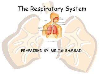
- 1. The Respiratory System PREPAIRED BY: MR.J.G SAMBAD
- 6. Divisions of the Respiratory System Upper respiratory tract (outside thorax) Nose Nasal Cavity Sinuses Pharynx Larynx
- 7. Divisions of the Respiratory System Lower respiratory tract (within thorax) Trachea Bronchial Tree Lungs
- 8. Structures of the Upper Respiratory Tract Nose - warms and moistens air Palantine bone separates nasal cavity from mouth. • Cleft palate - Palantine bone does not form correctly, difficulty in swallowing and speaking. Septum - separates right and left nostrils • rich blood supply = nose bleeds. Sinuses - 4 air containing spaces – open or drain into nose - (lowers weight of skull).
- 14. Structures of the Lower Respiratory Tract • Larynx - voice box – Root of tongue to upper end of trachea. – Made of cartilage – 2 pairs of folds • Vestibular - false vocal cords • True vocal cords
- 15. Structures of the Lower Respiratory Tract larynx cont… • Thyroid cartilage - adam’s apple - larger in males due to testosterone. • Epiglottis - flap of skin (hatch) on trachea, moves when swallowing and speaking. – closes off trachea when swallowing food
- 16. Structures of the Lower Respiratory Tract • Trachea (windpipe) – Larynx to bronchi – Consists of smooth cartilage and C shaped rings of cartilage. – Tracheostomy - cutting of an opening in trachea to allow breathing.
- 18. TRACHEA • Structures associated with the trachea: Superiorly : the larynx. Inferiorly :the right & left bronchi. Anteriorly :Isthmus of Thyroid gland & the arch of the aorta & the sternum. Posteriorly :the esophagus separates the trachea from the vertebral column. Laterally :the lungs • Structures associated with the trachea: Superiorly : the larynx. Inferiorly :the right & left bronchi. Anteriorly :Isthmus of Thyroid gland & the arch of the aorta & the sternum. Posteriorly :the esophagus separates the trachea from the vertebral column. Laterally :the lungs
- 19. STRUCTURE OF THE TRACHEA • It is composed of 3 layers of tissues & held by between 16-20 incomplete rings of hyaline cartilage. • Rings are incomplete posteriorly. • Connective tissues & involuntary muscle join the cartilages & form the posterior wall which is in contact with the esophagus. • It is composed of 3 layers of tissues & held by between 16-20 incomplete rings of hyaline cartilage. • Rings are incomplete posteriorly. • Connective tissues & involuntary muscle join the cartilages & form the posterior wall which is in contact with the esophagus.
- 20. Con.. • Three layers of tissue: I. The outer layer: consists of fibrous & elastic tissue & encloses the cartilages. II.The middle layer :consists of bands of smooth muscles in a helical manner. There is some areolar tissue. III.The inner lining :consists of ciliated columnar epithelium, containing mucus secreting Goblet cells. • Three layers of tissue: I. The outer layer: consists of fibrous & elastic tissue & encloses the cartilages. II.The middle layer :consists of bands of smooth muscles in a helical manner. There is some areolar tissue. III.The inner lining :consists of ciliated columnar epithelium, containing mucus secreting Goblet cells.
- 21. Structures of the Lower Respiratory Tract • Bronchi – Tubes that branch off trachea and enter into lungs – Ciliated – Branches: Primary bronchi —secondary bronchi— tertiary bronchi— bronchioles – Bronchioles branch into microscopic alveolar ducts. Terminate into alveolar sacs – Gas exchange with blood occurs in sacs.
- 22. Structures of the Lower Respiratory Tract
- 23. Structures of the Lower Respiratory Tract • Lungs – Extend from diaphragm to clavicles – Divided into lobes by fissures. – Visceral pleura adheres to the lungs. • Pleurisy = inflammation of the pleural lining
- 29. Inside the lungs.. • Bronchi & bronchioles: Two primary bronchi are formed when trachea divides at the level of 5th thoracic vertebra. The right bronchus: - wider, shorter & more vertical than the left one. - Approx 2.5 cm The left bronchus: - About 5cm long - Narrower.
- 30. BRONCHIAL TREE The bronchi divide as follows: Bronchi bronchioles terminal bronchioles respiratory bronchioles alveolar ducts alveoli. The bronchi divide as follows: Bronchi bronchioles terminal bronchioles respiratory bronchioles alveolar ducts alveoli. s
- 32. BLOOD SUPPLY The pulmonary trunk divided into the right and left pulmonary arteries, carrying deoxygenated blood to each lung. Within the lungs each pulmonary artery divides into many branches, which eventually end in dense capillary network around the alveoli. The walls of merge into a network of pulmonary venules.which in turn from two pulmonary veins carrying oxygenated blood from two pulmonary veins carrying oxygenated blood from each lung back to the left atrium of the heart.
- 33. Respiratory Physiology • Pulmonary Ventilation = breathing – Mechanism • Movement of gases through a pressure gradient - hi to low. • When atmospheric pressure (760 mmHg) is greater than lung pressure ---- air flows in = inspiration. • When lung pressure is greater than atmospheric pressure ---- air flows out = expiration.
- 34. Respiratory Physiology • Pressure gradients are established by changes in thoracic cavity. – increase size in thorax = a decrease in pressure --- air moves in. – Decrease size in thorax = increase in pressure --- air moves out.
- 36. Inspiration -contraction of diaphragm and intercostal muscles
- 37. Expiration • relaxation of diaphragm and intercostal muscles
- 38. Volumes of Air Exchange • Tidal volume - amount of air exhaled normally after a typical inspiration. Normal - about 500 ml • Expiratory Reserve volume - additional amount of air forcibly expired after tidal expiration (1000 - 1200 ml). • Inspiratory Reserve volume - (deep breath) amount of air that can be forcibly inhaled over and above normal. • Residual volume - amount of air that stays trapped in the alveoli (about 1.2 liters).
- 39. Volumes of Air Exchange • Vital capacity - the largest volume of air an individual can move in and out of the lungs. • Vital capacity = sum of IRV+TV+ERV • Depends of many factors • size of thoracic cavity • posture • volume of blood in lungs congestive heart failure, emphysema, disease, etc…
- 40. Volumes of Air Exchange • Eupnea - normal quiet breathing, 12-17 breaths per minute. • Hyperpnea - increase in breathing to meet an increased demand by body for oxygen. • Hyperventilation - increase in pulmonary ventilation in excess of the need for oxygen. – Someone hysterical Breathe into – exertion paper bag. • Hypoventilation - decrease in pulmonary ventilation. • Apnea - temporary cessation of breathing at the end of normal expiration.
- 41. Heimlich Maneuver • Lifesaving technique that is used to open a windpipe that is suddenly obstructed. • Air already in lungs used to expel object.
- 42. Heimlich Maneuver • Technique - Conscious victim – Ask the victim if he/she can talk – Stand behind victim and wrap your arms around their waist. – Make a fist with one hand and grasp it with the other hand. – Place thumb side of fist below xiphoid process and above navel. – Thrust your fist in and upward - about 4 times. • DO NOT press on ribs or sternum
- 43. Heimlich Maneuver – Technique - Unconscious victim • Catch victim if they begin to fall - place on floor face up. • Straddle hips • Place one hand on top of other on the victims abdomen - above navel and below xiphoid process. • Forceful upward thrusts with heel of hand - several times if necessary.
- 44. Alveoli • Structure of alveoli – Alveolar duct – Alveolar sac – Alveolus • Gas exchange takes place within the alveoli in the respiratory membrane • Squamous epithelial lining alveolar walls • Covered with pulmonary capillaries on external surfaces
- 45. Respiratory Membrane (Air- Blood Barrier) Figure 13.6
- 46. Gas Exchange • Gas crosses the respiratory membrane by diffusion – Oxygen enters the blood – Carbon dioxide enters the alveoli • Macrophages add protection • Surfactant coats gas-exposed alveolar surfaces
- 47. Events of Respiration • Pulmonary ventilation – moving air in and out of the lungs • External respiration – gas exchange between pulmonary blood and alveoli
- 48. Events of Respiration • Respiratory gas transport – transport of oxygen and carbon dioxide via the bloodstream • Internal respiration – gas exchange between blood and tissue cells in systemic capillaries
- 49. Mechanics of Breathing (Pulmonary Ventilation) • Mechanical process • Depends on volume changes in the thoracic cavity • Volume changes lead to pressure changes, which lead to equalize pressure of flow of gases • 2 phases – Inspiration – flow of air into lung – Expiration – air leaving lung
- 50. Inspiration • Diaphragm and intercostal muscles contract • The size of the thoracic cavity increases • External air is pulled into the lungs due to an increase in intrapulmonary volume
- 51. Expiration • Passive process dependent up on natural lung elasticity • As muscles relax, air is pushed out of the lungs • Forced expiration can occur mostly by contracting internal intercostal muscles to depress the rib cage
- 53. Pressure Differences in the Thoracic Cavity • Normal pressure within the pleural space is always negative (intrapleural pressure) • Differences in lung and pleural space pressures keep lungs from collapsing
- 54. Nonrespiratory Air Movements • Caused by reflexes or voluntary actions • Examples – Cough and sneeze – clears lungs of debris – Laughing – Crying – Yawn – Hiccup
- 55. Respiratory Sounds • Sounds are monitored with a stethoscope • Bronchial sounds – produced by air rushing through trachea and bronchi • Vesicular breathing sounds – soft sounds of air filling alveoli
- 56. External Respiration • Oxygen movement into the blood – The alveoli always has more oxygen than the blood – Oxygen moves by diffusion towards the area of lower concentration – Pulmonary capillary blood gains oxygen
- 57. External Respiration • Carbon dioxide movement out of the blood – Blood returning from tissues has higher concentrations of carbon dioxide than air in the alveoli – Pulmonary capillary blood gives up carbon dioxide • Blood leaving the lungs is oxygen-rich and carbon dioxide-poor
- 58. Gas Transport in the Blood • Oxygen transport in the blood – Inside red blood cells attached to hemoglobin (oxyhemoglobin [HbO2]) – A small amount is carried dissolved in the plasma • Carbon dioxide transport in the blood – Most is transported in the plasma as bicarbonate ion (HCO3–) – A small amount is carried inside red blood cells on hemoglobin, but at different binding sites than those of oxygen
- 59. Internal Respiration • Exchange of gases between blood and body cells • An opposite reaction to what occurs in the lungs – Carbon dioxide diffuses out of tissue to blood – Oxygen diffuses from blood into tissue
- 61. Neural Regulation of Respiration • Activity of respiratory muscles is transmitted to the brain by the phrenic and intercostal nerves • Neural centers that control rate & depth are located in the medulla • The pons appears to smooth out respiratory rate • Normal respiratory rate (eupnea) is 12–15 min. • Hypernia is increased respiratory rate often due to extra oxygen needs
- 62. Factors Influencing Respiratory Rate and Depth • Physical factors – Increased body temperature – Exercise – Talking – Coughing • Volition (conscious control) • Emotional factors
- 63. Factors Influencing Respiratory Rate and Depth • Chemical factors – Carbon dioxide levels • Level of carbon dioxide in the blood is the main regulatory chemical for respiration • Increased carbon dioxide increases respiration • Changes in carbon dioxide act directly on the medulla oblongata
- 64. Factors Influencing Respiratory Rate and Depth • Chemical factors (continued) – Oxygen levels • Changes in oxygen concentration in the blood are detected by chemoreceptors in the aorta and carotid artery • Information is sent to the medulla oblongata
- 65. Respiratory Rate Changes Throughout Life Respiration rate: • Newborns – 40 to 80 min. • Infants – 30 min. • Age 5 – 25 min. • Adults – 12 to 18 min • Rate often increases with old age