Electrolyte
•Transferir como PPTX, PDF•
69 gostaram•47,218 visualizações

it is very essential role in management of body homeostasis mechanism and acid base balance.

Recomendados
Mais conteúdo relacionado
Mais procurados
Mais procurados (20)
Semelhante a Electrolyte
Semelhante a Electrolyte (20)
Mais de MR. JAGDISH SAMBAD
Mais de MR. JAGDISH SAMBAD (20)
Último
Último (20)
Electrolyte
- 2. Objectives What is electrolyte? Which are imp electrolytes in our body? What are the functions of particular electrolyte? Regulation in body Sample collection Method of estimation Errors which can affect the result Pathological alteration
- 3. What are they?? Electrolytes are minerals found in body fluids that carry an electrical charge and are essential to keeping the heart, nerves and muscles functioning properly. As such, it is important to maintain a precise and constant balance of electrolytes. CATION - positively charged electrolyte ANION - negatively charged electrolyte Cations = Anions for homeostatsis to exist in each fluid compartment. Commonly measured in mEq/L.
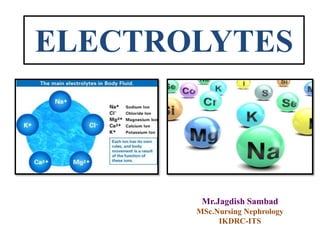
- 4. Which are they?? Cations Anions Sodium Potassium Calcium Magnesium Cations Anions Sodium (Na+) Chloride(Cl-) Potassium (K+) Biocarbonate (HCO-) Calcium (Ca++) Phosphorus (PO4 -) Magnesium (Mg+)
- 6. • Most prevalent cation in the ECF. • Total body Sodium is about 5000 mEq in an adult. • Daily requirement of sodium is about 100 mEq. • In normal individuals, the kidney strives to achieve Na+ balance – that is, to have Na+ excretion equal to Na+ ingestion. A. Sodium (Na+) Introduction
- 7. Maintain balance of extracellular fluid, thereby it controls the movements of the water between fluid compartments. Transmission of nerve impulses Neuro muscular and myocardial impulse transmission A. Sodium (Na+) FUNCTIONS
- 8. A. Hormones increasing sodium reabsorption: 1. Renin: Released from the juxtaglomerular apparatus of the kidney Release is stimulated by: raised sympathetic tone, falling plasma volume, and certain prostaglandins, such as PGE2 No direct effects promoting Na+ retention, it controls the renin- angiotensin-aldosterone axis 2. Angiotensin II: Levels rise as result of renin release In turn, it stimulates the release of aldosterone Also increases tone in the efferent glomerular arteriole. The net effect is to enhance Na+ reabsorption from the proximal tubule. A. Sodium (Na+) REGULATION
- 9. 3. Aldosterone: Steroid hormone released from the adrenal cortex End product of the RAAS system Acts on the distal tubule and collecting duct to increase Na+ and water reabsorption (proportionately more Na+ than water) 4. Arginine vasopressin (AVP), anti-diuretic hormone (ADH): Neuron cell bodies in supra-optic and paraventricular nuclei of the Hypothalamus, Stored in posterior pituitary. Passive absorption of water from the collecting ducts along with a small degree of Na+ re-absorption, concentrating the urine A. Sodium (Na+) REGULATION
- 10. RAAS SYSTEM A. Sodium (Na+) RAAS SYSTEM
- 11. B.Hormones increasing sodium excretion: 1.Atrial Natriuretic Peptide (ANP): A small peptide produced from the atrial wall as a result of atrial stretching. Increase Na (and hence water) excretion by increasing GFR and blocking Na reabsorption in PCT 2.Brain Natriuretic Peptide (ANP): secreted by the hypothalamus, termed brain natriuretic peptides (BNP) Have a similar roles. A. Sodium (Na+) REGULATION
- 12. Serum: venous blood sample in gel vaccutainer Urine: 24 hrs urine collection No preservative is required Store at 2-8 degree Sample collection Method of estimation Ions selective electrode A. Sodium (Na+)
- 14. Potentiometric measurements: Potentio-metry is to determine the difference in potential between a working (an indicator) electrode and a counter (a reference) electrode. - Cathode is the working / indicator electrode. - Anode is the counter / reference electrode. Indicator Electrode: Electrode that responds to analyte and donates / accepts electrons Reference Electrode: Second ½ cell at a constant potential ell voltage is difference between the indicator and reference electrode Ecell = Ec ─ Ea Where : Ec is the reduction potential at the cathode. : Ea is the reduction potential at the anode.
- 16. Serum: 135 – 145 mEq/L Urine: 24 hr urine sample: 40-220 mEq/day Note: For random, it is better to do FeNa . Also used in calculation of Anion gap & Osmolality Biological reference range Panic value Serum level >160 and <120 mEq/L A. Sodium (Na+)
- 17. Errors affecting Na+ result A. Pseudohyponatraemia : 1. Sample collection from IV site, thus the sample is diluted by the hypotonic fluid (5% dextrose).- confirmed by dilution effect on other parameters 2. High plasma glucose level: increase 100 mg/dl – lowering Na 1.6 mmol/L, after 400 mg/dl, every 100 mg/dl lowering 2.4 mmol/L Corrected Sodium = [0.016 x (serum glucose – 100)] + serum Na 3. Increased viscosity due to the Hyperproteinemia, Hyperlipidemia due subsequent decreased watery portion of plasma can thus cause false low sodium concentrations A. Sodium (Na+)
- 18. B. Pseudohypernatraemia : 1. Sample collection from IV site - confirmed by measurements of CL- and K+ 2. Drugs – SSRI, sodium valproate etc. Errors affecting Na+ result A. Sodium (Na+)
- 19. S. Sodium level < 135 mEq/L < 280 mOsm/Kg > 280 mOsm/Kg (N or increase) True Hyponatremia Pseudohyponatremia Hypo- volemic Eu- volemic Hyper- volemic Cause Investigation Paraproteinemia s. protein, electrophoresis Hyperlipidemia S. Cholestrol,TG Hyperglycemia S. Glucose A. HyponatremiaA. Sodium (Na+)
- 20. < 20 mEq/L Extrarenal causes vomiting Diarrhea Third spacing of fluid Burns Decrease total body water Decrease total body sodium (more deficit) Measure urinary sodium >20 mEq/L Renal causes Diuretics Salt losing nephropathy Mineralocorticoids Def. Osmotic diuresis Note : In vomiting, to compensate metabolic alkalosis urinary Na > 20 mEq/L, and urinary cl- < 10 mmol/L A. HyponatremiaA. Sodium (Na+) A. Hypovolemic hyponatremia
- 21. Increase total body water No change in total body sodium Urinary sodium > 20 mEq/L Urine osmolality>100 moSm/Kg Causes Glucocorticoids deficiency SIADH Hypothyroidism Drugs A. Sodium (Na+) B. Euvolemic hyponatremia A. Hyponatremia
- 22. Increase total body water (More) Increase total body sodium Measure urinary sodium < 20 mEq/L Other causes Cirrhosis Cardiac failure >20 mEq/L Renal causes AKI CKD A. Sodium (Na+) A. Hyponatremia C. Hypervolemic hyponatremia
- 23. A. Sodium (Na+) B. Hypernatremia S. Sodium level > 145 mEq/L
- 24. Hypo-volemic Eu-volemic Hyper-volemic Hypo- natremia Na - Na - N Na - H2O - H2O- H2O - Hyper- natremia Na - Na – N Na - H2O - H2O H2O - Sumarry of Natremic disorders
- 26. • Major intracellular cation. • Total body K+ content in a normal adult 3000- 4000mEq. • 98% Intracellular , 2% in ECF B. Potassium (K+) Introduction
- 27. Regulates neuromuscular excitability and muscle contraction As coenzyme - glycogen & protein synthesis Correction of acid base imbalances FUNCTIONS B. Potassium (K+)
- 28. The primary mechanism maintaining this balance : the Na-K – ATP phase pump Na-K ATPhase pump actively transports Na+ out of the cell and K+ into the cell in a 3:2 ratio Renal excretion – Major route of excess K+ Approx 90% of K+ excretion occurs in the urine less than 10% excreted through sweat or stool. Regulation B. Potassium (K+)
- 30. Serum: venous blood sample in gel vaccutainer Urine: 24 hrs urine collection No preservative is required Store at 2-8 degree Sample collection Method of estimation Ions Selective Electrode B. Potassium (K+)
- 31. Serum: 3.5 – 5.5 mEq/L Urine: • 24 hr urine sample: 25-125 mEq/day • Random urine: 13-101 mmol/g creat Note: Also used in calculation of anion gap Biological reference range Panic value Serum level <2.0 and >7.0 mEq/L B. Potassium (K+)
- 32. Errors affecting K+ result A. Pseudohyperkalemia : 1. Cleaning of puncture site: The site for venipuncture is to be cleaned with povidone iodine. 2. Tourniquet application: • It should be released once the first blood flow was established. • More than 1 min of tourniquet : haemo-concentration 3. Clenching fist for longer time causes elevation potassium. B. Potassium (K+)
- 33. B. Potassium (K+) 4. Needle or syringe: Size of syringe / pushing of blood in vaccutainer. 5. Order of drawing tubes: The blood collected in a specific order avoids cross contamination of additives. Examples of the common carryover problems observed are the EDTA which is easily determined by marked hyperkalemia, hypocalcemia, hypomagnesemia and hypozincanemia. 6. Centrifugation and vigorous shaking of sample.
- 34. 1. Sterile-blood culture 2. Coagulation (light blue)/ Citrate 3. Non-additive (red top)/ Plain 4. Gel separator tube (red or gold)/ Plain 5. Heparin tube (green top) 6. EDTA (lavender/purple top) 7. fluoride tube (grey top) 8. All other tubes Order of blood collection by CLSI
- 35. 7. Storage : store after separation of serum 8. Lymphocytosis and thrombocytosis: Hyper cellularity 9. Fluid containing potassium B. Potassium (K+)
- 36. B. Pseudohypokalemia : 1. Sample collection from IV site, thus the sample is diluted by the hypotonic fluid (5% dextrose).- confirmed by dilutional effect on other parameters 2. High plasma glucose level: Dilution effect 3. Increased viscosity Errors affecting K+ result B. Potassium (K+)
- 37. A. hypokalemiaB. Potassium (K+) S. Potassium level < 3.5 mEq/L
- 38. 1. Decreased intake: Food is the main source of potassium A. hypokalemiaB. Potassium (K+)
- 39. 2. Excessive loss : a. GIT loss: • Persistent vomiting • Diarrhea • Gastric suction and Fistula b. Hypovolemia: Increased aldosterone Hypo-K+ A. hypokalemiaB. Potassium (K+)
- 40. c. Renal loss: I. Hyperaldosteronism: Primary hyperaldosteronism : Adrenal tumors Secondary hyperaldosteronism: Reduced effective arterial volume in CCF Liver cirrhosis Nephrotic syndrome A. HypokalemiaB. Potassium (K+)
- 41. A. hypokalemiaB. Potassium (K+) II. Diuretics : Thiazines, Furosemide
- 42. A. hypokalemiaB. Potassium (K+) III. Renal Tubular Acidosis type-1/2: Type 1: dRTA : Decreased H+ Excretion in DCT Type 2: pRTA: Decreased reabsorption of HCO3 -
- 43. 3. Shifting of K+ from ECF to ICF A. hypokalemiaB. Potassium (K+) a. In alkalosis, H+ moves out of the cells, at the same time, K+ moves into the cells. For each 0.1 unit increase of pH in ECF, the [K+] of serum decreases 0.7 mmol/L. b. Insulin: stimulates Na+-K+ ATPase. c. Hyperthyroidism: Over-dose thyroxin stimulates Na+-K+ ATPase.
- 44. Serum potassium level of >5.5 mEq/L • Common Causes: 1. Intra- to extracellular shift: Acidosis – Uptake of H+, efflux of K+ Hyperosmolality- Hypertonic dextrose, mannitol, - Solvent Drag effect β2-Adrenergic antagonists (non-cardioselective agents): Suppresses catecholamine stimulated renin release- in turn aldosterone synthesis Digoxin and related glycosides : Inhibits Na/K ATPase Succinylcholine: Depolarizes Muscle cells, Efflux of K Rapid tumor lysis / Rhabdomyolysis B. HyperkalemiaB. Potassium (K+)
- 45. 2. Inhibition of the renin-angiotensin-aldosterone axis: (↑ risk of hyperkalemia when these drugs are used in combination) Angiotensin-converting enzyme (ACE) inhibitors Renin inhibitors; aliskiren Angiotensin receptor blockers (ARBs) Blockade of the mineralocorticoid receptor: spironolactone Blockade of the epithelial sodium channel (ENaC): amiloride, triamterene B. HyperkalemiaB. Potassium (K+)
- 46. 4. Renal resistance to mineralocorticoids: Tubulointerstitial diseases: SLE, amyloidosis, sickle cell anemia, obstructive uropathy, post– acute tubular necrosis Hereditary: pseudohypoaldosteronism type I; defects in the mineralocorticoid receptor or the epithelial sodium channel (ENaC) 5. Advanced renal insufficiency: Chronic kidney disease End-stage renal disease Acute oliguric kidney injury B. HyperkalemiaB. Potassium (K+)
- 48. Most abundant mineral in human beings. Total calcium in an average adult is about 1,000 gm : 99% in bones & teeth. Rest in various tissues and body fluids. Calcium is present in bones mainly in the form of calcium phosphate. C. Calcium (Ca++) Introduction
- 49. About 50% of this is bound to protein (protein-bound or non-diffusible calcium). About 5% with organic anions e.g. citrate, (diffusible) The remaining 45%: free ionized calcium (freely diffusible) Ionized calcium: Active form C. Calcium (Ca++)
- 50. Functions Formation of bones and teeth Excitability and conductivity of nerves Neuromuscular transmission Excitability and contractility of myocardium Coagulation of blood Action of hormones C. Calcium
- 51. Calcium regulating factors Vitamin D PTH Phosphorus level EMB-RCG C. Calcium
- 52. Cholecalciferol Hydroxylase 25-Hydroxycholecalciferol Parathormone Parathyroid glands 1, 25-Dihydroxycholecalciferol Induction Calcium-binding protein Ca -dependent ATPase Alkaline phosphatase ++ Calcium absorption Release into circulation Plasma calcium LIVER KIDNEY INTESTINAL MUCOSA Hydroxylase INTESTINAL MUCOSA – C. Calcium
- 53. Serum: venous blood sample in gel vaccutainer Urine: 24 hrs urine collection No preservative is required Store at 2-8 degree Sample collection Method of estimation C. Calcium 1. T. Calcium: Arsenzo- III Spectrophotometric method 2. Ionized Calcium: ISE Ca+ + + Arsenazo III Ca- Arsenazo III complex (purple)
- 54. T. Serum: 8.5-10.5 mg/dL Ionized Calcium: 0.9 – 1.3 mmol/L Urine: 24 hr urine sample: 100-250 mg/day Biological reference range C. Calcium
- 55. Free calcium is useful index - provides the better indication of calcium biologically active and tightly regulated by calcium binding hormones. Calcium is predominantly transported bound to serum proteins, total calcium levels are greatly influenced by protein concentration especially albumin. Earlier no proper technique to measure. This problem is now solved due to availability of ion-selective electrodes, for free calcium measurements . What is better Ionized or Total ???
- 56. Corrected calcium : Total calcium + 0.8 (4.0 – S. Alb) For this patient = 8.1 + 0.8 (4 – 3.2) = 8.1 + 0.64=8.75 Corrected total calcium ? ?
- 57. A. Pseudohypercalcemia: 1. Cleaning the venipuncture site: 2. Tourniquet application: 3. Clenching fist 4. Needle or syringe: 5. Order of drawing tubes: 6. Centrifugation and vigorous shaking of sample Errors affecting Ca++ level C. Calcium
- 58. 7. Storage: 8. Sample type: 9. Lymphocytosis and thrombocytosis Errors affecting Ca++ level C. Calcium B. Pseudohypocalcemia: 1. Sample collection from IV site 2. High plasma glucose level 3. Increased viscosity
- 59. Hyperparathyroidism Hypervitaminosis D Bone cancer Multiple myeloma Leukaemia Polycythaemia Milk-alkali syndrome Sarcoidosis Idiopathic infantile hypercalcaemia A. Hypercalcemia C. Calcium B. Hypocalcemia Hypoparathyroidism Rickets Osteomalacia Chronic renal failure Nephrotic syndrome
- 61. The total amount of chlorine in an average adult is about 80 gm Chlorine, in the form of chloride ions, is the chief anion of extracellular compartment Normal serum chloride level is 98-107 mEq/L (355-375 mg/dl) D. Chloride
- 62. The chloride content of cerebrospinal fluid is 120 to 130 mEq/L The interstitial fluid contains about 110 mEq/L The intracellular fluid contains only about 4 mEq/L D. Chloride
- 63. Functions Maintenance of osmotic pressure Maintenance of pH Formation of hydrochloric acid D. Chloride
- 64. Serum: venous blood sample in gel vaccutainer Urine: 24 hrs urine collection No preservative is required Store at 2-8 degree Sample collection D. Chloride Method of estimation By Ion Selective Electrode
- 65. Abnormal serum chloride levels Changes in serum chloride level are parallel to those in serum sodium level. Serum chloride level is raised (hyperchloraemia) in dehydration, respiratory alkalosis, metabolic acidosis and adrenocortical hyperactivity. Serum chloride level is decreased (hypochloraemia) in severe vomiting, prolonged gastric suction, respiratory acidosis, metabolic alkalosis and Addison’s disease. D. Chloride
- 67. Classification of Diuretics (According to the site of action) DRUGS USED IN RENAL DISORDERS Drugs that modify Drugs that modify salt excretion water excretion PCT TAL DCT CCT Osmotic diuretics ADH ADH agonist antagonist K+-sparing diuretics Thiazides Loop diuretics Carbonic anhydrase inhibitors
- 69. 1. PROXIMAL CONVULATED TUBULE 1. Direct entry of Na+ 2. Na+/ K+ pump @ BL membrane : Transfer of Na+ and K+ along with glucose, amino acids and other organic anions & PO4 -3 3. Exchange of H+ @ Luminal Membrane: Proximal tubule cells secrete H+ with the help of carbonic anhydrase. 4. Large amount of HCO3 -, amino acid, acetate along with Cl-
- 71. Diuretic acting on PCT 1. Carbonic anhydrase inhibitors: Acetazolamide Mechanism : Inhibit CAH enzyme in tubular cell & lumen Effects on serum electrolytes: • Hypo Na+ • Low HCO3 - Metabolic acidosis
- 72. Loop of Henle
- 73. 2a. LOOP OF HENLE (DESCEDING LOOP OF HENLE) • The filtrate entering: isotonic • Water absorption thereafter • Tubular fluid becomes concentrated (Hypertonic) (Three fold increase in salt concentration). 2b. LOOP OF HENLE (ASCENDING LOOP OF HENLE) 1. ASCENDING LOOP OF HENLE (Medullary part lined by cuboidal cells). • Unique, Impermeable to water
- 74. • Active reabsorption of Na+, K+ & Cl- is mediated by a Na+/K+/2Cl- Cotransporter. • Mg++ and Ca++ enter the interstitial fluid. • 25% to 30% of tubular NaCl returns to the Fluid (blood). 2. ASCENDING LOOP OF HENLE (Cortical part lined by flattened cells). • Impermeable to water • Salt reabsorption continues through Na+ electrochemical gradient transport
- 76. Diuretic acting on Thick Asc. Loop Henle 1. Na+ K+/ 2CL- symporter inhibitor Furosemide Effects on electrolytes: • Hypo Na + • Hypo K + • Hypo Cl- -Hypochloremic Alkalosis • Hypo Mg ++ • Hypo Ca++ Note: Magnesium and Calcium reabsorption in the thick ascending limb is dependent on the positive lumen voltage gradient set up by potassium recycling through renal outer medullary potassium channel, loop diuretics also inhibit their reabsorption
- 79. 3. DISTAL CONVULATED TUBULE • Impermeable to water. • 10% of NaCl is reabsorbed • Na+/Cl- transporter (inhibited by Thiazide) • Calcium reabsorption: Na+/Ca++ exchanger into the interstitial fluid. • Ca++ excretion: PTH
- 80. Diuretic acting on DCT 1. Na+/ CL- Symporter Thiazide Effects on electrolytes: • Hypo Na + • Hypo Cl- -Hypochloremic Alkalosis • Hyper Ca++ Note: By lowering the sodium concentration within the epithelial cells, thiazides increase the activity of the Na+/Ca2+ anti-porter on the basolateral membrane to transport more Ca2+ into the interstitium. This, in turn, decreases the intracellular Ca2+.
- 83. 4. COLLECTING TUBULE & DUCT • Aquaporins: Water Absorption – ADH dependent • Na+ absorption @ Luminal membrane: aldosterone dependent (inhibited by amiloride) • Na+/K+-ATPase @ BL membrane: aldosterone dependent
- 85. A. PCT 1. Na+: Direct entry Na+/ K+ pump @ BL membrane 2. Glucose, amino acids, PO4 -3 & organic anions : Active transport along with sodium 3. HCO3 -, acetate, Cl-: Passive diffusion B. Loop of Henle: a. Descending loop: 1. Water b. Ascending loop: 1. Active reabsorption of Na+, K+ & Cl- : Na+/K+/2Cl- Cotransporter. 2. Mg++ and Ca++: Gradient 3. Na+ electrochemical gradient transport Summary of sites of renal reabsorption of filtrate
- 86. C. DCT 1. Na+/Cl- transporter (inhibited by Thiazide) 2. Calcium reabsorption: Na+/Ca++ exchanger into the interstitial fluid. D. Collecting Duct 1. Aquaporins: Water Absorption – ADH dependent 2. Na+ absorption @ Luminal membrane: aldosterone dependent (inhibited by amiloride) 3. Na+/ K+-ATPase @ BL membrane: aldosterone dependent