SlideShare uma empresa Scribd logo
1 de 140
MAGNETOM Flash
       The Magazine of MRI


       Issue Number 3/2011
       RSNA Edition


How I do it
Cerebral MR
Spectroscopy at
1.5T and 3T
Page 6


Clinical
Surgical Planning
and Guidance for
Pediatric Epilepsy
Page 18

MRI of the
Lumbosacral Plexus
Page 26

3T MRI of Cartilage
using 3D DESS
Page 33

Simultaneous MR/PET –
Clinical Reality?
Page 74
Editorial


            Matthias Lichy, M.D.
                  Editor-in-Chief


                                    Dear Reader,
                                     The first time of travelling to Chicago to                       Sharing knowledge and showing how MR
                                     attend the annual meeting of the Radiologi-                      imaging technology influences patient
                                     cal Society of North America left me truly                       diagnosis are what MAGNETOM Flash maga-
                                     overwhelmed, not least by the huge number                        zine is all about. This issue is no different.
                                     of people sharing their knowledge and
                                     experience in all fields of medical imaging.                     Here you can read, for example, about how
                                                                                                      the Children’s Hospital in Atlanta deals
                                     There are only a few windows in the                              with imaging for surgical therapy planning of
                                     radiologist’s calendar in which to exchange                      epilepsy in children*; about imaging proto-
                                     ideas with so many colleagues from all over                      cols for imaging of the lumbosacral plexus at
                                     the world. For this reason, the first week                       Johns Hopkins Hospital; and about the clini-
                                     of December has now become a significant                         cal reality of simultaneous MR/PET at the
                                     week in all our calendars.                                       University Hospital of Tübingen. Of course,
                                                                                                      these are just three of many exciting and
                                     However, even more than this, the RSNA is                        informative articles awaiting you on the
                                     the largest platform for learning about the                      following pages. They have inspired me, and
                                     latest developments in imaging technology                        I am sure they will inspire you.
                                     (and this applies for different imaging
                                     modalities) and how this technology influ-                       Enjoy reading the latest issue of Flash!
                                     ences our daily patient care. For several
                                     years now we have distributed a new issue of
                                     the MAGNETOM Flash at the RSNA (we even
                                     call it the “RSNA issue”).                                       Matthias Lichy, M.D.


                                    *MR scanning has not been established as safe for imaging fetuses and infants under two years of age. The responsible physician
                                     must evaluate the benefit of the MRI examination in comparison to other imaging procedures.


2 MAGNETOM Flash · 3/2011 · www.siemens.com/magnetom-world
Editorial


Editorial Board
We appreciate your comments.
Please contact us at magnetomworld.med@siemens.com


Antje Hellwich                     Wellesley Were                     Dr. Sunil Kumar S.L.               Christiane Bernhardt
Associate Editor                   MR Business Development            Senior Manager Applications,       Head Outbound Marketing
                                   Manager Australia and New          Canada                             Erlangen, Germany
                                   Zealand


Milind Dhamankar, M.D.             Michelle Kessler                   Gary R. McNeal, MS (BME)           Peter Kreisler, PhD
Sr. Director, MR Product           US Installed Base Manager          Advanced Application Specialist,   Collaborations & Applications,
Marketing, Malvern, PA, USA        Malvern, PA, USA                   Cardiovascular MR Imaging          Erlangen, Germany
                                                                      Hoffman Estates, IL, USA


Review Board
Okan Ekinci, MD, Center of Clinical Competence – Cardiology
Jens-Christoph Georgi, PhD, Global Marketing Manager Biograph mMR
Christian Geppert, PhD, Application Development Breast Applications
Wilhelm Horger, Application Development Oncology
Jürgen Kampmeier, PhD, MR/PET
Berthold Kiefer, PhD, Oncological and Interventional Applications
Lars Lauer, PhD, Orthopedic Applications
Heiko Meyer, PhD, Neuro Applications
Edgar Müller, Cardiovascular Applications
Silke Quick, Global Marketing Manager Women’s Health
Ignacio Vallines, PhD, Global Marketing Manager Neurology and Orthopedics
Heike Weh, Clinical Data Manager
Lisa Chuah, PhD, Collaboration Manager MR Asean


                                                                                MAGNETOM Flash · 3/2011 · www.siemens.com/magnetom-world 3
Content


    Content                                                       6
                                                                  MR Spectroscopy
                                                                                                   18
                                                                                                   Pediatric Epilepsy


Further clinical information                                                                  Clinical Neurology
                                                                                            6 Case Reports: Cerebral Magnetic
                                                Visit the MAGNETOM
                                                                                              Resonance Spectroscopy at 1.5T
                                                World Internet pages at
                                                                                              and 3T
                                                www.siemens.com/
                                                                                              Marc Agzarian, Angela Walls
                                                magnetom-world
                                                for further clinical                       14 Case Series: Presurgical Planning
                                                information and talks                         with fMRI/DTI
                                                by international                              Jay J. Pillai, Domenico Zacà
                                                experts.
                                                                                           18 Neuroimaging as a Clinical Tool in
                                                                                              Surgical Planning and Guidance for
                                                                                              Pediatric Epilepsy
                                                                                              Binjian Sun, et al.


                                                                                              Clinical
                                                                                              Orthopedic Imaging
                                                                                           26 High-Resolution 3T MR Neurography
                                                                                              of the Lumbosacral Plexus
                                                                                              Avneesh Chhabra, et al.

The information presented in MAGNETOM Flash is for illustration only and is not            33 3T MR Imaging of Cartilage using 3D
intended to be relied upon by the reader for instruction as to the practice of medicine.      Dual Echo Steady State (DESS)
Any health care practitioner reading this information is reminded that they must use
their own learning, training and expertise in dealing with their individual patients.
                                                                                              Rashmi S. Thakkar, et al.
This material does not substitute for that duty and is not intended by Siemens Medical
Solutions to be used for any purpose in that regard. The treating physician bears the
                                                                                           37 The Importance of MRI of the Wrist in
sole responsibility for the diagnosis and treatment of patients, including drugs and
doses prescribed in connection with such use. The Operating Instructions must always          Patients with Rheumatoid Arthritis
be strictly followed when operating the MR System. The source for the technical data          Filippo Del Grande, et al.
is the corresponding data sheets.

MR scanning has not been established as safe for imaging fetuses and infants under
two years of age. The responsible physician must evaluate the benefit of the MRI
examination in comparison to other imaging procedures.


4 MAGNETOM Flash · 3/2011 · www.siemens.com/magnetom-world
Content


60                                           74                                                   84
Multi-Station Examinations                   Simultaneous MR/PET                                  Abdominal SWI


       How I do it                                Clinical Oncology
    42 Configuration and Use of the MR          74 Simultaneous MR/PET –                104 Assessment of Peripheral Arterial
       Knee Dot Engine                            Clinical Reality?                        Disease Using High Resolution
       Kai Reiter, Sebastian Auer                 Nina Schwenzer, et al.                   Dynamic MR Angiography and 4D
                                                                                           Visualization Software
    50 Simplifying the Workflow of Long                                                     Edward T. Martin, Gary R. McNeal
       Bone Imaging for Short Bore Systems        Clinical Abdominal
       Russell Grossen                                                                 108 Treadmill Exercise Stress CMR
                                                  Imaging                                  Orlando P. Simonetti, et al.

       Technology                              84 Abdominal Susceptibility-
                                                  Weighted Imaging: A New
    56 From Local Imaging to Understanding        Application for Liver Disease
                                                                                            Business
       the Extent of Diseases. Clinical           Yongming Dai, et al.                 120 Productivity Gains in Neurological
       Need and Technical Advances in                                                      and Oncological Reading
       Multi-Region MRI                        88 Dixon Sequence: Liver MRI
                                                                                           Hannes Lücking, et al.
       Heinz-Peter Schlemmer, et al.              and syngo.via Layouts
                                                  Jean-Paul Abécassis,
                                                  Denis Parienté
       Clinical                                                                             How I do it
       Pediatric Imaging                          Clinical
                                                                                       124 The Networked Scanner Workflow
                                                                                           Anja Fernandez Carmona,
   60 Multi-Station Examinations in
      Pediatric MRI
                                                  Cardiovascular                           Klaus Mayer

      Markus Lentschig                            Imaging
   64 Pediatric Whole-Body MR Imaging          92 CMR Imaging of Profound
      Status Quo and Practical Aspects in         Macrovascular Obstruction
      Daily Routine                               Anurag Sahu, Gary McNeal
      Jürgen Schäfer, et al.
                                              100 MRI of Giant Cell (Temporal)
                                                  Arteritis, GCA
                                                  Thorsten A. Bley, et al.


                                                                     MAGNETOM Flash · 3/2011 · www.siemens.com/magnetom-world 5
Clinical Neurology


Case Reports:
Cerebral Magnetic Resonance
Spectroscopy at 1.5T and 3T
Marc Agzarian, BMBS (Hons), FRANZCR; Angela Walls, M.MedSc, B.App.Sc.Med Rad.

Division of Medical Imaging, Flinders Medical Centre, Bedford Park, South Australia, Australia


Introduction
This is a pictorial review of cerebral
magnetic resonance spectroscopy (MRS)            Case 1:
using a 1.5T MAGNETOM Aera system                Right thalamic glioblastoma multiforme
with software version syngo MR D11 and
a 20-channel head and neck coil and a
                                            1A                                          1B
3T MAGNETOM Trio system with software
version syngo MR B15 and a 12-channel
head matrix coil at Flinders Medical
Centre, South Australia, Australia.
Magnetic resonance spectroscopy refers
to the process of performing a chemical
analysis in-vivo by the Fourier transform
of the magnetic resonance signal from a
voxel of tissue. Hydrogen nuclei (1H pro-
tons) are used for clinical cerebral MRS.
Proton spins in different molecules have
slightly different Larmor frequencies.
These differences are known as the
chemical shift and are expressed in parts
                                                 1A T2w TSE axial.                      1B   T1w SE post-gadolinium axial.
per million (ppm). Chemical shifts are
usually referenced to the Larmor            1C                                          1D
frequency of tetramethylsilane (TMS)
which is given a chemical shift of zero
ppm. All in-vivo molecules of interest
have chemical shifts that increase their
Larmor frequencies relative to TMS and
are thus shown on the spectrogram to
the left of zero.
Cerebral MRS can be acquired using two
techniques, point resolved spectroscopy
sequence (PRESS) and stimulated echo
acquisition mode (STEAM). PRESS has
become the most commonly used tech-          1C Multi-voxel PRESS MRS (TE 135 ms)       1D Multi-voxel PRESS MRS (TE 135 ms) within
nique as it has a higher signal-to-noise     within the lesion in the right thalamus.   normally appearing left thalamus.
ratio than STEAM. Both techniques can


6 MAGNETOM Flash · 3/2011 · www.siemens.com/magnetom-world
Neurology Clinical


be used in either single-voxel or multi-      the essential requirement for MRS is         The key normal resonant peaks in cere-
voxel acquisitions. Single-voxel acquisi-     exceptionally good magnetic field            bral MRS are N-acetyl-aspartate (NAA) at
tions provide a better quality spectrogram    homogeneity.                                 2 ppm, creatine (Cr) at 3 ppm and 3.9
from just one region of interest at a time    Cerebral MRS is usually acquired at one      ppm and choline (Cho) at 3.2 ppm.
whereas multi-voxel acquisitions provide      or more of three different echo times,       Whilst there is some regional variation in
a lesser quality spectrogram from a grid      short (TE 30 – 35 ms), intermediate          the brain, the NAA peak is normally the
of voxels acquired simultaneously.            (TE 135 – 144 ms) and long (TE 270 –         highest in the spectrum.
Multi-voxel MRS can be performed using        288 ms). The short echo time is better
PRESS or STEAM in 2D or 3D with addi-         at resolving the peaks of myoinositol,       Tips for performing
tional phase encoding steps. Multi-voxel      glutamine, glutamate and lipids whilst       cerebral MRS
MRS is also known as chemical shift           the long echo time tends to have less        On older generation MRI scanners,
imaging (CSI) or magnetic resonance           baseline noise. The intermediate echo        achieving high quality MRS was a diffi-
spectroscopic imaging (MRSI). Regardless      time is used to confirm the presence         cult feat. With improved magnetic field
of the technique and acquisition used,        of lactate as the lactate peak inverts.      homogeneities, higher field strengths
                                                                                           and optimised software, performing MRS
                                                                                           is no longer to be feared! At Flinders Med-
                                                                                           ical Centre we routinely perform cerebral
                                                                                           MRS using 2D multi-voxel PRESS CSI at
                                                                                           an intermediate echo time (TE 135 ms).
                                                                                           We have developed the following tips
                                                                                           that have proven to work for a variety of
                                                                                           pathologies and reduce operator depen-
Patient history                                                                            dance:
A 61-year-old female presented with a two week history of dysarthria and left              ■ Modify the volume of interest (VOI)
sided weakness. Initial computed tomography (CT) scan revealed a faintly                      size to best suit the lesion. We often
enhancing lesion within the right thalamus. MRI was performed to further char-                use a 4 x 4 VOI to closely target a
acterize the lesion. The diagnosis of glioblastoma multiforme was confirmed                   single lesion. Be sure to include some
with stereotactic biopsy.                                                                     normal brain inside the VOI to aid in
                                                                                              lesion diagnosis. A smaller VOI results
Sequence details                                                                              in an improved shim and spectrum.
Sagittal T1, axial PD, T2, T2 FLAIR, DWI, T1 and T1 post-gadolinium, coronal T2            ■ Ensure the VOI avoids structures that
and T1 post-gadolinium, 3D T1 MPRAGE post-gadolinium and multi-voxel PRESS                    will contaminate the baseline, such as
MRS (TE 135 ms) were performed on our Siemens 1.5T MAGNETOM Aera scanner.                     skull, scalp, air, cerebrospinal fluid and
                                                                                              major cerebral vessels.
Imaging findings                                                                            ■ Do not be afraid to perform the spec-
A solitary, high T2 signal, intra-axial mass lesion is demonstrated within the                troscopy in the coronal plane, particu-
right thalamus with mild surrounding vasogenic oedema (Fig. 1A). Multi-focal                  larly nearing the vertex.
contrast enhancement is present (Fig. 1B). The MRS at an intermediate echo                 ■ Ensure the VOI is planned according
time (TE 135 ms) within the lesion clearly demonstrates a significant elevation               to an imaging slice. If not, perform
of choline at 3.2 ppm relative to NAA at 2 ppm (Fig. 1C). This is in contrast to the          a T2 localizer in the best plane and
normal MRS obtained from the left thalamus (Fig. 1D).                                         angle for the target lesion. Use ‘copy
                                                                                              image position’ to ensure you have
Discussion                                                                                    anatomical images during post-pro-
Neoplasms display a characteristic MRS pattern, with an elevation of choline                  cessing.
relative to NAA. This is the reverse of the normal situation in which NAA is the           ■ Remember to use spatial saturation
highest peak in the spectrum. Choline is a component of phospholipids and                     bands to cover the skull, scalp and
the increase in choline seen in neoplasms has been postulated to be the result                vessels. We use six saturation bands
of increased cell membrane turnover secondary to rapid cellular proliferation.                positioned in 3D on all sides of the VOI.
A choline: NAA ratio of greater than 2.2 is highly suggestive of a neoplasm.                  We found that if the saturation bands
                                                                                              were positioned too close to the VOI
                                                                                              we would get some baseline distor-
                                                                                              tion. Leaving a buffer of one voxel’s


                                                                          MAGNETOM Flash · 3/2011 · www.siemens.com/magnetom-world 7
Clinical Neurology


    width between the saturation bands             This can be preset in your CSI protocol.      cols’. Here we have preset exactly
    and the VOI improves this significantly.   ■   Always perform MRS at the isocentre           what we wish our spectra to show,
■   Ensure ‘fully excited volume’ is               of the magnet.                                including range, scale and peak
    selected. This option helps to attain      ■   Ensure that your patient is comfort-          information. This significantly reduces
    homogenous excitation of all metabo-           able, as their stillness is imperative.       variability between operators.
    lites within the VOI, whilst suppressing   ■   Post-processing is made easy through
    interference from outside the VOI.             site defined defaults located in ‘proto-


                                                                           2A


Case 2:
Right cerebellar abscess


Patient history
A 62-year-old male presented with a three week history of
headache, atxia and diplopia. Initial CT scan revealed a ring
enhancing lesion within the right cerebellar hemisphere. MRI
was performed to further characterize the lesion. The diagnosis
of a cerebral abscess was confirmed by the presence of pus at
the time of operation and growth of Staphylococcus aureus on
culture.

Sequence details
Sagittal T1 FLAIR, axial PD, T2, T2 FLAIR, DWI, T1 and T1 post-
gadolinium, coronal T2 and T1 post-gadolinium, 3D T1 MPRAGE
post-gadolinium and multi-voxel PRESS MRS (TE 135 ms) were
performed on our Siemens 3T MAGNETOM Trio scanner.
                                                                           2A   T2w TSE axial.
Imaging findings
A solitary, ring enhancing, high T2 signal, intra-axial mass               2B
lesion is demonstrated within the right cerebellar hemisphere
with surrounding vasogenic oedema (Figs. 2A, B). Uniformly
restricted diffusion is present within the lesion, being hyper-
intense on DWI and hypointense on the ADC map (Figs. 2C,
D). The MRS at an intermediate echo time (TE 135 ms) clearly
demonstrates an elevated lipid and lacate peak at 1.3 ppm
(Fig. 2E). Importantly, there is no significant elevation of
choline at 3.2 ppm relative to NAA at 2 ppm.

Discussion
Cerebral abscess characteristically demonstrates a significantly
elevated lipid and lactate peak without an elevation of choline
relative to NAA on MRS. This allows MRS to reliably distinguish
between cerebral abscesses and neoplasms. The uniform
restricted diffusion is also typical of cerebral abscess. Thus,
the combination of MRS and DWI allows the diagnosis of a
cerebral abscess to be made with a high degree of certainty.


                                                                           2B T1w SE post intravenous gadolinium axial.


8 MAGNETOM Flash · 3/2011 · www.siemens.com/magnetom-world
Neurology Clinical


MRS sequence details
The MRS sequence parameters for          voxel size 10 x 10 x 15 mm, VOI 8 x 8, 4     csi_se_270), TR 1700 ms, TE 135 ms or
case 1, performed on our 1.5T            averages, acquisition time 7:12 min.         270 ms, 16 x 16 matrix, FOV 160 mm,
MAGNETOM Aera were: 2D multi-voxel       The MRS sequence parameters for              slice thickness 15 mm, voxel size 10 x 10
PRESS 1H spectroscopy (csi_se_135),      cases 2, 3 and 4, performed on our 3T        x 15 mm, VOI 4 x 4, 3 averages, acquisi-
TR 1500 ms, TE 135 ms, 16 x 16 matrix,   MAGNETOM Trio were: 2D multi-voxel           tion time 6:53 min.
FOV 160 mm, slice thickness 15 mm,       PRESS 1H spectroscopy (csi_se_135 or


2C                                                             2D


2C DWI axial.                                                  2D Corresponding ADC map.


2E


                                                                               2E Multi-voxel PRESS MRS
                                                                              (TE 135 ms) within the lesion
                                                                              in the right cerebellum.


                                                                    MAGNETOM Flash · 3/2011 · www.siemens.com/magnetom-world 9
Clinical Neurology


3A                                                                      3B


  3A    T2w TSE axial.                                                  3B T1w SE post intravenous gadolinium axial.


     Case 3:
     Right cerebellar glioblastoma multiforme

     Patient history                                                                               Discussion
     A 69-year-old male presented three days        B). No restricted diffusion is present         Magnetic resonance spectroscopy can
     after a fall with progressively worsening      within the lesion, being hypointense on        be helpful in distinguishing a solitary
     headache. Initial CT scan revealed a ring      DWI and hyperintense on the ADC map,           primary cerebral neoplasm from a sol-
     enhancing lesion within the right cere-        in keeping with T2 shine through (Figs.        itary cerebral metastatic deposit. The
     bellar hemisphere. MRI was performed           3C, D). The MRS within the lesion, at a        spectroscopic evaluation of the brain
     to further characterize the lesion. The        long echo time (TE 270 ms) clearly dem-        parenchyma outside of the lesion is
     diagnosis of glioblastoma multiforme           onstrates a significant elevation of cho-      the key in making this distinction. If,
     was confirmed on excision biopsy.              line at 3.2 ppm relative to NAA at 2 ppm       as in this case, there is an elevation of
                                                    (Fig. 3E). An elevated lactate peak is also    choline relative to NAA outside of the
     Sequence details                               present at 1.3 ppm. The presence of lac-       lesion, it is far more likely that the
     Sagittal T1 FLAIR, axial PD, T2, T2 FLAIR,     tate is confirmed with inversion of the        lesion represents a primary cerebral
     DWI, T1 and T1 post-gadolinium, coronal        peak when an intermediate echo time            neoplasm. It is postulated that this is
     T2 and T1 post-gadolinium, 3D T1               (TE 135 ms) is used (Fig. 3F). Importantly,    due to the infiltrative growth pattern
     MPRAGE post-gadolinium and multi-voxel         MRS, at a long echo time (TE 270 ms)           of higher grade primary cerebral neo-
     PRESS MRS (TE 135 ms and TE 270 ms)            from white matter adjacent the lesion          plasms. The lack of restricted diffusion
     were performed on our Siemens 3T               and largely outside of the surrounding         in this case is in contrast to case 2.
     MAGNETOM Trio scanner.                         vasogenic oedema, continues to demon-
                                                    strate an abnormal elevation of choline
     Imaging findings                                relative to NAA (Fig. 3G).
     A solitary, slightly irregular, ring enhanc-
     ing, high T2 signal, intra-axial mass
     lesion is demonstrated within the right
     cerebellar hemisphere with mild sur-
     rounding vasogenic oedema (Figs. 3A,


10 MAGNETOM Flash · 3/2011 · www.siemens.com/magnetom-world
Neurology Clinical


3C                                                                      3D


3C DWI axial.                                                           3D Corresponding ADC map.


3E                                             3G


 3E Multi-voxel PRESS MRS (TE 270 ms) within
the lesion in the right cerebellum.


3F


 3F Multi-voxel PRESS MRS (TE 135 ms) within   3G Multi-voxel PRESS MRS (TE 270 ms) adjacent the lesion in the right cerebellum.
the lesion in the right cerebellum.


                                                                             MAGNETOM Flash · 3/2011 · www.siemens.com/magnetom-world 11
Clinical Neurology


                                               4A

Case 4:
Right frontal lobe metastases


Patient history
A 50-year-old male presented with a two
week history of progressively worsening
headache. Initial CT scan revealed a ring
enhancing lesion within the right frontal
lobe. MRI was performed to further char-
acterize the lesion. The diagnosis of met-
astatic adenocarcinoma was confirmed
on excision biopsy.

Sequence details
Sagittal T1 FLAIR, axial PD, T2, T2 FLAIR,
DWI, T1 and T1 post-gadolinium, coronal
T2 and T1 post-gadolinium, 3D T1
MPRAGE post-gadolinium and multi-voxel
PRESS MRS (TE 135 ms) were performed
on our Siemens 3T MAGNETOM Trio
scanner.

Imaging findings
                                               4A   T2w TSE axial.
A solitary ring enhancing, heterogeneous
T2 signal, intra-axial mass lesion is demon-
strated within the right frontal lobe with                                                    Discussion
extensive surrounding vasogenic oedema         echo time (TE 135 ms) from the white           In this case, the MRS from the brain
(Figs. 4A, B). The MRS within the lesion,      matter adjacent the lesion, within the         parenchyma outside of the lesion does
at an intermediate echo time (TE 135 ms)       surrounding vasogenic oedema, does             not demonstrate an elevation of choline
clearly demonstrates a significant eleva-      not demonstrate an abnormal elevation          relative to NAA, consistent with a soli-
tion of choline at 3.2 ppm relative to NAA     of choline relative to NAA (Fig. 4D). A        tary metastatic deposit. This is in contrast
at 2 ppm (Fig. 4C). An elevated lipid and      subsequently performed CT chest                to case 3.
lactate peak is also present at 1.3 ppm.       revealed a lobulated mass within the
Importantly, MRS, at an intermediate           right upper lobe (Fig. 4E).


Case studies discussion                                                                         References
                                                                                              1 Horska A, Barker PB. Imaging of brain tumors:
Magnetic resonance spectroscopy is a           radiation necrosis from recurrent tumor,         MR spectroscopy and metabolic imaging.
powerful technique that adds metabolic         diagnosing metabolic and mitochondrial           Neuroimag Clin N Am 2010;20:293-310. Review.
information to routine MRI brain exami-        disorders such as MELAS (mitochondrial         2 Al-Okaili RN, Krejza J, Wang S, Woo JH, Melhem
nations. As shown in these four cases,         encephalopathy lactic acidosis and               ER. Advanced MR imaging techniques in the
                                                                                                diagnosis of intraaxial brain tumors in adults.
this additional information is useful in       stroke like episodes) and guiding the
                                                                                                Radiographics 2006;26:S173–S189. Review.
differentiating cerebral neoplasms from        biopsy of lesions. Recent improvements         3 Barker PB, Bizzi A, De Stafano N, Gullapalli RP,
abscesses and distinguishing solitary          in scanner hardware and software allow           Lin DDM. Clinical MR spectroscopy: techniques
primary cerebral neoplasms from                MRS to be performed reliably and                 and applications. Cambridge University Press,
solitary cerebral metastatic deposits.         quickly, facilitating its incorporation into     Cambridge, 2010.

MRS is also beneficial in distinguishing       routine clinical practice.


12 MAGNETOM Flash · 3/2011 · www.siemens.com/magnetom-world
Neurology Clinical


4B                                                                4C


4B T1w SE post intravenous gadolinium axial.                      4C Multi-voxel PRESS MRS (TE 135 ms) within the lesion in the right frontal lobe.


4D                                                                                         4E


4D Multi-voxel PRESS MRS (TE 135 ms) adjacent the lesion in the right frontal lobe.        4E   CT chest.


                                                                                                            Contact
                                                                                                            Marc Agzarian BMBS(Hons), FRANZCR
                                                                                                            Head of MRI
4 Gillard JH, Waldman AD, Barker PB. Clinical MR     7 Weybright P, Sundgren PC, Maly P, Hassan DG,         Division of Medical Imaging
  neuroimaging: diffusion, perfusion and spectros-     Nan B, Rohrer S, Junck L. Differentiation            Flinders Medical Centre
  copy. Cambridge University Press, Cambridge,         between brain tumor recurrence and radiation         Bedford Park, South Australia, 5041
  2004.                                                injury Using MR spectroscopy. AJR                    Australia
5 McRobbie DW, Moore EA, Graves MJ, Prince MR.         2005;185:1471–1476.                                  Phone: +61 8 8204 4405
  MRI from picture to proton, 2nd edn. Cambridge     8 Burtscher IM, Skagerberg G, Geijer B, Englund E,     marc.agzarian@health.sa.gov.au
  University Press, Cambridge, 2007.                   Stahlberg F, Holtas S. Proton MR spectroscopy
                                                                                                            Angela Walls M.MedSc, B.App.Sc.Med Rad.
6 Meyerand ME, Pipas JM, Mamourian A, Tosteson         and preoperative diagnostic accuracy: an evalua-
                                                                                                            Head MRI Radiographer
  TD, Dunn JF. Classification of biopsy-confirmed      tion of intracranial mass lesions characterized by
                                                                                                            Division of Medical Imaging
  brain tumors using single-voxel MR spectroscopy.     stereotactic biopsy findings. AJNR Am J Neurora-
                                                                                                            Flinders Medical Centre
  AJNR Am J Neuroradiol 1999;20:117–123.               diol 2000;21:84–93.
                                                                                                            Bedford Park, South Australia, 5041
                                                                                                            Australia
                                                                                                            Phone: +61 8 8204 5750
                                                                                                            angela.walls@health.sa.gov.au


                                                                                   MAGNETOM Flash · 3/2011 · www.siemens.com/magnetom-world 13
Clinical Neurology


Case Series: Presurgical Planning
with fMRI/DTI
Jay J. Pillai, M.D.; Domenico Zacà, Ph.D.

Johns Hopkins University, School of Medicine, Neuroradiology Division, Russell H. Morgan Department of
Radiology and Radiological Science, Baltimore, MD, USA


Background
Blood Oxygen Level Dependent func-           the location of critical gray matter and   been available using only MR structural
tional Magnetic Resonance Imaging            white matter areas at risk of being        images. Below the fMRI imaging protocol
(BOLD fMRI) is clinically indicated mainly   resected during lesion removal and pro-    used at our institution on a Siemens 3T
for mapping presurgically motor, sen-        vides also information on language         MAGNETOM Trio scanner is reported.
sory and language areas in neurosurgi-       lateralization.                            Selected parameters of the sequences
cal patients with brain tumors and other     This case series aims to demonstrate       of interest for this case series were as
potentially resectable lesions (e.g.         how fMRI and DTI can influence the         follows:
epilepsy). BOLD fMRI together with Dif-      surgical decision making process provid-
fusion Tensor Imaging (DTI) determines       ing information that would have not


1
                                                                                                           1 Multiplanar
                                                                                                          view in Neuro3D of
                                                                                                          fused T1 MPRAGE,
                                                                                                          bilateral finger
1A                                                                                                        tapping activation
                                                                                                          maps and DTI
                                                                                                          (FA-weighted) color
                                                                                                          directional maps.
                                                                                                          On the axial image
                                                                                                          two critical foci
                                                                                                          of activation are
                                                                                                          visible located pos-
                                                                                                          terior medial and
                                                                                                          lateral to the bor-
                                                                                                          der of the lesion
                                                                                                          (blue arrow). On
                                                                                                          the sagittal and
                                                                                                          coronal plane infil-
                                                                                                          tration of the corti-
                                                                                                          cal spinal tract by
                                                                                                          the tumor is well
                                                                                                          depicted.


14 MAGNETOM Flash · 3/2011 · www.siemens.com/magnetom-world
Neurology Clinical


■   T1-weighted pre-, post-contrast           BOLD activation maps were calculated          resection was considered as a viable
    3D MPRAGE                                 based on General Linear Model (GLM)           treatment option. Clinical fMRI/DTI
    TR/TE/TI: 2300/3.5/900 ms; FA: 9°;        analysis and displayed as thresholded         examination was requested primarily for
    voxel size: 1 x 1 x 1 mm3;                t-statistic maps. DTI data were processed     language and motor mapping.
    BW: 240 Hz/pix                            using the diffusion ellipsoid model and       Postprocessed DTI, Fractional Anisotropy
■   T2-weighted 3D FLAIR                      the display of the preferred diffusion        (FA)-weighted color directional maps
    TR/TE/TI: 6000/366/2100 ms; FA: 90°;      direction of the voxels was color coded       and BOLD activation maps were created
    voxel size: 1 x 1 x 4 mm3;                according to the following scheme:            and fused with structural images by
    BW: 673 Hz/pix                            ■ Red: right – left                           using the Neuro3D Package. Imaging
■   EPI BOLD 2D for functional MRI            ■ Blue: superior – inferior                   findings show critical motor activation at
    TR/TE: 2000/30 ms; FA: 90°; voxel size:   ■ Green: anterior – posterior                 the posterior margins of the lesion and
    3.6 x 3.6 x 4 mm3; BW: 2232 Hz/pix        The Neuro3D Package was used for mul-         involvement by the tumor of the cortico-
■   EPI Diffusion Tensor Imaging 2D           tiplanar reconstruction and 3D visualiza-     spinal tract (Fig. 1).
    2 b-values (0-800), 20 directions         tion.                                         A decision in favor of surgical resection
    TR/TE: 6600/90 ms; FA: 90°; voxel size:                                                 of this lesion was made. The results of
    2.4 x 2.4 x 3 mm3; BW: 1408 Hz/pix        Case 1                                        brain mapping by fMRI and DTI directed
                                              This 51-year-old female patient had a         a superior approach as visible on post-
Material and methods                          diagnosis of a left frontal lobe lesion ten   surgical coronal T1 contrast-enhanced
All images shown in this case series          years earlier. After mild episodes of loss    images (Fig. 2).
were acquired at 3 Tesla (MAGNETOM            of speech follow up MRI examination
Trio) using software version syngo MR         showed slight increase in tumor size          Case 2
B17. Images were postprocessed using          and demonstrated progressive contrast         A 67-year-old female who presented
the software version syngo MR B17.            enhancement of this lesion, surgical          with facial seizures underwent a


2
                                                                                                            2 Coronal T1 contrast-
                                                                                                           enhanced images
                                                                                                           acquired after surgery
                                                                                                           visualize the surgical
                                                                                                           approach taken by the
                                                                                                           neurosurgeon to
                                                                                                           remove the lesion in
                                                                                                           order to minimize the
                                                                                                           risk of postoperative
                                                                                                           neurological deficits.


                                                                        MAGNETOM Flash · 3/2011 · www.siemens.com/magnetom-world 15
Clinical Neurology


              3


                   3 Multiplanar reconstruction of left hand activation map fused with T2 FLAIR images localizes the right primary motor
                  cortex that was infiltrated by the mass.


              4


                   4 Coronal reconstruction of the FA-weighted color directional maps fused with T1 MPRAGE images delineates the
                  infiltration by the tumor of the cortical spinal tract (yellow arrow).


16 MAGNETOM Flash · 3/2011 · www.siemens.com/magnetom-world
Neurology Clinical


structural MRI scan that revealed a            5
non-enhancing T2 hyperintense right
parietal cystic lesion. The neurosurgeon
requested a functional MRI and DTI scan
for assessment of the tumor surround-
ing motor function to plan possible
surgery.
GLM based activation maps from an
alternating hand opening/closing task
demonstrated that the right primary
motor cortex (PMC) was quite involved
by the lesion (Fig. 3). In addition, the
reconstructed coronal DTI, FA-weighted,
color directional maps demonstrate
infiltration by the tumor of the fibers of
the corticospinal tract (CST) subserving
the face and hand representation areas
of the right PMC (Fig. 4).
 The surgical decision was to perform
awake craniotomy with intraoperative
cortical stimulation in order to monitor
constantly the left side at risk of a tempo-
rary or a permanent neurologic deficit.
In the operating room the lesion removal
was stopped when the patient experi-
enced weakness on the distal left lower
extremity upon stimulation. The surgical
resection was therefore subtotal as
demonstrated on the post-surgical axial
T1 contrast-enhanced images (Fig. 5).

Conclusion
This short case series shows how fMRI               5 Axial post-surgical T1-weighted images demonstrate the surgical trajectory taken
coupled with DTI can assist neurosur-              for the resection of the lesion.
geons in the surgical planning of brain
tumor patients who are at risk of devel-
oping post-operative neurological defi-
cits. With increasing experience in using
                                                 References
and interpreting functional imaging by         1 Medina, L. S., et al. Seizure disorders: Func-       Contact
neuroradiologists and neurosurgeons              tional MR imaging for diagnostic evaluation          Jay J. Pillai, M.D.
and with improved standardization in             and surgical treatment – prospective study.          Neuroradiology Division
                                                 Radiology 236, 247-53 (2005).                        Russell H. Morgan Department of
data acquisition and analysis methods,
                                               2 Petrella, J. R., et al. Preoperative Functional      Radiology and Radiological Science
fMRI has the potential to become a                                                                    The Johns Hopkins University School
                                                 MR Imaging Localization of Language and
routinely used imaging technique for             Motor Areas: Effect on Therapeutic Decision          of Medicine & The Johns Hopkins Hospital
neurosurgical treatment planning.                Making in Patients with Potentially Resect-          600 N. Wolfe Street
                                                 able Brain Tumors. Radiology 240, 793-802            Phipps B-100
Acknowledgement                                  (2006).                                              Baltimore, MD 21287
                                               3 Pillai, J. J. The Evolution of Clinical Functional   USA
This work was funded in part by a                Imaging during the Past Two Decades and Its          Phone: +1 410.955.2353
research grant from Siemens Medical              Current Impact on Neurosurgical Planning.            Fax: +1 410.614.1213
Solutions, Inc.                                  AJNR Am J Neuroradiol 31(2), 219-25                  jpillai1@jhmi.edu
                                                 (2010).


                                                                                MAGNETOM Flash · 3/2011 · www.siemens.com/magnetom-world 17
Clinical Neurology


     Neuroimaging as a Clinical Tool in
     Surgical Planning and Guidance for
     Pediatric Epilepsy
     Binjian Sun, Ph.D.; Robert Flamini, M.D.; Susan Palasis, M.D.; Richard A. Jones, Ph.D.

     Children’s Healthcare of Atlanta, Department of Radiology, Atlanta, GA, USA


     Epilepsy is the fourth most common               anti-epilepsy drugs on cognitive and             free post-surgery rate was 45% for non-
     neurological disorder in all ages. Accord-       psychosocial development and to                  lesional patients and 81% for lesional
     ing to the Epilepsy Foundation, 1% of            increase the chances of post-operative           patients. This highlights the importance
     the population is expected to develop            neurological reorganization due to the           of successfully locating, and characteriz-
     epilepsy before 20 years of age. Extra-          inherent functional plasticity of the            ing, the epileptogenic zone (EZ), which
     temporal cortical resection is the most          pediatric brain. Tellez-Zenteno et al. sur-      is defined as the volume of brain tissue
     common curative epilepsy surgery in              veyed 3557 (697 non-lesional, 2860               responsible for the generation of sei-
     infants* and children, whereas temporal          lesional) temporal and extra-temporal            zures and whose removal consequently
     lobectomy is the most common in adults.          epilepsy data from children for whom             leads to freedom from seizures. It is
     Children with intractable epilepsy may           structural MRI was used to define the            important to characterize the structural
     benefit from early surgical intervention         patient as being lesional or non-lesional        lesion because the spatial relationship
     to avoid the potential negative effects of       and who then underwent surgery [1].              of the lesion to the EZ varies between
     continued seizures and prolonged use of          The meta-analysis revealed the seizure           different types of lesions. For most brain


1A


     1A Inversion recovery and T2-weighted images acquired with high spatial resolution on a 3T MAGNETOM Trio scanner. A subtle lesion (red arrows)
     can be seen that is hypo- and hyper-intense on the IR and T2-weighted images respectively.


     18 MAGNETOM Flash · 3/2011 · www.siemens.com/magnetom-world
Clinical Neurology


     tumors, removal of the lesion, including           functioning following TL resection [5].            Localization of
     the immediate margins of tissue around             Hence thorough and meticulous pre-                 epileptogenic focus
     the lesion, will lead to freedom from              surgical assessments are necessary to
     seizures in a high percentage of patients.         address both aspects of the surgical               The brain lesions associated with phar-
     However, in the case of focal cortical             outcome, i.e., post-surgical seizure               macoresistant epilepsy are often subtle
     dysplasia, removal of the lesion and its           activity and neurological functioning.             and require special expertise for their
     margins frequently does not lead to                Neuroimaging is now routinely pre-                 detection and characterization. In addi-
     freedom from seizures, implying that the           scribed for TLE patients who are poten-            tion, the use of a dedicated epilepsy pro-
     EZ extends beyond the visible lesion in            tial candidates for temporal lobectomy.            tocol, rather than a standard brain pro-
     this case. As with any surgery there are           The neuroimaging assessment includes               tocol, has been shown to offer improved
     potential risks as well as benefits, in            video EEG monitoring, structural neuro-            lesion detection [6]. Dedicated TL epi-
     particular that the epileptogenic focus            imaging (MRI), functional cerebral imag-           lepsy protocols include high spatial reso-
     may share, or be in close proximity, to            ing such as fMRI, Single Photon Emission           lution thin slice, T1 and T2-weighted
     eloquent cortex. Thus it is not uncom-             Computed Tomography (SPECT) and                    coronal and axial imaging with the coro-
     mon for patients to suffer from varying            Positron Emission Tomography (PET).                nal images being acquired in an oblique
     degrees of neurocognitive decline post-            The results of these evaluations can be            plane perpendicular to the long axis of
     surgery. For patients with temporal lobe           used individually, or collectively, to:            the hippocampus when evaluating TLE.
     epilepsy (TLE) the decline is predomi-             1) Localize and characterize the epilepto-         It has also been shown that the use of a
     nately manifested as verbal or visual                 genic focus and thus help the physician         3T magnet and phased array coils further
     memory impairment [2–5]. For exam-                    to determine if surgical resection of the       improves the sensitivity of lesion detec-
     ple, one study showed that TLE patients               extratemporal cortex is appropriate.            tion, when compared to 1.5T results [7].
     suffered an average of 11% verbal mem-             2) Identify both the laterality of func-           Figure 1 illustrates two cases where
     ory decline following TL resection [4].               tions such as language and the loca-            epileptogenic foci were identified on
     Another, more recent, study revealed                  tion of patient-specific, functionally          high resolution anatomical MR images.
     that although group level comparison                  important, cortical regions and white           To date, most studies have shown that
     shows no significant verbal intellectual              matter tracts that should, if possible,         interictal diffusion imaging is not helpful
     loss, roughly 10% of TLE children experi-             be spared by surgery.                           for lesion localization [8]. Changes in
     enced a significant decline in verbal                                                                 the diffusion tensor have been detected


1B


     1B Inversion recovery and post-gadolinium T1 images identifying a small cystic lesion (yellow circles) in temporal lobe.


                                                                                     MAGNETOM Flash · 3/2011 · www.siemens.com/magnetom-world 19
2
Clinical Neurology


but in general do not reach statistical
significance even at the group level [9].
This may be, in part, due to partial
volume effects from the poor spatial
resolution typically used for diffusion
studies. The development of higher
resolution techniques for diffusion
imaging may change this in the future.
In a small portion of our epilepsy surgery
population, PET is used to assist the local-
ization of epileptogenic focus. Due to the
poor resolution of PET images, CT and/or
MRI are also performed on this group of
                                                    2 Fused PET-CT (left) and PET-MR images of a patient with tuberous sclerosis. The PET images
patients. The PET images are then coreg-           detected regions of hypometabolism in three of the multiple sub-cortical tubers (only one is shown
istered with CT and MR results in order            (red arrows)) allowing these to be identified as the possible seizure foci.
to allow the anatomical location of areas
of poor perfusion to be more accurately
identified. Figure 2 shows a case where
an epileptogenic lesion was identified on
the fused PET-CT and PET-MR images.
Another technique that is used more fre-       coregistered to and then represented as             SISCOM study, the results of these studies
quently at our institution for the localiza-   a color overlay on a T1-weighted volu-              are used to guide the grid placement. In
tion of the EZ is SISCOM (Subtraction          metric MR image in order to better local-           our hospital, we used the Stealth station
Ictal SPECT Co-registered to MRI). This        ize the lesion. One study showed that               navigation system by Medtronic to
technique combines SPECT studies of a          the localization of epileptic foci is 88.2%         guide the electrode grid placement. For
tracer that reflects cerebral perfusion,       for SISCOM but only 39.2% for side-by-              the structural images, the procedure is
with the studies being acquired both           side inspection of SPECT images [11].               straightforward. For the SISCOM images,
ictally (during a seizure) and an interic-     Figure 3 shows the SPECT data, the pro-             the procedure is a little more involved
tally (between seizures) [10, 11]. Con-        cessed difference image and the fused               and requires the following steps:
ventionally, side-by-side visual analysis of   SISCOM images for a patient with TLE.               1) Register the differential SPECT image
ictal and interictal SPECT images can be       Using the information from SISCOM and/                 to the anatomical MRI.
used for lesion identification. However,       or video-EEG to further refine the epi-             2) Color code the differential image.
the interpretation of the images can be        lepsy protocol can allow an experienced             3) Fuse the color coded SISCOM to the
quite difficult due to differences in the      radiologist to detect focal cortical dys-              MR image on the navigation system.
injected dose, tracer uptake and decay,        plasia in patients who were considered              After grid placement, the patients are
patient head position, and slice position-     non-lesional on the standard epilepsy               observed using continuous EEG video
ing. To address these problems, the            protocol [12]. While SISCOM can be a                monitoring in order to detect seizures.
SISCOM technique registers the two             very useful technique, it is rather time            Once a seizure is identified, the elec-
SPECT scans using a mutual information         and labor intensive as the injection must           trodes showing the strongest activity at
based registration algorithm (we do an         be performed promptly after the seizure             the time of the seizure can be localized.
initial crude manual registration in order     for the technique to work properly. The             In order to identify the spatial location of
to avoid problems with local maxima            seizure must also be of sufficient dura-            these electrodes a CT scan of the elec-
of the registration algorithm) and then        tion and of the type that typically occurs          trode grid is obtained, preferably per-
normalizes the two SPECT scans prior to        in that patient (video EEG is required to           formed immediately post-surgery in
computing a difference image that              verify this).                                       order to minimize the swelling caused by
represents a semi-quantitative map of          For patients scheduled for TL resection,            post-surgical edema. Co-registering of
the cerebral blood flow changes occur-         intracranial EEG recordings are per-                the CT scan to a structural MRI volume is
ring during the seizure [10]. Unfortu-         formed in some cases in order to provide            performed using the fusion tool on the
nately, the poor spatial resolution and        additional information of the location of           MultiModality Workplace (Leonardo).
contrast of the SPECT images complicate        the epileptogenic focus, or to better               The neurologists and radiologists can
the determination of the exact anatomi-        define functional cortex that needs to be           then determine the spatial location of
cal location of any seizure focus detected     spared. If lesions have been identified             the electrodes closest to the source of
on the differential image. For this reason,    by structural MRI or an area of abnormal            the seizure activity. Figure 4 illustrates a
the differential image is subsequently         perfusion has been identified on the                case of CT grid and MRI co-registration.


20 MAGNETOM Flash · 3/2011 · www.siemens.com/magnetom-world
Clinical Neurology


Identification of laterality and                    a series of language and memory related          be performed under local, rather than
functional cortex                                  tests during and after the Amytal effect.        general, anaesthetic.
                                                   The results of the test are then used to         Recently, fMRI has emerged as a non-
Traditionally, a neuropsychological eval-          estimate the potential functional effects        invasive alternative method for lateraliz-
uation called the Wada test is performed           of the surgery in language and memory,           ing the language and motor networks,
to determine which side of the brain is            and help in planning the surgical                and to a lesser degree – memory, in
responsible for certain vital cognitive            approach. When localization of function          cooperative children with epilepsy
functions, specifically language and               to a particular area of the brain is             [13–15]. Brain mapping with fMRI has
memory. The Wada test consists of a                required electro-cortical stimulation            also revolutionized the evaluation of
standard neuropsychological assessment             (ECS) was traditionally used. This test          patients undergoing epilepsy surgery by
performed in conjunction with intraca-             requires that electrodes are placed on           providing a non-invasive method for
rotid injections of sodium amytal. The             the surface of the brain and direct              identifying critical brain areas to spare
drug is injected into one hemisphere at            stimulation of the electrodes on the             during resection and offering a powerful
a time inactivating language and/or                exposed brain is then performed to map           tool for studying neural plasticity. There
memory function in that hemisphere in              regionally specific function. As the             are numerous advantages of fMRI over
order to evaluate the contralateral                patient has to interact with the surgeon         other conventional modes in terms of
hemisphere. The patient is engaged in              for this test the actual mapping has to          language mapping: fMRI is non-invasive,
                                                                                                    does not carry the risk of either the
                                                                                                    WADA test or ECS, and different para-
                                                                                                    digms can be utilized to map different
3A                                                  3B                                              cognitive functions. Unlike the WADA
                                                                                                    test and ECS, fMRI can be readily
                                                                                                    repeated to confirm findings, and can
                                                                                                    both lateralize and localize different
                                                                                                    aspects of language functions. Correla-
                                                                                                    tion of fMRI results with those obtained
                                                                                                    using ECS show good, but not perfect,
                                                                                                    agreement. For language paradigms,
                                                                                                    when allowing for up to 10 mm between
                                                                                                    the activated areas using the two tech-
                                                                                                    niques, the sensitivity of fMRI to detect
                                                                                                    the language areas was between 67 and
                                                                                                    100% depending on the paradigm [16].
                                                                                                    It should be noted that the intrinsic
                                                                                                    resolution of fMRI studies is typically in
                                                                                                    excess of 3 mm and the smoothing used
3C                                                  3D                                              in the post-processing increases this to
                                                                                                    5 mm or more.
                                                                                                    For the pediatric population, impaired
                                                                                                    language is the most common adverse
                                                                                                    consequence of TL surgery. At our insti-
                                                                                                    tution, language mapping is performed
                                                                                                    using one or more of three novel fMRI
                                                                                                    paradigms. The language paradigms
                                                                                                    were developed in conjunction with the
                                                                                                    Children’s National Medical Center and
                                                                                                    are designed to target both expressive
                                                                                                    and receptive language processing.
                                                                                                    They were specifically developed for the
                                                                                                    assessment of the BOLD response in
                                                                                                    the language cortex of children and
  3 SISCOM result for a pediatric patient with TLE (3A). Ictal SPECT (3B). Interictal SPECT (3C).
                                                                                                    young adults under clinical conditions.
  Differential SPECT and (3D) SISCOM result.                                                        Specifically, the following three tasks are
                                                                                                    employed, each using a block design:


                                                                                 MAGNETOM Flash · 3/2011 · www.siemens.com/magnetom-world 21
Clinical Neurology


4


     4 This case illustrates CT grid and MRI co-registration. Registration and initial visualization were achieved with 3D task card on a
    Siemens MultiModality Workplace (Leonardo). The electrodes can be seen as yellow/white area on the fused image due to their high
    intensity (i.e. high attenuation) on the CT images, areas of bone can be seen in red due to the relatively high attenuation of bone.
    The 3D representation was generated using in-house software which segments the CT scan to remove the bone and brain tissue and
    then provides an overview of the position of the grids on the surface of the brain.


22 MAGNETOM Flash · 3/2011 · www.siemens.com/magnetom-world
Clinical Neurology


5


    5 A case illustrating CT grid, fMRI (ADDT) activation and MRI co-registration. The CT electrodes are highlighted in green and the fMRI
    activations are rendered in red.


a) Auditory category decision task (AU              to press a single button for correct                surgeons so that an optimized surgery
CAT): A category is presented followed              answers or upon hearing a beep (70%                 plan can be developed. In some cases,
by a series of words. Participants are              true and 30% false for task condition,              the fMRI results along with CT grid and/
instructed to press a button if a word              matching number of button pushes for                or SISCOM results are all uploaded to the
belongs to the specified category;                  the corresponding control condition)                navigation system and used for realtime
b) Auditory description decision task               during the course of the experiment.                surgery guidance. For such purposes,
(ADDT): A description of an object is               The responses are collected and inspec-             the fMRI data is processed with FSL
presented to the participants. The par-             ted for the purposes of task monitoring             (www.fmrib.ox.ac.uk/fsl) and the statis-
ticipants are required to press a button            and evaluation of response accuracy.                tical parametric maps are exported in
when the description is consistent with             The paradigms are also graded for skill             DICOM format, which is required by our
the matched object;                                 and the appropriate version is selected             navigation system. Briefly, the fMRI data
c) Listening to stories (Listening): Stories        for each participant based on his/her               is pre-processed using the standard
based on Gray Oral Reading Test are                 neuropsychological test score. All MRI              options and then analyzed using a gen-
presented with pseudorandom inserted                scans are conducted on a Siemens 3T                 eral linear model, the resulting spatial
beeping sounds. Participants are instruc-           MAGNETOM Trio scanner equipped with                 statistical maps are then projected back
ted to press a button when a beep is                8-channel receive only head coil. A gra-            to the corresponding anatomical MRIs
heard while listening to the story. The             dient echo EPI with TR = 3 sec and TE =             and fed to the navigation software.
participants are told that they will be             36 milliseconds and a nominal spatial               Figure 5 shows a case where we simulta-
questioned on the story after the scan in           resolution of 3.0 mm3 is used for the               neously overlaid a CT grid scan (seg-
order to try and ensure that the partici-           fMRI studies. Oblique axial slicing is cho-         mented to exclude brain tissue and
pant pays attention to the story.                   sen for the fMRI acquisitions in order to           bones) and the ADDT fMRI result on the
The rest condition for all three paradigms          increase coverage and reduce suscepti-              anatomical MRI image. Diffusion tensor
consists of reverse speech of the experi-           bility effects. Inline fMRI analysis is done        imaging (DTI) can also be used to map
mental condition with intermittent                  within the BOLD task card. Pediatric neu-           the major white matter pathways which
beeps as cues for button presses. All par-          roradiologists and neuropsychologists               project into the temporal lobe and this
adigms comprise five 30 second task                 use these results to determine the later-           information can be used by the surgeons
blocks and five 30 second control (rest)            ality of language and to locate the corti-          to try and spare these regions and hence
blocks, resulting in a total duration of            cal regions implicated in language. The             preserve the communication with other
five minutes. Participants are instructed           results are communicated to the neuro-              regions of the brain. To achieve this goal,


                                                                                  MAGNETOM Flash · 3/2011 · www.siemens.com/magnetom-world 23
Clinical Neurology


white matter tracts connecting vital cor-         References
tical regions (as identified by fMRI) are       1 Tellez-Zenteno, J.F., et al., Surgical outcomes in       11 O’Brien, T.J., et al., Subtraction ictal SPECT
                                                  lesional and non-lesional epilepsy: a systematic            co-registered to MRI improves clinical usefulness
traced on DTI images, and those tracts
                                                  review and meta-analysis. Epilepsy Res, 2010.               of SPECT in localizing the surgical seizure focus.
are then uploaded onto the navigation             89(2–3): p. 310-8.                                          Neurology, 1998. 50(2): p. 445-54.
system for surgical guidance. The DTI           2 Binder, J.R., et al., A comparison of two fMRI           12 Ruggieri, P.M., et al., Neuroimaging of the corti-
data is acquired axially using a 30 direc-        methods for predicting verbal memory decline                cal dysplasias. Neurology, 2004. 62(6 Suppl 3):
tions EPI sequence (b = 1000 sec/mm2),            after left temporal lobectomy: language                     p. S27-9.
                                                  lateralization versus hippocampal activation             13 Binder, J.R., et al., Determination of language
5 b = 0 scans, TR/TE = 8900/96 ms,
                                                  asymmetry. Epilepsia, 2010. 51(4): p. 618-26.               dominance using functional MRI: a comparison
a parallel imaging factor of 2 and a            3 Helmstaedter, C. and C.E. Elger, Cognitive con-             with the Wada test. Neurology, 1996. 46(4):
nominal spatial resolution of 2 mm3.              sequences of two-thirds anterior temporal                   p. 978-84.
Our radiologists first review the DTI             lobectomy on verbal memory in 144 patients:              14 Gaillard, W.D., C.B. Grandin, and B. Xu, Develop-
                                                  a three-month follow-up study. Epilepsia, 1996.             mental aspects of pediatric fMRI: considerations
results produced by Neuro3D on the
                                                  37(2): p. 171-80.                                           for image acquisition, analysis, and interpreta-
Siemens console and determine if DTI            4 Ojemann, G.A. and C.B. Dodrill, Verbal memory               tion. Neuroimage, 2001. 13(2): p. 239-49.
guidance is appropriate. If so whole              deficits after left temporal lobectomy for               15 Saykin, A.J., et al., Language before and after
brain tractography using the FACT algo-           epilepsy. Mechanism and intraoperative predic-              temporal lobectomy: specificity of acute changes
rithm is performed. The relevant fiber            tion. J. Neurosurg, 1985. 62(1): p. 101-7.                  and relation to early risk factors. Epilepsia,
                                                5 Westerveld, M., et al., Temporal lobectomy in               1995. 36(11): p. 1071-7.
tracts are generated using the fMRI
                                                  children: cognitive outcome. J Neurosurg,                16 FitzGerald, D.B., et al., Location of language in
results as seed/target points. Binary             2000. 92(1): p. 24-30.                                      the cortex: a comparison between functional
masks are then created from the fiber           6 Von Oertzen, J., et al., Standard magnetic                  MR imaging and electrocortical stimulation.
tracts (voxels in the path of the tracts          resonance imaging is inadequate for patients                AJNR Am J Neuroradiol, 1997. 18(8): p. 1529-39.
are assigned value 1 while others are             with refractory focal epilepsy. J Neurol
                                                  Neurosurg Psychiatry, 2002. 73(6): p. 643-7.
assigned value 0). The resulting binary
                                                7 Knake, S., et al., 3T phased array MRI improves
images are exported as DICOM images               the presurgical evaluation in focal epilepsies:
and sent to the navigation system (in             a prospective study. Neurology, 2005. 65(7):                Contact
a similar manner to the SISCOM and                p. 1026-31.                                                 Richard A. Jones
                                                8 Wehner, T., et al., The value of interictal                 Department of Radiology
fMRI results) where they can be used to
                                                  diffusion-weighted imaging in lateralizing                  Children’s Healthcare of Atlanta
highlight the fiber tracts to be avoided          temporal lobe epilepsy. Neurology,                          1001 Johnson Ferry Road
during surgery.                                   2007. 68(2): p. 122-7.                                      Atlanta, GA 30342
These imaging techniques are now a              9 Widjaja, E., et al., Subcortical alterations in tissue      USA
routine part of the clinical workup for all       microstructure adjacent to focal cortical dyspla-           Richard.Jones@choa.org
                                                  sia: detection at diffusion-tensor MR imaging by
of the epilepsy patients at our institution.
                                                  using magnetoencephalographic dipole cluster
They have greatly improved our ability            localization. Radiology, 2009. 251(1): p. 206-15.
to detect focal lesions, to minimize the       10 Nehlig, A., et al., Ictal and interictal perfusion       *MR scanning has not been established as safe for
                                                                                                            imaging fetuses and infants under two years of
detrimental effects of epilepsy surgery           variations measured by SISCOM analysis in
                                                                                                            age. The responsible physician must evaluate the
and to provide improved guidance to the           typical childhood absence seizures. Epileptic
                                                                                                            benefit of the MRI examination in comparison to
                                                  Disord, 2004. 6(4): p. 247-53.
neurosurgeon.                                                                                               other imaging procedures.


24 MAGNETOM Flash · 3/2011 · www.siemens.com/magnetom-world
How-I-do-it


Try them on your system
Trial licenses for most of the                    1
applications featured in this issue of
MAGNETOM Flash are available free
of charge for a period of 90 days:
Please contact your local Siemens rep-
resentative for system requirements
and ordering details or visit us online*
at www.siemens.com/discoverMR
for further details, product overviews,
image galleries, step-by-step videos,
case studies and general requirement
information.
                                                 1    Multi-voxel MR Spectroscopy, page 6.


 2                                                                           3


2    Functional MR imaging (syngo BOLD) and Diffusion Tensor Imaging         3   Networked Scanner: Simultaneously work with MAGNETOM Skyra/Aera
     (syngo DTI), page 14.                                                       and syngo.via on two screens, page 124.


 4


4    Susceptibility-weighted imaging (syngo SWI), page 84.


                                                                           *Direct link for US customers: www.siemens.com/WebShop
                                                                              MAGNETOM UK · 2/2011 · www.siemens.com/magnetom-world 25
                                                                            Direct link forFlashcustomers: www.siemens.co.uk/mrwebshop
Clinical Orthopedic Imaging


High-Resolution 3T MR Neurography
of the Lumbosacral Plexus
Avneesh Chhabra, M.D.; Aaron Flammang, MBA, BS; John A. Carrino, M.D., M.P.H.

Russell H. Morgan Department of Radiology and Radiological Science,
Johns Hopkins Medical Institutions, Baltimore, MD, USA


1A                                            1B                                                 1C


     1A–C   Lumbosacral Plexus 3T MR Neurography evaluation: Isotropic multiplanar reformats from 3D T2 SPACE (1A–1C).


Abstract
The lumbosacral (LS) plexus is a series of         trum of pathologies along with their               ings and electrodiagnostic study results.
nerve convergences and ramifications               respective imaging findings using                  However, differentiation of LS plexopathy
that provide motor and sensor innerva-             3 Tesla MR Neurography.                            from spine related abnormality and
tion to pelvis and lower extremities. LS                                                              definition to type, location and extent
plexopathy is a serious condition caused           Introduction                                       of pathology often remains a diagnostic
by a variety of pathologies. Magnetic              The lumbosacral (LS) plexus is com-                challenge due to deep location of the
Resonance Neurography is an important              prised of an intricate architecture of             nerves and variable regional muscle
modality for evaluation of LS plexopathy           nerves that supply the pelvis and lower            innervation [1, 2]. With current high
due to variable clinical presentations             extremity. It can be subject to a variety          resolution 3 Tesla (T) Magnetic Reso-
and deep location leading to suboptimal            of pathologies, which may result in LS             nance Neurography (MRN) techniques,
accessibility of the plexus to electrodiag-        plexopathy, a clinical syndrome that               diagnostic evaluation of large LS plexus
nostic studies. This article describes the         includes motor and sensory distur-                 branches, such as sciatic and femoral
role of MR Neurography in evaluation of            bances. The diagnosis of LS plexopathy             nerves, as well as smaller segments,
the LS plexus and illustrates the spec-            has traditionally relied on clinical find-         such as nerve roots convergences and


26 MAGNETOM Flash · 3/2011 · www.siemens.com/magnetom-world
Orthopedic Imaging Clinical


1D                                                                         1E


1F                                                                         1G


      1D–G Coronal reconstructed 3D STIR SPACE (1D) showing sciatic nerves (arrows). Axial T1w (1E) and T2 SPAIR (1F) images showing bilateral
     femoral nerves (arrows) and lateral femoral cutaneous nerves (arrowheads). MIP coronal 3D STIR SPACE (1G) image showing bilateral LS plexus
     nerve roots, femoral nerves (arrowheads) and sciatic nerves (arrows).


     peripheral nerves is feasible, aiding in          ventral rami of lumbar (L1–4 +/– T12),             and lateral femoral cutaneous nerves
     pre-surgical evaluation and appropriate           sacral (S1–4 and LS trunk, L4–S1) and              (L2, 3)]. All branches exit lateral to the
     patient management [3, 4]. This article           the lower sacrococcygeal (S2–4 and C1)             psoas muscle and course under the
     provides a pertinent discussion of the            nerves, respectively [5]. The plexus is            inguinal ligament, except the obturator
     LS plexus anatomy, current role of MRN            formed lateral to the intervertebral               nerve and LS trunk, which exit medial to
     in evaluation of plexopathy and                   foramina and various branches course               the psoas muscle. The sacral plexus gives
     describes the respective imaging find-            through the psoas major muscle. The                rise to anterior branches, namely the
     ings of various pathologies using 3T MR           ventral rami are further split into ante-          tibial part of the sciatic nerve (L4–S3),
     Neurography.                                      rior and posterior divisions. The anterior         pudendal nerve (S1–4) and medial part
                                                       divisions give rise to the iliohypogastric         of the posterior femoral cutaneous
     Anatomic considerations                           (L1), ilioinguinal (L1), genitofemoral             nerve (S1–3) and, posterior branches,
     The LS plexus is comprised of lumbar              (L1-L2) and obturator nerves (L2–4).               namely the common peroneal part of the
     plexus, sacral plexus and the pudendal            The posterior divisions combine to form            sciatic nerve (L4–S2), superior (L4–S1)
     plexus, with contributions from the               the posterior branches [femoral (L2–4)             and inferior gluteal nerves (L5–S2),


                                                                                   MAGNETOM Flash · 3/2011 · www.siemens.com/magnetom-world 27
Clinical Orthopedic Imaging


  Table 1: S The 3T MR imaging protocol employed in our institution of the evaluation of the
  lumbosacral plexus.


            Sequence                   Slice          Field-of-view           Voxel size (mm3)             TR/ TE (ms)            Turbo factor
                                                           (cm)


           Axial T1 TSE                  BL                  33                       0.64                    800/12                     6

          Axial T2 SPAIR                 BL                  33                       1.00                   4500/80                    17

        Coronal PD SPAIR                 BL                36-38                      0.6                    4980/38                     7

          Coronal T1 TSE                 BL                36-38                      0.5                     550/10                     3


     3D Coronal STIR SPACE               BL                36-38                      1.45                  1500/103                    61


                                      Lumbar
      3D Sagittal T2 SPACE                                   28                       1.45                   1000/99                    69
                                       spine

        Coronal 3D VIBE *                BL                36-38                      0.58                  4.39/2.01                    –


Abbreviations are: TSE = Turbo Spin Echo, SPAIR = spectral adiabatic inversion recovery, STIR = short tau inversion recovery, 3D SPACE = three-dimen-
sional Sampling Perfection with Application optimized Contrasts using constantly varying flip angle Evolutions, VIBE = Volume Interpolated Breath-
hold Exam (* optional), BL = bilateral


lateral part of the posterior femoral              clinical and laboratory findings. MRN               guidance during perineural and intra-
cutaneous nerve (S1–3) and the nerve               may be used in the latter case to confirm           muscular medication injection.
to the piriformis muscle (L5, S2). The             lumbar plexitis / plexopathy in clinically
above two plexuses connect via the LS              confusing presentation and underlying               Clinical findings
trunk to form the LS plexus [2, 5].                known systemic condition. In addition, a            LS plexopathy most often presents with
                                                   primary or idiopathic form of LS plexop-            asymmetric weakness, pain and/or par-
Pathologic conditions and                          athy may occur, possibly due to altered             esthesias in the lower extremities involv-
indications of MRN                                 immunological response and is consid-               ing multiple contiguous LS nerve root
The LS plexus is relatively protected by           ered analogous to idiopathic brachial               distributions. Generally, unilateral local-
the axial skeleton and entrapment neu-             plexopathy [6–8]. The entity has a favor-           ization of symptoms indicates a local
ropathy is much less common than bra-              able outcome and spontaneous recov-                 pathology, whereas bilateral symptoms
chial plexopathy. It is considered as the          ery, whereas its diagnosis is based upon            suggest a systemic process. The clinical
counterpart of the brachial plexus in the          exclusion of other etiologies and some-             picture often varies depending upon the
lower body and is affected by similar              times may require confirmation with                 location and degree of plexus involve-
types of diseases. The LS plexus may be            MRN in case of indeterminate electrodi-             ment. In cases of involvement of the
involved by local processes in the viscin-         agnostic results. An important indication           upper nerve roots, patients predomi-
ity of the plexus, such as extrinsic com-          of MRN is in patients under consider-               nantly present with femoral and obtura-
pression by space occupying lesions,               ation for surgery for peripheral nerve              tor nerve symptoms. LS trunk and upper
injury or infiltration by tumor / infec-           lesions (piriformis syndrome/meralgia               sacral plexus lesions result in foot drop
tious process – which is an indication for         paresthetica), post abdominal surgery               depending on the extent of involvement
MRN; or in systemic conditions, such               entrapment of ilioinguinal/genitofemo-              and weakness of knee flexion or hip
as metabolic, autoimmune, vasculitis,              ral nerves, or after injury to a large              abduction. Sensory symptoms may vary
ischemic or inflammatory disorders –               branch (sciatic, femoral, obturator).               based on the individual nerves involved,
which are usually diagnosed based on               Finally, MRN is increasingly used for               which may include numbness or


28 MAGNETOM Flash · 3/2011 · www.siemens.com/magnetom-world
Orthopedic Imaging Clinical


2A                                                            2B           2B
                                                                                                                             2 Normal vs
                                                                                                                            abnormal LS
                                                                                                                            Plexus. Coronal
                                                                                                                            MIP 3D STIR SPACE
                                                                                                                            images in a nor-
                                                                                                                            mal subject (2A)
                                                                                                                            and another sub-
                                                                                                                            ject with schwan-
                                                                                                                            nomatosis (arrows
                                                                                                                            in 2B).


3A                                                                         3B


   3 Piriformis syndrome – large LS plexus branch nerve abnormality. 33-year-old man with right buttock and pelvic pain, suspected piriformis
  syndrome. Coronal T1w (3A) and coronal MIP 3D STIR SPACE (3B) images through the pelvis show split right sciatic nerve by an accessory slip of
  the right piriformis muscle (arrow). Notice abnormal T2 hyperintensity of the sciatic nerve in keeping with entrapment neuropathy.


dysesthesia in the anterolateral thigh            MRN technique and normal                          tures under interrogation, it is essential
from lateral femoral cutaneous nerve              appearances                                       to use high resolution imaging with a
involvement; in the mons and labia                Compared to 1.5T systems 3 Tesla                  combination of 2D (dimensional) and 3D
majora from genitofemoral nerve                   (MAGNETOM Verio and MAGNETOM Trio,                isotropic spin echo type imaging for
involvement and in the lower abdomen              Siemens, Erlangen, Germany) imaging               optimal assessment. In the presence of
and inguinal area, upper medial thigh or          is preferred for most MRN examinations            known metal* in the area of imaging,
pelvis from damage to the ilioinguinal,           by the authors due to high quality scans          1.5T imaging is preferred. Table 1 and
iliohypogastric, or pudendal nerves,              obtained by these systems due to better           Fig. 1 show the 3T MRN imaging protocol
respectively. Rarely, there may be associ-        signal-to-noise ratio and contrast reso-          employed at Johns Hopkins for the eval-
ated bowel and bladder incontinence as            lution available in short imaging times.          uation of the LS plexus. For fascicular
well as sexual dysfunction [6–8].                 Due to the relatively small nerve struc-          architecture and subtle signal intensity


                                                                                MAGNETOM Flash · 3/2011 · www.siemens.com/magnetom-world 29
Magnetom flash 48 *SIEMENS*

Mais conteúdo relacionado

Semelhante a Magnetom flash 48 *SIEMENS*

Semelhante a Magnetom flash 48 *SIEMENS* (13)

ISMRM Edition – Issue 58
ISMRM Edition – Issue 58ISMRM Edition – Issue 58
ISMRM Edition – Issue 58
 
ISMRM Edition - Issue 46
ISMRM Edition - Issue 46ISMRM Edition - Issue 46
ISMRM Edition - Issue 46
 
Magnetom flash 45
Magnetom flash 45Magnetom flash 45
Magnetom flash 45
 
ASTRO Edition - Issue 54
ASTRO Edition - Issue 54 ASTRO Edition - Issue 54
ASTRO Edition - Issue 54
 
RAD_NewsletterWinter2011.pd
RAD_NewsletterWinter2011.pdRAD_NewsletterWinter2011.pd
RAD_NewsletterWinter2011.pd
 
Magnetom flash 55
Magnetom flash 55Magnetom flash 55
Magnetom flash 55
 
[In Clinical Practice] Usiakimi Igbaseimokumo - Brain CT Scans in Clinical Pr...
[In Clinical Practice] Usiakimi Igbaseimokumo - Brain CT Scans in Clinical Pr...[In Clinical Practice] Usiakimi Igbaseimokumo - Brain CT Scans in Clinical Pr...
[In Clinical Practice] Usiakimi Igbaseimokumo - Brain CT Scans in Clinical Pr...
 
Shopeeg
ShopeegShopeeg
Shopeeg
 
Fetal MRI
Fetal MRIFetal MRI
Fetal MRI
 
Magnetom Flash 50 - Revista MRI
Magnetom Flash 50 - Revista MRIMagnetom Flash 50 - Revista MRI
Magnetom Flash 50 - Revista MRI
 
Paper of mri
Paper of mriPaper of mri
Paper of mri
 
New sources of data for neurosurgical planning
New sources of data for neurosurgical planningNew sources of data for neurosurgical planning
New sources of data for neurosurgical planning
 
Report[1]
Report[1]Report[1]
Report[1]
 

Mais de Jhon Arriaga Cordova (20)

Resonancia no ecoplanar en colesteatoma.pptx
Resonancia no ecoplanar en colesteatoma.pptxResonancia no ecoplanar en colesteatoma.pptx
Resonancia no ecoplanar en colesteatoma.pptx
 
Oído.pptx
Oído.pptxOído.pptx
Oído.pptx
 
Toshiba Visions 23
Toshiba Visions 23Toshiba Visions 23
Toshiba Visions 23
 
FieldStrength 49
FieldStrength 49FieldStrength 49
FieldStrength 49
 
FieldStrength 50 advanced neuro
FieldStrength 50 advanced neuroFieldStrength 50 advanced neuro
FieldStrength 50 advanced neuro
 
Ct somatom sessions-33
Ct somatom sessions-33Ct somatom sessions-33
Ct somatom sessions-33
 
Somatom sessions 32
Somatom sessions 32Somatom sessions 32
Somatom sessions 32
 
Somatom sessions 31
Somatom sessions 31Somatom sessions 31
Somatom sessions 31
 
Somatom sessions 30
Somatom sessions 30Somatom sessions 30
Somatom sessions 30
 
Somatom sessions 26
Somatom sessions 26Somatom sessions 26
Somatom sessions 26
 
Somatom sessions 25
Somatom sessions 25Somatom sessions 25
Somatom sessions 25
 
Somatom sessions 24
Somatom sessions 24Somatom sessions 24
Somatom sessions 24
 
Somatom session 29
Somatom session 29Somatom session 29
Somatom session 29
 
Somatom session 29
Somatom session 29Somatom session 29
Somatom session 29
 
Somatom session 28
Somatom session 28Somatom session 28
Somatom session 28
 
Somatom session 27
Somatom session 27Somatom session 27
Somatom session 27
 
Somatom session 27
Somatom session 27Somatom session 27
Somatom session 27
 
Neurography Supplement to Issue 50
Neurography Supplement to Issue 50Neurography Supplement to Issue 50
Neurography Supplement to Issue 50
 
SCMR Edition – Issue 56
SCMR Edition – Issue 56SCMR Edition – Issue 56
SCMR Edition – Issue 56
 
MR Angiography Edition – Issue 53
MR Angiography Edition – Issue 53MR Angiography Edition – Issue 53
MR Angiography Edition – Issue 53
 

Último

Best Rate (Hyderabad) Call Girls Jahanuma ⟟ 8250192130 ⟟ High Class Call Girl...
Best Rate (Hyderabad) Call Girls Jahanuma ⟟ 8250192130 ⟟ High Class Call Girl...Best Rate (Hyderabad) Call Girls Jahanuma ⟟ 8250192130 ⟟ High Class Call Girl...
Best Rate (Hyderabad) Call Girls Jahanuma ⟟ 8250192130 ⟟ High Class Call Girl...astropune
 
Call Girls Agra Just Call 8250077686 Top Class Call Girl Service Available
Call Girls Agra Just Call 8250077686 Top Class Call Girl Service AvailableCall Girls Agra Just Call 8250077686 Top Class Call Girl Service Available
Call Girls Agra Just Call 8250077686 Top Class Call Girl Service AvailableDipal Arora
 
Premium Call Girls In Jaipur {8445551418} ❤️VVIP SEEMA Call Girl in Jaipur Ra...
Premium Call Girls In Jaipur {8445551418} ❤️VVIP SEEMA Call Girl in Jaipur Ra...Premium Call Girls In Jaipur {8445551418} ❤️VVIP SEEMA Call Girl in Jaipur Ra...
Premium Call Girls In Jaipur {8445551418} ❤️VVIP SEEMA Call Girl in Jaipur Ra...parulsinha
 
Call Girls Bangalore Just Call 8250077686 Top Class Call Girl Service Available
Call Girls Bangalore Just Call 8250077686 Top Class Call Girl Service AvailableCall Girls Bangalore Just Call 8250077686 Top Class Call Girl Service Available
Call Girls Bangalore Just Call 8250077686 Top Class Call Girl Service AvailableDipal Arora
 
Night 7k to 12k Navi Mumbai Call Girl Photo 👉 BOOK NOW 9833363713 👈 ♀️ night ...
Night 7k to 12k Navi Mumbai Call Girl Photo 👉 BOOK NOW 9833363713 👈 ♀️ night ...Night 7k to 12k Navi Mumbai Call Girl Photo 👉 BOOK NOW 9833363713 👈 ♀️ night ...
Night 7k to 12k Navi Mumbai Call Girl Photo 👉 BOOK NOW 9833363713 👈 ♀️ night ...aartirawatdelhi
 
Call Girls Gwalior Just Call 8617370543 Top Class Call Girl Service Available
Call Girls Gwalior Just Call 8617370543 Top Class Call Girl Service AvailableCall Girls Gwalior Just Call 8617370543 Top Class Call Girl Service Available
Call Girls Gwalior Just Call 8617370543 Top Class Call Girl Service AvailableDipal Arora
 
Call Girls Tirupati Just Call 8250077686 Top Class Call Girl Service Available
Call Girls Tirupati Just Call 8250077686 Top Class Call Girl Service AvailableCall Girls Tirupati Just Call 8250077686 Top Class Call Girl Service Available
Call Girls Tirupati Just Call 8250077686 Top Class Call Girl Service AvailableDipal Arora
 
Top Quality Call Girl Service Kalyanpur 6378878445 Available Call Girls Any Time
Top Quality Call Girl Service Kalyanpur 6378878445 Available Call Girls Any TimeTop Quality Call Girl Service Kalyanpur 6378878445 Available Call Girls Any Time
Top Quality Call Girl Service Kalyanpur 6378878445 Available Call Girls Any TimeCall Girls Delhi
 
Premium Call Girls Cottonpet Whatsapp 7001035870 Independent Escort Service
Premium Call Girls Cottonpet Whatsapp 7001035870 Independent Escort ServicePremium Call Girls Cottonpet Whatsapp 7001035870 Independent Escort Service
Premium Call Girls Cottonpet Whatsapp 7001035870 Independent Escort Servicevidya singh
 
College Call Girls in Haridwar 9667172968 Short 4000 Night 10000 Best call gi...
College Call Girls in Haridwar 9667172968 Short 4000 Night 10000 Best call gi...College Call Girls in Haridwar 9667172968 Short 4000 Night 10000 Best call gi...
College Call Girls in Haridwar 9667172968 Short 4000 Night 10000 Best call gi...perfect solution
 
Call Girls Jabalpur Just Call 8250077686 Top Class Call Girl Service Available
Call Girls Jabalpur Just Call 8250077686 Top Class Call Girl Service AvailableCall Girls Jabalpur Just Call 8250077686 Top Class Call Girl Service Available
Call Girls Jabalpur Just Call 8250077686 Top Class Call Girl Service AvailableDipal Arora
 
Call Girls Ooty Just Call 8250077686 Top Class Call Girl Service Available
Call Girls Ooty Just Call 8250077686 Top Class Call Girl Service AvailableCall Girls Ooty Just Call 8250077686 Top Class Call Girl Service Available
Call Girls Ooty Just Call 8250077686 Top Class Call Girl Service AvailableDipal Arora
 
Call Girls Cuttack Just Call 9907093804 Top Class Call Girl Service Available
Call Girls Cuttack Just Call 9907093804 Top Class Call Girl Service AvailableCall Girls Cuttack Just Call 9907093804 Top Class Call Girl Service Available
Call Girls Cuttack Just Call 9907093804 Top Class Call Girl Service AvailableDipal Arora
 
Call Girls Bhubaneswar Just Call 9907093804 Top Class Call Girl Service Avail...
Call Girls Bhubaneswar Just Call 9907093804 Top Class Call Girl Service Avail...Call Girls Bhubaneswar Just Call 9907093804 Top Class Call Girl Service Avail...
Call Girls Bhubaneswar Just Call 9907093804 Top Class Call Girl Service Avail...Dipal Arora
 
Top Rated Bangalore Call Girls Mg Road ⟟ 9332606886 ⟟ Call Me For Genuine S...
Top Rated Bangalore Call Girls Mg Road ⟟   9332606886 ⟟ Call Me For Genuine S...Top Rated Bangalore Call Girls Mg Road ⟟   9332606886 ⟟ Call Me For Genuine S...
Top Rated Bangalore Call Girls Mg Road ⟟ 9332606886 ⟟ Call Me For Genuine S...narwatsonia7
 
Call Girls Kochi Just Call 8250077686 Top Class Call Girl Service Available
Call Girls Kochi Just Call 8250077686 Top Class Call Girl Service AvailableCall Girls Kochi Just Call 8250077686 Top Class Call Girl Service Available
Call Girls Kochi Just Call 8250077686 Top Class Call Girl Service AvailableDipal Arora
 
Night 7k to 12k Chennai City Center Call Girls 👉👉 7427069034⭐⭐ 100% Genuine E...
Night 7k to 12k Chennai City Center Call Girls 👉👉 7427069034⭐⭐ 100% Genuine E...Night 7k to 12k Chennai City Center Call Girls 👉👉 7427069034⭐⭐ 100% Genuine E...
Night 7k to 12k Chennai City Center Call Girls 👉👉 7427069034⭐⭐ 100% Genuine E...hotbabesbook
 
(👑VVIP ISHAAN ) Russian Call Girls Service Navi Mumbai🖕9920874524🖕Independent...
(👑VVIP ISHAAN ) Russian Call Girls Service Navi Mumbai🖕9920874524🖕Independent...(👑VVIP ISHAAN ) Russian Call Girls Service Navi Mumbai🖕9920874524🖕Independent...
(👑VVIP ISHAAN ) Russian Call Girls Service Navi Mumbai🖕9920874524🖕Independent...Taniya Sharma
 
Book Paid Powai Call Girls Mumbai 𖠋 9930245274 𖠋Low Budget Full Independent H...
Book Paid Powai Call Girls Mumbai 𖠋 9930245274 𖠋Low Budget Full Independent H...Book Paid Powai Call Girls Mumbai 𖠋 9930245274 𖠋Low Budget Full Independent H...
Book Paid Powai Call Girls Mumbai 𖠋 9930245274 𖠋Low Budget Full Independent H...Call Girls in Nagpur High Profile
 
Russian Call Girls Service Jaipur {8445551418} ❤️PALLAVI VIP Jaipur Call Gir...
Russian Call Girls Service  Jaipur {8445551418} ❤️PALLAVI VIP Jaipur Call Gir...Russian Call Girls Service  Jaipur {8445551418} ❤️PALLAVI VIP Jaipur Call Gir...
Russian Call Girls Service Jaipur {8445551418} ❤️PALLAVI VIP Jaipur Call Gir...parulsinha
 

Último (20)

Best Rate (Hyderabad) Call Girls Jahanuma ⟟ 8250192130 ⟟ High Class Call Girl...
Best Rate (Hyderabad) Call Girls Jahanuma ⟟ 8250192130 ⟟ High Class Call Girl...Best Rate (Hyderabad) Call Girls Jahanuma ⟟ 8250192130 ⟟ High Class Call Girl...
Best Rate (Hyderabad) Call Girls Jahanuma ⟟ 8250192130 ⟟ High Class Call Girl...
 
Call Girls Agra Just Call 8250077686 Top Class Call Girl Service Available
Call Girls Agra Just Call 8250077686 Top Class Call Girl Service AvailableCall Girls Agra Just Call 8250077686 Top Class Call Girl Service Available
Call Girls Agra Just Call 8250077686 Top Class Call Girl Service Available
 
Premium Call Girls In Jaipur {8445551418} ❤️VVIP SEEMA Call Girl in Jaipur Ra...
Premium Call Girls In Jaipur {8445551418} ❤️VVIP SEEMA Call Girl in Jaipur Ra...Premium Call Girls In Jaipur {8445551418} ❤️VVIP SEEMA Call Girl in Jaipur Ra...
Premium Call Girls In Jaipur {8445551418} ❤️VVIP SEEMA Call Girl in Jaipur Ra...
 
Call Girls Bangalore Just Call 8250077686 Top Class Call Girl Service Available
Call Girls Bangalore Just Call 8250077686 Top Class Call Girl Service AvailableCall Girls Bangalore Just Call 8250077686 Top Class Call Girl Service Available
Call Girls Bangalore Just Call 8250077686 Top Class Call Girl Service Available
 
Night 7k to 12k Navi Mumbai Call Girl Photo 👉 BOOK NOW 9833363713 👈 ♀️ night ...
Night 7k to 12k Navi Mumbai Call Girl Photo 👉 BOOK NOW 9833363713 👈 ♀️ night ...Night 7k to 12k Navi Mumbai Call Girl Photo 👉 BOOK NOW 9833363713 👈 ♀️ night ...
Night 7k to 12k Navi Mumbai Call Girl Photo 👉 BOOK NOW 9833363713 👈 ♀️ night ...
 
Call Girls Gwalior Just Call 8617370543 Top Class Call Girl Service Available
Call Girls Gwalior Just Call 8617370543 Top Class Call Girl Service AvailableCall Girls Gwalior Just Call 8617370543 Top Class Call Girl Service Available
Call Girls Gwalior Just Call 8617370543 Top Class Call Girl Service Available
 
Call Girls Tirupati Just Call 8250077686 Top Class Call Girl Service Available
Call Girls Tirupati Just Call 8250077686 Top Class Call Girl Service AvailableCall Girls Tirupati Just Call 8250077686 Top Class Call Girl Service Available
Call Girls Tirupati Just Call 8250077686 Top Class Call Girl Service Available
 
Top Quality Call Girl Service Kalyanpur 6378878445 Available Call Girls Any Time
Top Quality Call Girl Service Kalyanpur 6378878445 Available Call Girls Any TimeTop Quality Call Girl Service Kalyanpur 6378878445 Available Call Girls Any Time
Top Quality Call Girl Service Kalyanpur 6378878445 Available Call Girls Any Time
 
Premium Call Girls Cottonpet Whatsapp 7001035870 Independent Escort Service
Premium Call Girls Cottonpet Whatsapp 7001035870 Independent Escort ServicePremium Call Girls Cottonpet Whatsapp 7001035870 Independent Escort Service
Premium Call Girls Cottonpet Whatsapp 7001035870 Independent Escort Service
 
College Call Girls in Haridwar 9667172968 Short 4000 Night 10000 Best call gi...
College Call Girls in Haridwar 9667172968 Short 4000 Night 10000 Best call gi...College Call Girls in Haridwar 9667172968 Short 4000 Night 10000 Best call gi...
College Call Girls in Haridwar 9667172968 Short 4000 Night 10000 Best call gi...
 
Call Girls Jabalpur Just Call 8250077686 Top Class Call Girl Service Available
Call Girls Jabalpur Just Call 8250077686 Top Class Call Girl Service AvailableCall Girls Jabalpur Just Call 8250077686 Top Class Call Girl Service Available
Call Girls Jabalpur Just Call 8250077686 Top Class Call Girl Service Available
 
Call Girls Ooty Just Call 8250077686 Top Class Call Girl Service Available
Call Girls Ooty Just Call 8250077686 Top Class Call Girl Service AvailableCall Girls Ooty Just Call 8250077686 Top Class Call Girl Service Available
Call Girls Ooty Just Call 8250077686 Top Class Call Girl Service Available
 
Call Girls Cuttack Just Call 9907093804 Top Class Call Girl Service Available
Call Girls Cuttack Just Call 9907093804 Top Class Call Girl Service AvailableCall Girls Cuttack Just Call 9907093804 Top Class Call Girl Service Available
Call Girls Cuttack Just Call 9907093804 Top Class Call Girl Service Available
 
Call Girls Bhubaneswar Just Call 9907093804 Top Class Call Girl Service Avail...
Call Girls Bhubaneswar Just Call 9907093804 Top Class Call Girl Service Avail...Call Girls Bhubaneswar Just Call 9907093804 Top Class Call Girl Service Avail...
Call Girls Bhubaneswar Just Call 9907093804 Top Class Call Girl Service Avail...
 
Top Rated Bangalore Call Girls Mg Road ⟟ 9332606886 ⟟ Call Me For Genuine S...
Top Rated Bangalore Call Girls Mg Road ⟟   9332606886 ⟟ Call Me For Genuine S...Top Rated Bangalore Call Girls Mg Road ⟟   9332606886 ⟟ Call Me For Genuine S...
Top Rated Bangalore Call Girls Mg Road ⟟ 9332606886 ⟟ Call Me For Genuine S...
 
Call Girls Kochi Just Call 8250077686 Top Class Call Girl Service Available
Call Girls Kochi Just Call 8250077686 Top Class Call Girl Service AvailableCall Girls Kochi Just Call 8250077686 Top Class Call Girl Service Available
Call Girls Kochi Just Call 8250077686 Top Class Call Girl Service Available
 
Night 7k to 12k Chennai City Center Call Girls 👉👉 7427069034⭐⭐ 100% Genuine E...
Night 7k to 12k Chennai City Center Call Girls 👉👉 7427069034⭐⭐ 100% Genuine E...Night 7k to 12k Chennai City Center Call Girls 👉👉 7427069034⭐⭐ 100% Genuine E...
Night 7k to 12k Chennai City Center Call Girls 👉👉 7427069034⭐⭐ 100% Genuine E...
 
(👑VVIP ISHAAN ) Russian Call Girls Service Navi Mumbai🖕9920874524🖕Independent...
(👑VVIP ISHAAN ) Russian Call Girls Service Navi Mumbai🖕9920874524🖕Independent...(👑VVIP ISHAAN ) Russian Call Girls Service Navi Mumbai🖕9920874524🖕Independent...
(👑VVIP ISHAAN ) Russian Call Girls Service Navi Mumbai🖕9920874524🖕Independent...
 
Book Paid Powai Call Girls Mumbai 𖠋 9930245274 𖠋Low Budget Full Independent H...
Book Paid Powai Call Girls Mumbai 𖠋 9930245274 𖠋Low Budget Full Independent H...Book Paid Powai Call Girls Mumbai 𖠋 9930245274 𖠋Low Budget Full Independent H...
Book Paid Powai Call Girls Mumbai 𖠋 9930245274 𖠋Low Budget Full Independent H...
 
Russian Call Girls Service Jaipur {8445551418} ❤️PALLAVI VIP Jaipur Call Gir...
Russian Call Girls Service  Jaipur {8445551418} ❤️PALLAVI VIP Jaipur Call Gir...Russian Call Girls Service  Jaipur {8445551418} ❤️PALLAVI VIP Jaipur Call Gir...
Russian Call Girls Service Jaipur {8445551418} ❤️PALLAVI VIP Jaipur Call Gir...
 

Magnetom flash 48 *SIEMENS*

  • 1. MAGNETOM Flash The Magazine of MRI Issue Number 3/2011 RSNA Edition How I do it Cerebral MR Spectroscopy at 1.5T and 3T Page 6 Clinical Surgical Planning and Guidance for Pediatric Epilepsy Page 18 MRI of the Lumbosacral Plexus Page 26 3T MRI of Cartilage using 3D DESS Page 33 Simultaneous MR/PET – Clinical Reality? Page 74
  • 2. Editorial Matthias Lichy, M.D. Editor-in-Chief Dear Reader, The first time of travelling to Chicago to Sharing knowledge and showing how MR attend the annual meeting of the Radiologi- imaging technology influences patient cal Society of North America left me truly diagnosis are what MAGNETOM Flash maga- overwhelmed, not least by the huge number zine is all about. This issue is no different. of people sharing their knowledge and experience in all fields of medical imaging. Here you can read, for example, about how the Children’s Hospital in Atlanta deals There are only a few windows in the with imaging for surgical therapy planning of radiologist’s calendar in which to exchange epilepsy in children*; about imaging proto- ideas with so many colleagues from all over cols for imaging of the lumbosacral plexus at the world. For this reason, the first week Johns Hopkins Hospital; and about the clini- of December has now become a significant cal reality of simultaneous MR/PET at the week in all our calendars. University Hospital of Tübingen. Of course, these are just three of many exciting and However, even more than this, the RSNA is informative articles awaiting you on the the largest platform for learning about the following pages. They have inspired me, and latest developments in imaging technology I am sure they will inspire you. (and this applies for different imaging modalities) and how this technology influ- Enjoy reading the latest issue of Flash! ences our daily patient care. For several years now we have distributed a new issue of the MAGNETOM Flash at the RSNA (we even call it the “RSNA issue”). Matthias Lichy, M.D. *MR scanning has not been established as safe for imaging fetuses and infants under two years of age. The responsible physician must evaluate the benefit of the MRI examination in comparison to other imaging procedures. 2 MAGNETOM Flash · 3/2011 · www.siemens.com/magnetom-world
  • 3. Editorial Editorial Board We appreciate your comments. Please contact us at magnetomworld.med@siemens.com Antje Hellwich Wellesley Were Dr. Sunil Kumar S.L. Christiane Bernhardt Associate Editor MR Business Development Senior Manager Applications, Head Outbound Marketing Manager Australia and New Canada Erlangen, Germany Zealand Milind Dhamankar, M.D. Michelle Kessler Gary R. McNeal, MS (BME) Peter Kreisler, PhD Sr. Director, MR Product US Installed Base Manager Advanced Application Specialist, Collaborations & Applications, Marketing, Malvern, PA, USA Malvern, PA, USA Cardiovascular MR Imaging Erlangen, Germany Hoffman Estates, IL, USA Review Board Okan Ekinci, MD, Center of Clinical Competence – Cardiology Jens-Christoph Georgi, PhD, Global Marketing Manager Biograph mMR Christian Geppert, PhD, Application Development Breast Applications Wilhelm Horger, Application Development Oncology Jürgen Kampmeier, PhD, MR/PET Berthold Kiefer, PhD, Oncological and Interventional Applications Lars Lauer, PhD, Orthopedic Applications Heiko Meyer, PhD, Neuro Applications Edgar Müller, Cardiovascular Applications Silke Quick, Global Marketing Manager Women’s Health Ignacio Vallines, PhD, Global Marketing Manager Neurology and Orthopedics Heike Weh, Clinical Data Manager Lisa Chuah, PhD, Collaboration Manager MR Asean MAGNETOM Flash · 3/2011 · www.siemens.com/magnetom-world 3
  • 4. Content Content 6 MR Spectroscopy 18 Pediatric Epilepsy Further clinical information Clinical Neurology 6 Case Reports: Cerebral Magnetic Visit the MAGNETOM Resonance Spectroscopy at 1.5T World Internet pages at and 3T www.siemens.com/ Marc Agzarian, Angela Walls magnetom-world for further clinical 14 Case Series: Presurgical Planning information and talks with fMRI/DTI by international Jay J. Pillai, Domenico Zacà experts. 18 Neuroimaging as a Clinical Tool in Surgical Planning and Guidance for Pediatric Epilepsy Binjian Sun, et al. Clinical Orthopedic Imaging 26 High-Resolution 3T MR Neurography of the Lumbosacral Plexus Avneesh Chhabra, et al. The information presented in MAGNETOM Flash is for illustration only and is not 33 3T MR Imaging of Cartilage using 3D intended to be relied upon by the reader for instruction as to the practice of medicine. Dual Echo Steady State (DESS) Any health care practitioner reading this information is reminded that they must use their own learning, training and expertise in dealing with their individual patients. Rashmi S. Thakkar, et al. This material does not substitute for that duty and is not intended by Siemens Medical Solutions to be used for any purpose in that regard. The treating physician bears the 37 The Importance of MRI of the Wrist in sole responsibility for the diagnosis and treatment of patients, including drugs and doses prescribed in connection with such use. The Operating Instructions must always Patients with Rheumatoid Arthritis be strictly followed when operating the MR System. The source for the technical data Filippo Del Grande, et al. is the corresponding data sheets. MR scanning has not been established as safe for imaging fetuses and infants under two years of age. The responsible physician must evaluate the benefit of the MRI examination in comparison to other imaging procedures. 4 MAGNETOM Flash · 3/2011 · www.siemens.com/magnetom-world
  • 5. Content 60 74 84 Multi-Station Examinations Simultaneous MR/PET Abdominal SWI How I do it Clinical Oncology 42 Configuration and Use of the MR 74 Simultaneous MR/PET – 104 Assessment of Peripheral Arterial Knee Dot Engine Clinical Reality? Disease Using High Resolution Kai Reiter, Sebastian Auer Nina Schwenzer, et al. Dynamic MR Angiography and 4D Visualization Software 50 Simplifying the Workflow of Long Edward T. Martin, Gary R. McNeal Bone Imaging for Short Bore Systems Clinical Abdominal Russell Grossen 108 Treadmill Exercise Stress CMR Imaging Orlando P. Simonetti, et al. Technology 84 Abdominal Susceptibility- Weighted Imaging: A New 56 From Local Imaging to Understanding Application for Liver Disease Business the Extent of Diseases. Clinical Yongming Dai, et al. 120 Productivity Gains in Neurological Need and Technical Advances in and Oncological Reading Multi-Region MRI 88 Dixon Sequence: Liver MRI Hannes Lücking, et al. Heinz-Peter Schlemmer, et al. and syngo.via Layouts Jean-Paul Abécassis, Denis Parienté Clinical How I do it Pediatric Imaging Clinical 124 The Networked Scanner Workflow Anja Fernandez Carmona, 60 Multi-Station Examinations in Pediatric MRI Cardiovascular Klaus Mayer Markus Lentschig Imaging 64 Pediatric Whole-Body MR Imaging 92 CMR Imaging of Profound Status Quo and Practical Aspects in Macrovascular Obstruction Daily Routine Anurag Sahu, Gary McNeal Jürgen Schäfer, et al. 100 MRI of Giant Cell (Temporal) Arteritis, GCA Thorsten A. Bley, et al. MAGNETOM Flash · 3/2011 · www.siemens.com/magnetom-world 5
  • 6. Clinical Neurology Case Reports: Cerebral Magnetic Resonance Spectroscopy at 1.5T and 3T Marc Agzarian, BMBS (Hons), FRANZCR; Angela Walls, M.MedSc, B.App.Sc.Med Rad. Division of Medical Imaging, Flinders Medical Centre, Bedford Park, South Australia, Australia Introduction This is a pictorial review of cerebral magnetic resonance spectroscopy (MRS) Case 1: using a 1.5T MAGNETOM Aera system Right thalamic glioblastoma multiforme with software version syngo MR D11 and a 20-channel head and neck coil and a 1A 1B 3T MAGNETOM Trio system with software version syngo MR B15 and a 12-channel head matrix coil at Flinders Medical Centre, South Australia, Australia. Magnetic resonance spectroscopy refers to the process of performing a chemical analysis in-vivo by the Fourier transform of the magnetic resonance signal from a voxel of tissue. Hydrogen nuclei (1H pro- tons) are used for clinical cerebral MRS. Proton spins in different molecules have slightly different Larmor frequencies. These differences are known as the chemical shift and are expressed in parts 1A T2w TSE axial. 1B T1w SE post-gadolinium axial. per million (ppm). Chemical shifts are usually referenced to the Larmor 1C 1D frequency of tetramethylsilane (TMS) which is given a chemical shift of zero ppm. All in-vivo molecules of interest have chemical shifts that increase their Larmor frequencies relative to TMS and are thus shown on the spectrogram to the left of zero. Cerebral MRS can be acquired using two techniques, point resolved spectroscopy sequence (PRESS) and stimulated echo acquisition mode (STEAM). PRESS has become the most commonly used tech- 1C Multi-voxel PRESS MRS (TE 135 ms) 1D Multi-voxel PRESS MRS (TE 135 ms) within nique as it has a higher signal-to-noise within the lesion in the right thalamus. normally appearing left thalamus. ratio than STEAM. Both techniques can 6 MAGNETOM Flash · 3/2011 · www.siemens.com/magnetom-world
  • 7. Neurology Clinical be used in either single-voxel or multi- the essential requirement for MRS is The key normal resonant peaks in cere- voxel acquisitions. Single-voxel acquisi- exceptionally good magnetic field bral MRS are N-acetyl-aspartate (NAA) at tions provide a better quality spectrogram homogeneity. 2 ppm, creatine (Cr) at 3 ppm and 3.9 from just one region of interest at a time Cerebral MRS is usually acquired at one ppm and choline (Cho) at 3.2 ppm. whereas multi-voxel acquisitions provide or more of three different echo times, Whilst there is some regional variation in a lesser quality spectrogram from a grid short (TE 30 – 35 ms), intermediate the brain, the NAA peak is normally the of voxels acquired simultaneously. (TE 135 – 144 ms) and long (TE 270 – highest in the spectrum. Multi-voxel MRS can be performed using 288 ms). The short echo time is better PRESS or STEAM in 2D or 3D with addi- at resolving the peaks of myoinositol, Tips for performing tional phase encoding steps. Multi-voxel glutamine, glutamate and lipids whilst cerebral MRS MRS is also known as chemical shift the long echo time tends to have less On older generation MRI scanners, imaging (CSI) or magnetic resonance baseline noise. The intermediate echo achieving high quality MRS was a diffi- spectroscopic imaging (MRSI). Regardless time is used to confirm the presence cult feat. With improved magnetic field of the technique and acquisition used, of lactate as the lactate peak inverts. homogeneities, higher field strengths and optimised software, performing MRS is no longer to be feared! At Flinders Med- ical Centre we routinely perform cerebral MRS using 2D multi-voxel PRESS CSI at an intermediate echo time (TE 135 ms). We have developed the following tips that have proven to work for a variety of pathologies and reduce operator depen- Patient history dance: A 61-year-old female presented with a two week history of dysarthria and left ■ Modify the volume of interest (VOI) sided weakness. Initial computed tomography (CT) scan revealed a faintly size to best suit the lesion. We often enhancing lesion within the right thalamus. MRI was performed to further char- use a 4 x 4 VOI to closely target a acterize the lesion. The diagnosis of glioblastoma multiforme was confirmed single lesion. Be sure to include some with stereotactic biopsy. normal brain inside the VOI to aid in lesion diagnosis. A smaller VOI results Sequence details in an improved shim and spectrum. Sagittal T1, axial PD, T2, T2 FLAIR, DWI, T1 and T1 post-gadolinium, coronal T2 ■ Ensure the VOI avoids structures that and T1 post-gadolinium, 3D T1 MPRAGE post-gadolinium and multi-voxel PRESS will contaminate the baseline, such as MRS (TE 135 ms) were performed on our Siemens 1.5T MAGNETOM Aera scanner. skull, scalp, air, cerebrospinal fluid and major cerebral vessels. Imaging findings ■ Do not be afraid to perform the spec- A solitary, high T2 signal, intra-axial mass lesion is demonstrated within the troscopy in the coronal plane, particu- right thalamus with mild surrounding vasogenic oedema (Fig. 1A). Multi-focal larly nearing the vertex. contrast enhancement is present (Fig. 1B). The MRS at an intermediate echo ■ Ensure the VOI is planned according time (TE 135 ms) within the lesion clearly demonstrates a significant elevation to an imaging slice. If not, perform of choline at 3.2 ppm relative to NAA at 2 ppm (Fig. 1C). This is in contrast to the a T2 localizer in the best plane and normal MRS obtained from the left thalamus (Fig. 1D). angle for the target lesion. Use ‘copy image position’ to ensure you have Discussion anatomical images during post-pro- Neoplasms display a characteristic MRS pattern, with an elevation of choline cessing. relative to NAA. This is the reverse of the normal situation in which NAA is the ■ Remember to use spatial saturation highest peak in the spectrum. Choline is a component of phospholipids and bands to cover the skull, scalp and the increase in choline seen in neoplasms has been postulated to be the result vessels. We use six saturation bands of increased cell membrane turnover secondary to rapid cellular proliferation. positioned in 3D on all sides of the VOI. A choline: NAA ratio of greater than 2.2 is highly suggestive of a neoplasm. We found that if the saturation bands were positioned too close to the VOI we would get some baseline distor- tion. Leaving a buffer of one voxel’s MAGNETOM Flash · 3/2011 · www.siemens.com/magnetom-world 7
  • 8. Clinical Neurology width between the saturation bands This can be preset in your CSI protocol. cols’. Here we have preset exactly and the VOI improves this significantly. ■ Always perform MRS at the isocentre what we wish our spectra to show, ■ Ensure ‘fully excited volume’ is of the magnet. including range, scale and peak selected. This option helps to attain ■ Ensure that your patient is comfort- information. This significantly reduces homogenous excitation of all metabo- able, as their stillness is imperative. variability between operators. lites within the VOI, whilst suppressing ■ Post-processing is made easy through interference from outside the VOI. site defined defaults located in ‘proto- 2A Case 2: Right cerebellar abscess Patient history A 62-year-old male presented with a three week history of headache, atxia and diplopia. Initial CT scan revealed a ring enhancing lesion within the right cerebellar hemisphere. MRI was performed to further characterize the lesion. The diagnosis of a cerebral abscess was confirmed by the presence of pus at the time of operation and growth of Staphylococcus aureus on culture. Sequence details Sagittal T1 FLAIR, axial PD, T2, T2 FLAIR, DWI, T1 and T1 post- gadolinium, coronal T2 and T1 post-gadolinium, 3D T1 MPRAGE post-gadolinium and multi-voxel PRESS MRS (TE 135 ms) were performed on our Siemens 3T MAGNETOM Trio scanner. 2A T2w TSE axial. Imaging findings A solitary, ring enhancing, high T2 signal, intra-axial mass 2B lesion is demonstrated within the right cerebellar hemisphere with surrounding vasogenic oedema (Figs. 2A, B). Uniformly restricted diffusion is present within the lesion, being hyper- intense on DWI and hypointense on the ADC map (Figs. 2C, D). The MRS at an intermediate echo time (TE 135 ms) clearly demonstrates an elevated lipid and lacate peak at 1.3 ppm (Fig. 2E). Importantly, there is no significant elevation of choline at 3.2 ppm relative to NAA at 2 ppm. Discussion Cerebral abscess characteristically demonstrates a significantly elevated lipid and lactate peak without an elevation of choline relative to NAA on MRS. This allows MRS to reliably distinguish between cerebral abscesses and neoplasms. The uniform restricted diffusion is also typical of cerebral abscess. Thus, the combination of MRS and DWI allows the diagnosis of a cerebral abscess to be made with a high degree of certainty. 2B T1w SE post intravenous gadolinium axial. 8 MAGNETOM Flash · 3/2011 · www.siemens.com/magnetom-world
  • 9. Neurology Clinical MRS sequence details The MRS sequence parameters for voxel size 10 x 10 x 15 mm, VOI 8 x 8, 4 csi_se_270), TR 1700 ms, TE 135 ms or case 1, performed on our 1.5T averages, acquisition time 7:12 min. 270 ms, 16 x 16 matrix, FOV 160 mm, MAGNETOM Aera were: 2D multi-voxel The MRS sequence parameters for slice thickness 15 mm, voxel size 10 x 10 PRESS 1H spectroscopy (csi_se_135), cases 2, 3 and 4, performed on our 3T x 15 mm, VOI 4 x 4, 3 averages, acquisi- TR 1500 ms, TE 135 ms, 16 x 16 matrix, MAGNETOM Trio were: 2D multi-voxel tion time 6:53 min. FOV 160 mm, slice thickness 15 mm, PRESS 1H spectroscopy (csi_se_135 or 2C 2D 2C DWI axial. 2D Corresponding ADC map. 2E 2E Multi-voxel PRESS MRS (TE 135 ms) within the lesion in the right cerebellum. MAGNETOM Flash · 3/2011 · www.siemens.com/magnetom-world 9
  • 10. Clinical Neurology 3A 3B 3A T2w TSE axial. 3B T1w SE post intravenous gadolinium axial. Case 3: Right cerebellar glioblastoma multiforme Patient history Discussion A 69-year-old male presented three days B). No restricted diffusion is present Magnetic resonance spectroscopy can after a fall with progressively worsening within the lesion, being hypointense on be helpful in distinguishing a solitary headache. Initial CT scan revealed a ring DWI and hyperintense on the ADC map, primary cerebral neoplasm from a sol- enhancing lesion within the right cere- in keeping with T2 shine through (Figs. itary cerebral metastatic deposit. The bellar hemisphere. MRI was performed 3C, D). The MRS within the lesion, at a spectroscopic evaluation of the brain to further characterize the lesion. The long echo time (TE 270 ms) clearly dem- parenchyma outside of the lesion is diagnosis of glioblastoma multiforme onstrates a significant elevation of cho- the key in making this distinction. If, was confirmed on excision biopsy. line at 3.2 ppm relative to NAA at 2 ppm as in this case, there is an elevation of (Fig. 3E). An elevated lactate peak is also choline relative to NAA outside of the Sequence details present at 1.3 ppm. The presence of lac- lesion, it is far more likely that the Sagittal T1 FLAIR, axial PD, T2, T2 FLAIR, tate is confirmed with inversion of the lesion represents a primary cerebral DWI, T1 and T1 post-gadolinium, coronal peak when an intermediate echo time neoplasm. It is postulated that this is T2 and T1 post-gadolinium, 3D T1 (TE 135 ms) is used (Fig. 3F). Importantly, due to the infiltrative growth pattern MPRAGE post-gadolinium and multi-voxel MRS, at a long echo time (TE 270 ms) of higher grade primary cerebral neo- PRESS MRS (TE 135 ms and TE 270 ms) from white matter adjacent the lesion plasms. The lack of restricted diffusion were performed on our Siemens 3T and largely outside of the surrounding in this case is in contrast to case 2. MAGNETOM Trio scanner. vasogenic oedema, continues to demon- strate an abnormal elevation of choline Imaging findings relative to NAA (Fig. 3G). A solitary, slightly irregular, ring enhanc- ing, high T2 signal, intra-axial mass lesion is demonstrated within the right cerebellar hemisphere with mild sur- rounding vasogenic oedema (Figs. 3A, 10 MAGNETOM Flash · 3/2011 · www.siemens.com/magnetom-world
  • 11. Neurology Clinical 3C 3D 3C DWI axial. 3D Corresponding ADC map. 3E 3G 3E Multi-voxel PRESS MRS (TE 270 ms) within the lesion in the right cerebellum. 3F 3F Multi-voxel PRESS MRS (TE 135 ms) within 3G Multi-voxel PRESS MRS (TE 270 ms) adjacent the lesion in the right cerebellum. the lesion in the right cerebellum. MAGNETOM Flash · 3/2011 · www.siemens.com/magnetom-world 11
  • 12. Clinical Neurology 4A Case 4: Right frontal lobe metastases Patient history A 50-year-old male presented with a two week history of progressively worsening headache. Initial CT scan revealed a ring enhancing lesion within the right frontal lobe. MRI was performed to further char- acterize the lesion. The diagnosis of met- astatic adenocarcinoma was confirmed on excision biopsy. Sequence details Sagittal T1 FLAIR, axial PD, T2, T2 FLAIR, DWI, T1 and T1 post-gadolinium, coronal T2 and T1 post-gadolinium, 3D T1 MPRAGE post-gadolinium and multi-voxel PRESS MRS (TE 135 ms) were performed on our Siemens 3T MAGNETOM Trio scanner. Imaging findings 4A T2w TSE axial. A solitary ring enhancing, heterogeneous T2 signal, intra-axial mass lesion is demon- strated within the right frontal lobe with Discussion extensive surrounding vasogenic oedema echo time (TE 135 ms) from the white In this case, the MRS from the brain (Figs. 4A, B). The MRS within the lesion, matter adjacent the lesion, within the parenchyma outside of the lesion does at an intermediate echo time (TE 135 ms) surrounding vasogenic oedema, does not demonstrate an elevation of choline clearly demonstrates a significant eleva- not demonstrate an abnormal elevation relative to NAA, consistent with a soli- tion of choline at 3.2 ppm relative to NAA of choline relative to NAA (Fig. 4D). A tary metastatic deposit. This is in contrast at 2 ppm (Fig. 4C). An elevated lipid and subsequently performed CT chest to case 3. lactate peak is also present at 1.3 ppm. revealed a lobulated mass within the Importantly, MRS, at an intermediate right upper lobe (Fig. 4E). Case studies discussion References 1 Horska A, Barker PB. Imaging of brain tumors: Magnetic resonance spectroscopy is a radiation necrosis from recurrent tumor, MR spectroscopy and metabolic imaging. powerful technique that adds metabolic diagnosing metabolic and mitochondrial Neuroimag Clin N Am 2010;20:293-310. Review. information to routine MRI brain exami- disorders such as MELAS (mitochondrial 2 Al-Okaili RN, Krejza J, Wang S, Woo JH, Melhem nations. As shown in these four cases, encephalopathy lactic acidosis and ER. Advanced MR imaging techniques in the diagnosis of intraaxial brain tumors in adults. this additional information is useful in stroke like episodes) and guiding the Radiographics 2006;26:S173–S189. Review. differentiating cerebral neoplasms from biopsy of lesions. Recent improvements 3 Barker PB, Bizzi A, De Stafano N, Gullapalli RP, abscesses and distinguishing solitary in scanner hardware and software allow Lin DDM. Clinical MR spectroscopy: techniques primary cerebral neoplasms from MRS to be performed reliably and and applications. Cambridge University Press, solitary cerebral metastatic deposits. quickly, facilitating its incorporation into Cambridge, 2010. MRS is also beneficial in distinguishing routine clinical practice. 12 MAGNETOM Flash · 3/2011 · www.siemens.com/magnetom-world
  • 13. Neurology Clinical 4B 4C 4B T1w SE post intravenous gadolinium axial. 4C Multi-voxel PRESS MRS (TE 135 ms) within the lesion in the right frontal lobe. 4D 4E 4D Multi-voxel PRESS MRS (TE 135 ms) adjacent the lesion in the right frontal lobe. 4E CT chest. Contact Marc Agzarian BMBS(Hons), FRANZCR Head of MRI 4 Gillard JH, Waldman AD, Barker PB. Clinical MR 7 Weybright P, Sundgren PC, Maly P, Hassan DG, Division of Medical Imaging neuroimaging: diffusion, perfusion and spectros- Nan B, Rohrer S, Junck L. Differentiation Flinders Medical Centre copy. Cambridge University Press, Cambridge, between brain tumor recurrence and radiation Bedford Park, South Australia, 5041 2004. injury Using MR spectroscopy. AJR Australia 5 McRobbie DW, Moore EA, Graves MJ, Prince MR. 2005;185:1471–1476. Phone: +61 8 8204 4405 MRI from picture to proton, 2nd edn. Cambridge 8 Burtscher IM, Skagerberg G, Geijer B, Englund E, marc.agzarian@health.sa.gov.au University Press, Cambridge, 2007. Stahlberg F, Holtas S. Proton MR spectroscopy Angela Walls M.MedSc, B.App.Sc.Med Rad. 6 Meyerand ME, Pipas JM, Mamourian A, Tosteson and preoperative diagnostic accuracy: an evalua- Head MRI Radiographer TD, Dunn JF. Classification of biopsy-confirmed tion of intracranial mass lesions characterized by Division of Medical Imaging brain tumors using single-voxel MR spectroscopy. stereotactic biopsy findings. AJNR Am J Neurora- Flinders Medical Centre AJNR Am J Neuroradiol 1999;20:117–123. diol 2000;21:84–93. Bedford Park, South Australia, 5041 Australia Phone: +61 8 8204 5750 angela.walls@health.sa.gov.au MAGNETOM Flash · 3/2011 · www.siemens.com/magnetom-world 13
  • 14. Clinical Neurology Case Series: Presurgical Planning with fMRI/DTI Jay J. Pillai, M.D.; Domenico Zacà, Ph.D. Johns Hopkins University, School of Medicine, Neuroradiology Division, Russell H. Morgan Department of Radiology and Radiological Science, Baltimore, MD, USA Background Blood Oxygen Level Dependent func- the location of critical gray matter and been available using only MR structural tional Magnetic Resonance Imaging white matter areas at risk of being images. Below the fMRI imaging protocol (BOLD fMRI) is clinically indicated mainly resected during lesion removal and pro- used at our institution on a Siemens 3T for mapping presurgically motor, sen- vides also information on language MAGNETOM Trio scanner is reported. sory and language areas in neurosurgi- lateralization. Selected parameters of the sequences cal patients with brain tumors and other This case series aims to demonstrate of interest for this case series were as potentially resectable lesions (e.g. how fMRI and DTI can influence the follows: epilepsy). BOLD fMRI together with Dif- surgical decision making process provid- fusion Tensor Imaging (DTI) determines ing information that would have not 1 1 Multiplanar view in Neuro3D of fused T1 MPRAGE, bilateral finger 1A tapping activation maps and DTI (FA-weighted) color directional maps. On the axial image two critical foci of activation are visible located pos- terior medial and lateral to the bor- der of the lesion (blue arrow). On the sagittal and coronal plane infil- tration of the corti- cal spinal tract by the tumor is well depicted. 14 MAGNETOM Flash · 3/2011 · www.siemens.com/magnetom-world
  • 15. Neurology Clinical ■ T1-weighted pre-, post-contrast BOLD activation maps were calculated resection was considered as a viable 3D MPRAGE based on General Linear Model (GLM) treatment option. Clinical fMRI/DTI TR/TE/TI: 2300/3.5/900 ms; FA: 9°; analysis and displayed as thresholded examination was requested primarily for voxel size: 1 x 1 x 1 mm3; t-statistic maps. DTI data were processed language and motor mapping. BW: 240 Hz/pix using the diffusion ellipsoid model and Postprocessed DTI, Fractional Anisotropy ■ T2-weighted 3D FLAIR the display of the preferred diffusion (FA)-weighted color directional maps TR/TE/TI: 6000/366/2100 ms; FA: 90°; direction of the voxels was color coded and BOLD activation maps were created voxel size: 1 x 1 x 4 mm3; according to the following scheme: and fused with structural images by BW: 673 Hz/pix ■ Red: right – left using the Neuro3D Package. Imaging ■ EPI BOLD 2D for functional MRI ■ Blue: superior – inferior findings show critical motor activation at TR/TE: 2000/30 ms; FA: 90°; voxel size: ■ Green: anterior – posterior the posterior margins of the lesion and 3.6 x 3.6 x 4 mm3; BW: 2232 Hz/pix The Neuro3D Package was used for mul- involvement by the tumor of the cortico- ■ EPI Diffusion Tensor Imaging 2D tiplanar reconstruction and 3D visualiza- spinal tract (Fig. 1). 2 b-values (0-800), 20 directions tion. A decision in favor of surgical resection TR/TE: 6600/90 ms; FA: 90°; voxel size: of this lesion was made. The results of 2.4 x 2.4 x 3 mm3; BW: 1408 Hz/pix Case 1 brain mapping by fMRI and DTI directed This 51-year-old female patient had a a superior approach as visible on post- Material and methods diagnosis of a left frontal lobe lesion ten surgical coronal T1 contrast-enhanced All images shown in this case series years earlier. After mild episodes of loss images (Fig. 2). were acquired at 3 Tesla (MAGNETOM of speech follow up MRI examination Trio) using software version syngo MR showed slight increase in tumor size Case 2 B17. Images were postprocessed using and demonstrated progressive contrast A 67-year-old female who presented the software version syngo MR B17. enhancement of this lesion, surgical with facial seizures underwent a 2 2 Coronal T1 contrast- enhanced images acquired after surgery visualize the surgical approach taken by the neurosurgeon to remove the lesion in order to minimize the risk of postoperative neurological deficits. MAGNETOM Flash · 3/2011 · www.siemens.com/magnetom-world 15
  • 16. Clinical Neurology 3 3 Multiplanar reconstruction of left hand activation map fused with T2 FLAIR images localizes the right primary motor cortex that was infiltrated by the mass. 4 4 Coronal reconstruction of the FA-weighted color directional maps fused with T1 MPRAGE images delineates the infiltration by the tumor of the cortical spinal tract (yellow arrow). 16 MAGNETOM Flash · 3/2011 · www.siemens.com/magnetom-world
  • 17. Neurology Clinical structural MRI scan that revealed a 5 non-enhancing T2 hyperintense right parietal cystic lesion. The neurosurgeon requested a functional MRI and DTI scan for assessment of the tumor surround- ing motor function to plan possible surgery. GLM based activation maps from an alternating hand opening/closing task demonstrated that the right primary motor cortex (PMC) was quite involved by the lesion (Fig. 3). In addition, the reconstructed coronal DTI, FA-weighted, color directional maps demonstrate infiltration by the tumor of the fibers of the corticospinal tract (CST) subserving the face and hand representation areas of the right PMC (Fig. 4). The surgical decision was to perform awake craniotomy with intraoperative cortical stimulation in order to monitor constantly the left side at risk of a tempo- rary or a permanent neurologic deficit. In the operating room the lesion removal was stopped when the patient experi- enced weakness on the distal left lower extremity upon stimulation. The surgical resection was therefore subtotal as demonstrated on the post-surgical axial T1 contrast-enhanced images (Fig. 5). Conclusion This short case series shows how fMRI 5 Axial post-surgical T1-weighted images demonstrate the surgical trajectory taken coupled with DTI can assist neurosur- for the resection of the lesion. geons in the surgical planning of brain tumor patients who are at risk of devel- oping post-operative neurological defi- cits. With increasing experience in using References and interpreting functional imaging by 1 Medina, L. S., et al. Seizure disorders: Func- Contact neuroradiologists and neurosurgeons tional MR imaging for diagnostic evaluation Jay J. Pillai, M.D. and with improved standardization in and surgical treatment – prospective study. Neuroradiology Division Radiology 236, 247-53 (2005). Russell H. Morgan Department of data acquisition and analysis methods, 2 Petrella, J. R., et al. Preoperative Functional Radiology and Radiological Science fMRI has the potential to become a The Johns Hopkins University School MR Imaging Localization of Language and routinely used imaging technique for Motor Areas: Effect on Therapeutic Decision of Medicine & The Johns Hopkins Hospital neurosurgical treatment planning. Making in Patients with Potentially Resect- 600 N. Wolfe Street able Brain Tumors. Radiology 240, 793-802 Phipps B-100 Acknowledgement (2006). Baltimore, MD 21287 3 Pillai, J. J. The Evolution of Clinical Functional USA This work was funded in part by a Imaging during the Past Two Decades and Its Phone: +1 410.955.2353 research grant from Siemens Medical Current Impact on Neurosurgical Planning. Fax: +1 410.614.1213 Solutions, Inc. AJNR Am J Neuroradiol 31(2), 219-25 jpillai1@jhmi.edu (2010). MAGNETOM Flash · 3/2011 · www.siemens.com/magnetom-world 17
  • 18. Clinical Neurology Neuroimaging as a Clinical Tool in Surgical Planning and Guidance for Pediatric Epilepsy Binjian Sun, Ph.D.; Robert Flamini, M.D.; Susan Palasis, M.D.; Richard A. Jones, Ph.D. Children’s Healthcare of Atlanta, Department of Radiology, Atlanta, GA, USA Epilepsy is the fourth most common anti-epilepsy drugs on cognitive and free post-surgery rate was 45% for non- neurological disorder in all ages. Accord- psychosocial development and to lesional patients and 81% for lesional ing to the Epilepsy Foundation, 1% of increase the chances of post-operative patients. This highlights the importance the population is expected to develop neurological reorganization due to the of successfully locating, and characteriz- epilepsy before 20 years of age. Extra- inherent functional plasticity of the ing, the epileptogenic zone (EZ), which temporal cortical resection is the most pediatric brain. Tellez-Zenteno et al. sur- is defined as the volume of brain tissue common curative epilepsy surgery in veyed 3557 (697 non-lesional, 2860 responsible for the generation of sei- infants* and children, whereas temporal lesional) temporal and extra-temporal zures and whose removal consequently lobectomy is the most common in adults. epilepsy data from children for whom leads to freedom from seizures. It is Children with intractable epilepsy may structural MRI was used to define the important to characterize the structural benefit from early surgical intervention patient as being lesional or non-lesional lesion because the spatial relationship to avoid the potential negative effects of and who then underwent surgery [1]. of the lesion to the EZ varies between continued seizures and prolonged use of The meta-analysis revealed the seizure different types of lesions. For most brain 1A 1A Inversion recovery and T2-weighted images acquired with high spatial resolution on a 3T MAGNETOM Trio scanner. A subtle lesion (red arrows) can be seen that is hypo- and hyper-intense on the IR and T2-weighted images respectively. 18 MAGNETOM Flash · 3/2011 · www.siemens.com/magnetom-world
  • 19. Clinical Neurology tumors, removal of the lesion, including functioning following TL resection [5]. Localization of the immediate margins of tissue around Hence thorough and meticulous pre- epileptogenic focus the lesion, will lead to freedom from surgical assessments are necessary to seizures in a high percentage of patients. address both aspects of the surgical The brain lesions associated with phar- However, in the case of focal cortical outcome, i.e., post-surgical seizure macoresistant epilepsy are often subtle dysplasia, removal of the lesion and its activity and neurological functioning. and require special expertise for their margins frequently does not lead to Neuroimaging is now routinely pre- detection and characterization. In addi- freedom from seizures, implying that the scribed for TLE patients who are poten- tion, the use of a dedicated epilepsy pro- EZ extends beyond the visible lesion in tial candidates for temporal lobectomy. tocol, rather than a standard brain pro- this case. As with any surgery there are The neuroimaging assessment includes tocol, has been shown to offer improved potential risks as well as benefits, in video EEG monitoring, structural neuro- lesion detection [6]. Dedicated TL epi- particular that the epileptogenic focus imaging (MRI), functional cerebral imag- lepsy protocols include high spatial reso- may share, or be in close proximity, to ing such as fMRI, Single Photon Emission lution thin slice, T1 and T2-weighted eloquent cortex. Thus it is not uncom- Computed Tomography (SPECT) and coronal and axial imaging with the coro- mon for patients to suffer from varying Positron Emission Tomography (PET). nal images being acquired in an oblique degrees of neurocognitive decline post- The results of these evaluations can be plane perpendicular to the long axis of surgery. For patients with temporal lobe used individually, or collectively, to: the hippocampus when evaluating TLE. epilepsy (TLE) the decline is predomi- 1) Localize and characterize the epilepto- It has also been shown that the use of a nately manifested as verbal or visual genic focus and thus help the physician 3T magnet and phased array coils further memory impairment [2–5]. For exam- to determine if surgical resection of the improves the sensitivity of lesion detec- ple, one study showed that TLE patients extratemporal cortex is appropriate. tion, when compared to 1.5T results [7]. suffered an average of 11% verbal mem- 2) Identify both the laterality of func- Figure 1 illustrates two cases where ory decline following TL resection [4]. tions such as language and the loca- epileptogenic foci were identified on Another, more recent, study revealed tion of patient-specific, functionally high resolution anatomical MR images. that although group level comparison important, cortical regions and white To date, most studies have shown that shows no significant verbal intellectual matter tracts that should, if possible, interictal diffusion imaging is not helpful loss, roughly 10% of TLE children experi- be spared by surgery. for lesion localization [8]. Changes in enced a significant decline in verbal the diffusion tensor have been detected 1B 1B Inversion recovery and post-gadolinium T1 images identifying a small cystic lesion (yellow circles) in temporal lobe. MAGNETOM Flash · 3/2011 · www.siemens.com/magnetom-world 19
  • 20. 2 Clinical Neurology but in general do not reach statistical significance even at the group level [9]. This may be, in part, due to partial volume effects from the poor spatial resolution typically used for diffusion studies. The development of higher resolution techniques for diffusion imaging may change this in the future. In a small portion of our epilepsy surgery population, PET is used to assist the local- ization of epileptogenic focus. Due to the poor resolution of PET images, CT and/or MRI are also performed on this group of 2 Fused PET-CT (left) and PET-MR images of a patient with tuberous sclerosis. The PET images patients. The PET images are then coreg- detected regions of hypometabolism in three of the multiple sub-cortical tubers (only one is shown istered with CT and MR results in order (red arrows)) allowing these to be identified as the possible seizure foci. to allow the anatomical location of areas of poor perfusion to be more accurately identified. Figure 2 shows a case where an epileptogenic lesion was identified on the fused PET-CT and PET-MR images. Another technique that is used more fre- coregistered to and then represented as SISCOM study, the results of these studies quently at our institution for the localiza- a color overlay on a T1-weighted volu- are used to guide the grid placement. In tion of the EZ is SISCOM (Subtraction metric MR image in order to better local- our hospital, we used the Stealth station Ictal SPECT Co-registered to MRI). This ize the lesion. One study showed that navigation system by Medtronic to technique combines SPECT studies of a the localization of epileptic foci is 88.2% guide the electrode grid placement. For tracer that reflects cerebral perfusion, for SISCOM but only 39.2% for side-by- the structural images, the procedure is with the studies being acquired both side inspection of SPECT images [11]. straightforward. For the SISCOM images, ictally (during a seizure) and an interic- Figure 3 shows the SPECT data, the pro- the procedure is a little more involved tally (between seizures) [10, 11]. Con- cessed difference image and the fused and requires the following steps: ventionally, side-by-side visual analysis of SISCOM images for a patient with TLE. 1) Register the differential SPECT image ictal and interictal SPECT images can be Using the information from SISCOM and/ to the anatomical MRI. used for lesion identification. However, or video-EEG to further refine the epi- 2) Color code the differential image. the interpretation of the images can be lepsy protocol can allow an experienced 3) Fuse the color coded SISCOM to the quite difficult due to differences in the radiologist to detect focal cortical dys- MR image on the navigation system. injected dose, tracer uptake and decay, plasia in patients who were considered After grid placement, the patients are patient head position, and slice position- non-lesional on the standard epilepsy observed using continuous EEG video ing. To address these problems, the protocol [12]. While SISCOM can be a monitoring in order to detect seizures. SISCOM technique registers the two very useful technique, it is rather time Once a seizure is identified, the elec- SPECT scans using a mutual information and labor intensive as the injection must trodes showing the strongest activity at based registration algorithm (we do an be performed promptly after the seizure the time of the seizure can be localized. initial crude manual registration in order for the technique to work properly. The In order to identify the spatial location of to avoid problems with local maxima seizure must also be of sufficient dura- these electrodes a CT scan of the elec- of the registration algorithm) and then tion and of the type that typically occurs trode grid is obtained, preferably per- normalizes the two SPECT scans prior to in that patient (video EEG is required to formed immediately post-surgery in computing a difference image that verify this). order to minimize the swelling caused by represents a semi-quantitative map of For patients scheduled for TL resection, post-surgical edema. Co-registering of the cerebral blood flow changes occur- intracranial EEG recordings are per- the CT scan to a structural MRI volume is ring during the seizure [10]. Unfortu- formed in some cases in order to provide performed using the fusion tool on the nately, the poor spatial resolution and additional information of the location of MultiModality Workplace (Leonardo). contrast of the SPECT images complicate the epileptogenic focus, or to better The neurologists and radiologists can the determination of the exact anatomi- define functional cortex that needs to be then determine the spatial location of cal location of any seizure focus detected spared. If lesions have been identified the electrodes closest to the source of on the differential image. For this reason, by structural MRI or an area of abnormal the seizure activity. Figure 4 illustrates a the differential image is subsequently perfusion has been identified on the case of CT grid and MRI co-registration. 20 MAGNETOM Flash · 3/2011 · www.siemens.com/magnetom-world
  • 21. Clinical Neurology Identification of laterality and a series of language and memory related be performed under local, rather than functional cortex tests during and after the Amytal effect. general, anaesthetic. The results of the test are then used to Recently, fMRI has emerged as a non- Traditionally, a neuropsychological eval- estimate the potential functional effects invasive alternative method for lateraliz- uation called the Wada test is performed of the surgery in language and memory, ing the language and motor networks, to determine which side of the brain is and help in planning the surgical and to a lesser degree – memory, in responsible for certain vital cognitive approach. When localization of function cooperative children with epilepsy functions, specifically language and to a particular area of the brain is [13–15]. Brain mapping with fMRI has memory. The Wada test consists of a required electro-cortical stimulation also revolutionized the evaluation of standard neuropsychological assessment (ECS) was traditionally used. This test patients undergoing epilepsy surgery by performed in conjunction with intraca- requires that electrodes are placed on providing a non-invasive method for rotid injections of sodium amytal. The the surface of the brain and direct identifying critical brain areas to spare drug is injected into one hemisphere at stimulation of the electrodes on the during resection and offering a powerful a time inactivating language and/or exposed brain is then performed to map tool for studying neural plasticity. There memory function in that hemisphere in regionally specific function. As the are numerous advantages of fMRI over order to evaluate the contralateral patient has to interact with the surgeon other conventional modes in terms of hemisphere. The patient is engaged in for this test the actual mapping has to language mapping: fMRI is non-invasive, does not carry the risk of either the WADA test or ECS, and different para- digms can be utilized to map different 3A 3B cognitive functions. Unlike the WADA test and ECS, fMRI can be readily repeated to confirm findings, and can both lateralize and localize different aspects of language functions. Correla- tion of fMRI results with those obtained using ECS show good, but not perfect, agreement. For language paradigms, when allowing for up to 10 mm between the activated areas using the two tech- niques, the sensitivity of fMRI to detect the language areas was between 67 and 100% depending on the paradigm [16]. It should be noted that the intrinsic resolution of fMRI studies is typically in excess of 3 mm and the smoothing used 3C 3D in the post-processing increases this to 5 mm or more. For the pediatric population, impaired language is the most common adverse consequence of TL surgery. At our insti- tution, language mapping is performed using one or more of three novel fMRI paradigms. The language paradigms were developed in conjunction with the Children’s National Medical Center and are designed to target both expressive and receptive language processing. They were specifically developed for the assessment of the BOLD response in the language cortex of children and 3 SISCOM result for a pediatric patient with TLE (3A). Ictal SPECT (3B). Interictal SPECT (3C). young adults under clinical conditions. Differential SPECT and (3D) SISCOM result. Specifically, the following three tasks are employed, each using a block design: MAGNETOM Flash · 3/2011 · www.siemens.com/magnetom-world 21
  • 22. Clinical Neurology 4 4 This case illustrates CT grid and MRI co-registration. Registration and initial visualization were achieved with 3D task card on a Siemens MultiModality Workplace (Leonardo). The electrodes can be seen as yellow/white area on the fused image due to their high intensity (i.e. high attenuation) on the CT images, areas of bone can be seen in red due to the relatively high attenuation of bone. The 3D representation was generated using in-house software which segments the CT scan to remove the bone and brain tissue and then provides an overview of the position of the grids on the surface of the brain. 22 MAGNETOM Flash · 3/2011 · www.siemens.com/magnetom-world
  • 23. Clinical Neurology 5 5 A case illustrating CT grid, fMRI (ADDT) activation and MRI co-registration. The CT electrodes are highlighted in green and the fMRI activations are rendered in red. a) Auditory category decision task (AU to press a single button for correct surgeons so that an optimized surgery CAT): A category is presented followed answers or upon hearing a beep (70% plan can be developed. In some cases, by a series of words. Participants are true and 30% false for task condition, the fMRI results along with CT grid and/ instructed to press a button if a word matching number of button pushes for or SISCOM results are all uploaded to the belongs to the specified category; the corresponding control condition) navigation system and used for realtime b) Auditory description decision task during the course of the experiment. surgery guidance. For such purposes, (ADDT): A description of an object is The responses are collected and inspec- the fMRI data is processed with FSL presented to the participants. The par- ted for the purposes of task monitoring (www.fmrib.ox.ac.uk/fsl) and the statis- ticipants are required to press a button and evaluation of response accuracy. tical parametric maps are exported in when the description is consistent with The paradigms are also graded for skill DICOM format, which is required by our the matched object; and the appropriate version is selected navigation system. Briefly, the fMRI data c) Listening to stories (Listening): Stories for each participant based on his/her is pre-processed using the standard based on Gray Oral Reading Test are neuropsychological test score. All MRI options and then analyzed using a gen- presented with pseudorandom inserted scans are conducted on a Siemens 3T eral linear model, the resulting spatial beeping sounds. Participants are instruc- MAGNETOM Trio scanner equipped with statistical maps are then projected back ted to press a button when a beep is 8-channel receive only head coil. A gra- to the corresponding anatomical MRIs heard while listening to the story. The dient echo EPI with TR = 3 sec and TE = and fed to the navigation software. participants are told that they will be 36 milliseconds and a nominal spatial Figure 5 shows a case where we simulta- questioned on the story after the scan in resolution of 3.0 mm3 is used for the neously overlaid a CT grid scan (seg- order to try and ensure that the partici- fMRI studies. Oblique axial slicing is cho- mented to exclude brain tissue and pant pays attention to the story. sen for the fMRI acquisitions in order to bones) and the ADDT fMRI result on the The rest condition for all three paradigms increase coverage and reduce suscepti- anatomical MRI image. Diffusion tensor consists of reverse speech of the experi- bility effects. Inline fMRI analysis is done imaging (DTI) can also be used to map mental condition with intermittent within the BOLD task card. Pediatric neu- the major white matter pathways which beeps as cues for button presses. All par- roradiologists and neuropsychologists project into the temporal lobe and this adigms comprise five 30 second task use these results to determine the later- information can be used by the surgeons blocks and five 30 second control (rest) ality of language and to locate the corti- to try and spare these regions and hence blocks, resulting in a total duration of cal regions implicated in language. The preserve the communication with other five minutes. Participants are instructed results are communicated to the neuro- regions of the brain. To achieve this goal, MAGNETOM Flash · 3/2011 · www.siemens.com/magnetom-world 23
  • 24. Clinical Neurology white matter tracts connecting vital cor- References tical regions (as identified by fMRI) are 1 Tellez-Zenteno, J.F., et al., Surgical outcomes in 11 O’Brien, T.J., et al., Subtraction ictal SPECT lesional and non-lesional epilepsy: a systematic co-registered to MRI improves clinical usefulness traced on DTI images, and those tracts review and meta-analysis. Epilepsy Res, 2010. of SPECT in localizing the surgical seizure focus. are then uploaded onto the navigation 89(2–3): p. 310-8. Neurology, 1998. 50(2): p. 445-54. system for surgical guidance. The DTI 2 Binder, J.R., et al., A comparison of two fMRI 12 Ruggieri, P.M., et al., Neuroimaging of the corti- data is acquired axially using a 30 direc- methods for predicting verbal memory decline cal dysplasias. Neurology, 2004. 62(6 Suppl 3): tions EPI sequence (b = 1000 sec/mm2), after left temporal lobectomy: language p. S27-9. lateralization versus hippocampal activation 13 Binder, J.R., et al., Determination of language 5 b = 0 scans, TR/TE = 8900/96 ms, asymmetry. Epilepsia, 2010. 51(4): p. 618-26. dominance using functional MRI: a comparison a parallel imaging factor of 2 and a 3 Helmstaedter, C. and C.E. Elger, Cognitive con- with the Wada test. Neurology, 1996. 46(4): nominal spatial resolution of 2 mm3. sequences of two-thirds anterior temporal p. 978-84. Our radiologists first review the DTI lobectomy on verbal memory in 144 patients: 14 Gaillard, W.D., C.B. Grandin, and B. Xu, Develop- a three-month follow-up study. Epilepsia, 1996. mental aspects of pediatric fMRI: considerations results produced by Neuro3D on the 37(2): p. 171-80. for image acquisition, analysis, and interpreta- Siemens console and determine if DTI 4 Ojemann, G.A. and C.B. Dodrill, Verbal memory tion. Neuroimage, 2001. 13(2): p. 239-49. guidance is appropriate. If so whole deficits after left temporal lobectomy for 15 Saykin, A.J., et al., Language before and after brain tractography using the FACT algo- epilepsy. Mechanism and intraoperative predic- temporal lobectomy: specificity of acute changes rithm is performed. The relevant fiber tion. J. Neurosurg, 1985. 62(1): p. 101-7. and relation to early risk factors. Epilepsia, 5 Westerveld, M., et al., Temporal lobectomy in 1995. 36(11): p. 1071-7. tracts are generated using the fMRI children: cognitive outcome. J Neurosurg, 16 FitzGerald, D.B., et al., Location of language in results as seed/target points. Binary 2000. 92(1): p. 24-30. the cortex: a comparison between functional masks are then created from the fiber 6 Von Oertzen, J., et al., Standard magnetic MR imaging and electrocortical stimulation. tracts (voxels in the path of the tracts resonance imaging is inadequate for patients AJNR Am J Neuroradiol, 1997. 18(8): p. 1529-39. are assigned value 1 while others are with refractory focal epilepsy. J Neurol Neurosurg Psychiatry, 2002. 73(6): p. 643-7. assigned value 0). The resulting binary 7 Knake, S., et al., 3T phased array MRI improves images are exported as DICOM images the presurgical evaluation in focal epilepsies: and sent to the navigation system (in a prospective study. Neurology, 2005. 65(7): Contact a similar manner to the SISCOM and p. 1026-31. Richard A. Jones 8 Wehner, T., et al., The value of interictal Department of Radiology fMRI results) where they can be used to diffusion-weighted imaging in lateralizing Children’s Healthcare of Atlanta highlight the fiber tracts to be avoided temporal lobe epilepsy. Neurology, 1001 Johnson Ferry Road during surgery. 2007. 68(2): p. 122-7. Atlanta, GA 30342 These imaging techniques are now a 9 Widjaja, E., et al., Subcortical alterations in tissue USA routine part of the clinical workup for all microstructure adjacent to focal cortical dyspla- Richard.Jones@choa.org sia: detection at diffusion-tensor MR imaging by of the epilepsy patients at our institution. using magnetoencephalographic dipole cluster They have greatly improved our ability localization. Radiology, 2009. 251(1): p. 206-15. to detect focal lesions, to minimize the 10 Nehlig, A., et al., Ictal and interictal perfusion *MR scanning has not been established as safe for imaging fetuses and infants under two years of detrimental effects of epilepsy surgery variations measured by SISCOM analysis in age. The responsible physician must evaluate the and to provide improved guidance to the typical childhood absence seizures. Epileptic benefit of the MRI examination in comparison to Disord, 2004. 6(4): p. 247-53. neurosurgeon. other imaging procedures. 24 MAGNETOM Flash · 3/2011 · www.siemens.com/magnetom-world
  • 25. How-I-do-it Try them on your system Trial licenses for most of the 1 applications featured in this issue of MAGNETOM Flash are available free of charge for a period of 90 days: Please contact your local Siemens rep- resentative for system requirements and ordering details or visit us online* at www.siemens.com/discoverMR for further details, product overviews, image galleries, step-by-step videos, case studies and general requirement information. 1 Multi-voxel MR Spectroscopy, page 6. 2 3 2 Functional MR imaging (syngo BOLD) and Diffusion Tensor Imaging 3 Networked Scanner: Simultaneously work with MAGNETOM Skyra/Aera (syngo DTI), page 14. and syngo.via on two screens, page 124. 4 4 Susceptibility-weighted imaging (syngo SWI), page 84. *Direct link for US customers: www.siemens.com/WebShop MAGNETOM UK · 2/2011 · www.siemens.com/magnetom-world 25 Direct link forFlashcustomers: www.siemens.co.uk/mrwebshop
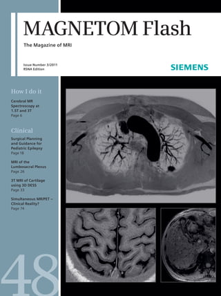
  • 26. Clinical Orthopedic Imaging High-Resolution 3T MR Neurography of the Lumbosacral Plexus Avneesh Chhabra, M.D.; Aaron Flammang, MBA, BS; John A. Carrino, M.D., M.P.H. Russell H. Morgan Department of Radiology and Radiological Science, Johns Hopkins Medical Institutions, Baltimore, MD, USA 1A 1B 1C 1A–C Lumbosacral Plexus 3T MR Neurography evaluation: Isotropic multiplanar reformats from 3D T2 SPACE (1A–1C). Abstract The lumbosacral (LS) plexus is a series of trum of pathologies along with their ings and electrodiagnostic study results. nerve convergences and ramifications respective imaging findings using However, differentiation of LS plexopathy that provide motor and sensor innerva- 3 Tesla MR Neurography. from spine related abnormality and tion to pelvis and lower extremities. LS definition to type, location and extent plexopathy is a serious condition caused Introduction of pathology often remains a diagnostic by a variety of pathologies. Magnetic The lumbosacral (LS) plexus is com- challenge due to deep location of the Resonance Neurography is an important prised of an intricate architecture of nerves and variable regional muscle modality for evaluation of LS plexopathy nerves that supply the pelvis and lower innervation [1, 2]. With current high due to variable clinical presentations extremity. It can be subject to a variety resolution 3 Tesla (T) Magnetic Reso- and deep location leading to suboptimal of pathologies, which may result in LS nance Neurography (MRN) techniques, accessibility of the plexus to electrodiag- plexopathy, a clinical syndrome that diagnostic evaluation of large LS plexus nostic studies. This article describes the includes motor and sensory distur- branches, such as sciatic and femoral role of MR Neurography in evaluation of bances. The diagnosis of LS plexopathy nerves, as well as smaller segments, the LS plexus and illustrates the spec- has traditionally relied on clinical find- such as nerve roots convergences and 26 MAGNETOM Flash · 3/2011 · www.siemens.com/magnetom-world
  • 27. Orthopedic Imaging Clinical 1D 1E 1F 1G 1D–G Coronal reconstructed 3D STIR SPACE (1D) showing sciatic nerves (arrows). Axial T1w (1E) and T2 SPAIR (1F) images showing bilateral femoral nerves (arrows) and lateral femoral cutaneous nerves (arrowheads). MIP coronal 3D STIR SPACE (1G) image showing bilateral LS plexus nerve roots, femoral nerves (arrowheads) and sciatic nerves (arrows). peripheral nerves is feasible, aiding in ventral rami of lumbar (L1–4 +/– T12), and lateral femoral cutaneous nerves pre-surgical evaluation and appropriate sacral (S1–4 and LS trunk, L4–S1) and (L2, 3)]. All branches exit lateral to the patient management [3, 4]. This article the lower sacrococcygeal (S2–4 and C1) psoas muscle and course under the provides a pertinent discussion of the nerves, respectively [5]. The plexus is inguinal ligament, except the obturator LS plexus anatomy, current role of MRN formed lateral to the intervertebral nerve and LS trunk, which exit medial to in evaluation of plexopathy and foramina and various branches course the psoas muscle. The sacral plexus gives describes the respective imaging find- through the psoas major muscle. The rise to anterior branches, namely the ings of various pathologies using 3T MR ventral rami are further split into ante- tibial part of the sciatic nerve (L4–S3), Neurography. rior and posterior divisions. The anterior pudendal nerve (S1–4) and medial part divisions give rise to the iliohypogastric of the posterior femoral cutaneous Anatomic considerations (L1), ilioinguinal (L1), genitofemoral nerve (S1–3) and, posterior branches, The LS plexus is comprised of lumbar (L1-L2) and obturator nerves (L2–4). namely the common peroneal part of the plexus, sacral plexus and the pudendal The posterior divisions combine to form sciatic nerve (L4–S2), superior (L4–S1) plexus, with contributions from the the posterior branches [femoral (L2–4) and inferior gluteal nerves (L5–S2), MAGNETOM Flash · 3/2011 · www.siemens.com/magnetom-world 27
  • 28. Clinical Orthopedic Imaging Table 1: S The 3T MR imaging protocol employed in our institution of the evaluation of the lumbosacral plexus. Sequence Slice Field-of-view Voxel size (mm3) TR/ TE (ms) Turbo factor (cm) Axial T1 TSE BL 33 0.64 800/12 6 Axial T2 SPAIR BL 33 1.00 4500/80 17 Coronal PD SPAIR BL 36-38 0.6 4980/38 7 Coronal T1 TSE BL 36-38 0.5 550/10 3 3D Coronal STIR SPACE BL 36-38 1.45 1500/103 61 Lumbar 3D Sagittal T2 SPACE 28 1.45 1000/99 69 spine Coronal 3D VIBE * BL 36-38 0.58 4.39/2.01 – Abbreviations are: TSE = Turbo Spin Echo, SPAIR = spectral adiabatic inversion recovery, STIR = short tau inversion recovery, 3D SPACE = three-dimen- sional Sampling Perfection with Application optimized Contrasts using constantly varying flip angle Evolutions, VIBE = Volume Interpolated Breath- hold Exam (* optional), BL = bilateral lateral part of the posterior femoral clinical and laboratory findings. MRN guidance during perineural and intra- cutaneous nerve (S1–3) and the nerve may be used in the latter case to confirm muscular medication injection. to the piriformis muscle (L5, S2). The lumbar plexitis / plexopathy in clinically above two plexuses connect via the LS confusing presentation and underlying Clinical findings trunk to form the LS plexus [2, 5]. known systemic condition. In addition, a LS plexopathy most often presents with primary or idiopathic form of LS plexop- asymmetric weakness, pain and/or par- Pathologic conditions and athy may occur, possibly due to altered esthesias in the lower extremities involv- indications of MRN immunological response and is consid- ing multiple contiguous LS nerve root The LS plexus is relatively protected by ered analogous to idiopathic brachial distributions. Generally, unilateral local- the axial skeleton and entrapment neu- plexopathy [6–8]. The entity has a favor- ization of symptoms indicates a local ropathy is much less common than bra- able outcome and spontaneous recov- pathology, whereas bilateral symptoms chial plexopathy. It is considered as the ery, whereas its diagnosis is based upon suggest a systemic process. The clinical counterpart of the brachial plexus in the exclusion of other etiologies and some- picture often varies depending upon the lower body and is affected by similar times may require confirmation with location and degree of plexus involve- types of diseases. The LS plexus may be MRN in case of indeterminate electrodi- ment. In cases of involvement of the involved by local processes in the viscin- agnostic results. An important indication upper nerve roots, patients predomi- ity of the plexus, such as extrinsic com- of MRN is in patients under consider- nantly present with femoral and obtura- pression by space occupying lesions, ation for surgery for peripheral nerve tor nerve symptoms. LS trunk and upper injury or infiltration by tumor / infec- lesions (piriformis syndrome/meralgia sacral plexus lesions result in foot drop tious process – which is an indication for paresthetica), post abdominal surgery depending on the extent of involvement MRN; or in systemic conditions, such entrapment of ilioinguinal/genitofemo- and weakness of knee flexion or hip as metabolic, autoimmune, vasculitis, ral nerves, or after injury to a large abduction. Sensory symptoms may vary ischemic or inflammatory disorders – branch (sciatic, femoral, obturator). based on the individual nerves involved, which are usually diagnosed based on Finally, MRN is increasingly used for which may include numbness or 28 MAGNETOM Flash · 3/2011 · www.siemens.com/magnetom-world
  • 29. Orthopedic Imaging Clinical 2A 2B 2B 2 Normal vs abnormal LS Plexus. Coronal MIP 3D STIR SPACE images in a nor- mal subject (2A) and another sub- ject with schwan- nomatosis (arrows in 2B). 3A 3B 3 Piriformis syndrome – large LS plexus branch nerve abnormality. 33-year-old man with right buttock and pelvic pain, suspected piriformis syndrome. Coronal T1w (3A) and coronal MIP 3D STIR SPACE (3B) images through the pelvis show split right sciatic nerve by an accessory slip of the right piriformis muscle (arrow). Notice abnormal T2 hyperintensity of the sciatic nerve in keeping with entrapment neuropathy. dysesthesia in the anterolateral thigh MRN technique and normal tures under interrogation, it is essential from lateral femoral cutaneous nerve appearances to use high resolution imaging with a involvement; in the mons and labia Compared to 1.5T systems 3 Tesla combination of 2D (dimensional) and 3D majora from genitofemoral nerve (MAGNETOM Verio and MAGNETOM Trio, isotropic spin echo type imaging for involvement and in the lower abdomen Siemens, Erlangen, Germany) imaging optimal assessment. In the presence of and inguinal area, upper medial thigh or is preferred for most MRN examinations known metal* in the area of imaging, pelvis from damage to the ilioinguinal, by the authors due to high quality scans 1.5T imaging is preferred. Table 1 and iliohypogastric, or pudendal nerves, obtained by these systems due to better Fig. 1 show the 3T MRN imaging protocol respectively. Rarely, there may be associ- signal-to-noise ratio and contrast reso- employed at Johns Hopkins for the eval- ated bowel and bladder incontinence as lution available in short imaging times. uation of the LS plexus. For fascicular well as sexual dysfunction [6–8]. Due to the relatively small nerve struc- architecture and subtle signal intensity MAGNETOM Flash · 3/2011 · www.siemens.com/magnetom-world 29