SlideShare uma empresa Scribd logo
1 de 5
Baixar para ler offline
International Research Journal of Engineering and Technology (IRJET) e-ISSN: 2395-0056
Volume: 07 Issue: 02 | Feb 2020 www.irjet.net p-ISSN: 2395-0072
© 2020, IRJET | Impact Factor value: 7.34 | ISO 9001:2008 Certified Journal | Page 2562
Breast Cancer Detection from Histopathology Images: A Review
Shehras V U 1, Sindhu R2
1PG Scholar, ECE Department, NSS College of Engineering, Palakkad, Kerala, India
2 Professor, ECE Department, NSS College of Engineering, Palakkad, Kerala, India
---------------------------------------------------------------------***---------------------------------------------------------------------
Abstract - Breast cancer is the most prevalent form of
cancer among women. Early detection of breast cancer will
increase survival rate. Automatic image analysis methods are
necessary to decreasetheworkloadamongpathologistsandto
improve the quality of interpretation. Nuclei detection is the
first stage of identifying the cells. Division of single cell and
the spreading of cancer from one part to others in the human
body can also detected from histopathological images. This
paper provides a review on breast cancer detection from
histopathology images.
Key Words: Histopathology images, Cell nuclei,
Metastasis,Mitosis,Convolutional Neural Network(CNN)
1. INTRODUCTION
Cancer is a cluster of diseases involving abnormal cell
growth with the potential to invade or spread to other parts
of the body known as malignant tumor. A benign tumor is
a tumor that does not spread around the body. Breast
cancer is a cancer that forms within the cells of the breasts.
Breast cancer can occur in both men and women, but it is
more commonly occur in women. Breast cancer is the most
common type of cancer diagnosed in women after skin
cancer.
Almost all cancers can spread. The original tumoriscalled
the primary tumor while the dispersed tumors are called
metastatic tumors. Mitosis is a method where single cell
divides in to two daughter cells. During mitosis, a cancerous
cell makes an exact copy of it and splits into two new cells
which are also cancerous. Metastasis iscausedbythespread
of cancer to other locations in the body. Most cancer deaths
are due to metastasis.
Early detection plays a key role in cancer detection and
can improve long-term survival rates. Medical imaging is a
very important technique for early cancer detection and
diagnosis. Manual interpretation of enormous number of
medical images can be tedious and time consuming and
easily causes human biasandmistakes.Therefore,Computer
Assisted diagnosis (CAD) systems were introduced to assist
doctors in interpreting medical images to improve their
efficiency.
A biopsy is the physical examination under which a piece
of sample tissue is taken out for microscopic examination.
The sample is then referred to the laboratory where
pathologist examines and analyses tissues under the
microscope. This microscopic examination and study of
biological cells and tissues are known as histopathology.
Histopathology is a Greek word in which Histo means
“tissues”, Patho means “disease” and logo refers to “study”.
Therefore, histopathology means the study of tissues forthe
identification of diseases.
A combination of Hematoxylin and Eosin are the most
commonly used stains in histopathology. Hematoxylin gets
bound to Deoxyribo Nucleic Acid(DNA)anditdyespurpleor
blue color to the nuclei. Eosin gets bound to proteins, so it
dyes pink color to other structures. These different stains
will help to identify the nuclei of the cells.
Fig-1: Hematoxylin and Eosin Images of breast cancer
(a): Benign (b): Malignant
Fig-1 shows the stained histopathology images to identify
benign and malignant tumor. A benign growth does not
usually threaten life but it interferes with vital structures,
tissues or organs. Benign growths aregenerally consistingof
masses of cells that closely resemble the normal cells
composing the tissue in which they are found. A malignant
growth is consisting of cells of a typical structure and
function when compared to the healthy cells surrounding
them. Malignanttumoractmaliciously,iscapableofinvading
other tissues and, if untreated, usually results in death.
2. HISTOPATHOLOGICAL IMAGE ANALYSIS
There are several breastcancerdetectiontechniquesfrom
histopathological images based on convolutional neural
networks. Fast and accuratetechniqueswhereintroducedto
improve the quality of interpretation in the field of
automatic histopathological image analysis. Automatic
detection of nuclei is the most importantintheidentification
of cells. Division of single nuclei into two new nuclei is
termed as mitosis. The division of single cell in to two new
daughter cells causes metastasis. Thenextsectiondealswith
the different breast cancer techniques.
International Research Journal of Engineering and Technology (IRJET) e-ISSN: 2395-0056
Volume: 07 Issue: 02 | Feb 2020 www.irjet.net p-ISSN: 2395-0072
© 2020, IRJET | Impact Factor value: 7.34 | ISO 9001:2008 Certified Journal | Page 2563
2.1 Breast Cancer Detection Techniques
M. M. Dundar et.al [1] developed a prototype system
for classifying breast microscopic tissues to distinguish
between Usual Ductal Hyperplasia (UDH) and actionable
subtypes Atypical Ductal Hyperplasia (ADH), and Ductal
Carcinoma In Situ (DCIS). The introduced system
automatically evaluates digitized slides of tissues forcertain
cytological criteria and classifies the tissues based on the
features derived. The developed system has great potential
in improving diagnostic accuracy and reproducibility.
H. Su et.al [2] proposed a method to apply a fast
scanning deep convolutional network (fCNN) to pixel wise
region segmentation. The aim of fCNN was to removes the
redundant computations in the original CNN without
affecting its performance. This method is robust, efficient
and scalable based on fast scanning deep neural network.
A method based on the extractionofimagepatchesfor
training CNN and from the combination of these patches
final classification was done by F.A. Spanhol et.al [3]. This
method aims to allow using high resolution images from
BreaKHis as input to existing CNN in order to avoid
adaptations of the model that led to a more complex and
computationally costly architecture. When compared to
previously reported results obtained by other machine
learning models trained with handcrafted textural
descriptors, a better performed CNN is introduced.
T. Wan et.al [4] presented a novel image analysis
based method for automatically identifying different breast
cancer grades such as low, intermediate, and high in
digitized histopathology. To segment cell nuclei in
histopathology images, an improved hybrid active contour
model based segmentation method was introduced. This
method provides an accuracy of 0.92forlowVshigh,0.77 for
low Vs intermediate, 0.76 for intermediate Vs high.
In 2018, a method for automatic classification of
breast cancer using neural network were introduced by S.
Kaymak et.al [5]. The proposed methodology representsthe
images using discrete Haar Wavelets which provides better
image content representation and then in putting them in to
neural networks. Classification of the images was achieved
using Back Propagation Neural Network (BPNN) and Radial
Basis Neural Network (RBFN).
B. Gecer et.al [6] proposed a system that classifies
whole slide images of breast biopsies in to five diagnostic
categories. A saliency detector by using a pipeline of four
sequential fully convolutional networks was used for multi
scale processing. For diagnosis, patch based multi class
convolutional network was used. Finally, a fusionofsaliency
detector and the fully convolutionalized classifier network
for pixel wise labelling of the whole slide.
In 2019, C. Kaushal et.al [7] introduced a Computer
Assisted Diagnosis (CAD) system to diagnose and detect
breast cancer from histopathology images which will helps
pathologists to detect the cancer earlier. CAD system will
help to reduce the time taken to diagnose the cancer as well
as computational complexity. Since it requires large amount
of data for training, data augmentation with the support of
various deep learning techniques had led to provide reliable
and accurate results.
R. Yan et.al [8] proposed a new hybrid model
combining both convolutional and recurrent deep neural
networks for breast cancer image classification which
integrates the advantages of the both convolutional and
recurrent deep neural networks. In this model, both the
short and long term spatial correlations were preserved
between the patches. This new hybrid model provides an
average accuracy of 91.3%.
When compared to manual detection system,
Computer Aideddiagnosissystemwill providebetterresults.
In deep learning, this is generally done by using
convolutional neural network for extracting features and
these features are classified usingfullyconnectednetwork.S.
Dabeer et.al [9] trained a convolutional neural network and
provided a prediction accuracy of up to 99.86%.
X. Li et.al [10] studied a practical, generalizable,and
self-interpretable solution to pathology image based cancer
diagnosis in 2019. The proposed method learns
discriminative patterns in weak-supervised manner from
Whole slide imaging (WSI) labels and explains its diagnosis
results via inferring locations of abnormalities in a
histopathology image.
Nuclei detectionistheimportantstageofidentifying
the cells from histopathological images. Accurate detection
and segmentation of nuclei can be done using convolutional
networks and auto encoder from high resolution images of
breast cancer.
2.2 Nuclei Detection
Y.Al-Kofahi et.al [11] presented a robust and accurate
method for the segmentation of nuclei. The image
foreground is automatically extracted using a graph-cuts-
based binarization. A novel method combining Multiscale
Laplacian of Gaussian filtering constrained by distance map
based adaptive scale selection were used to detect nuclear
seed points. An initial segmentation which is refined using a
second graph cuts based algorithm incorporating the
method of alpha expansions were performed using these
seed points.
P.Wang et.al [12] proposed an automatic quantitative
image analysis technique. To enhancetheimagequality,top-
bottom Hat transform were applied in nuclei segmentation.
To obtain the region of interest, Wavelet decompositionand
Multi scale region grown (WDMR) were used. To split
overlapped cells, a Double Strategy Splitting Model (DSS)
International Research Journal of Engineering and Technology (IRJET) e-ISSN: 2395-0056
Volume: 07 Issue: 02 | Feb 2020 www.irjet.net p-ISSN: 2395-0072
© 2020, IRJET | Impact Factor value: 7.34 | ISO 9001:2008 Certified Journal | Page 2564
containing adaptive mathematical morphology and
Curvature Scale Space (CSS) corner detection method were
applied for better accuracy and robustness.
J. Xu et.al [13] in 2015 introduced a Stacked Sparse Auto
Encoder (SSAE) basedalgorithmwhichisaninstanceofdeep
learning strategy for efficient nuclei detection on high
resolution histopathological images of breast cancer. The
SSAE learns high level features from pixel intensities in
order to identify distinguishing features of cell nuclei.
For the efficient and accurate detection of cell nuclei, H. Xu
et.al [14]presented an automatic technique based on
generalized Laplacian of Gaussian(gLoG)filterin2016. They
proposed computationally efficient nuclei seed detection
algorithm based on directionalsgLoGkernelsandmean-shift
clustering to remove false seeds in the image background.
In 2017, L. Hou et.al [15] proposed a sparse Convolutional
Auto Encoder (CAE) that uses visual characteristicsofnuclei
for simultaneous unsupervised nucleus detection and
feature extraction in histopathology tissue images. CAE
detects and encodes nuclei in image patches into sparse
feature maps that encode both the location and appearance
of nuclei.
In 2018, X. Li et.al [16] proposed a staged identification
framework for cell nuclei in colon cancer histopathology
images. A cascade residual fusion block was presented to
enhance the detection performance during the decoding
process. A multicropping module was designed for
effectively capturing contextual feature contentsaroundthe
centre of a nucleus for reducing the impact of uncertainty.
P. Naylor et.al [17] described a new method to
automatically segment nuclei from Haematoxylin and Eosin
(H&E) stained histopathology images with fully
convolutional networks. They addressed the problem of
segmenting touchingnuclei byformulatingthesegmentation
problem as a regression task of the distance map. They
showed that the fully convolutional networksarewell suited
for the task of nuclei segmentation.
The spreading of cancer cells is mainly due to the division
of single cell to two daughter cells. The cancer causing cells
will replace with the normal cells and led to the growth of
abnormal cells out of control.
2.3 Mitosis Detection
In 2016, A. Albayrak et.al [18] proposed a deep
learning based feature extraction method by Convolutional
Neural Network (CNN) for automated mitosis detection for
cancer diagnosis and grading by histopathological images.
After Pre processing, cell structures are found by combined
clustering based segmentation and blob analysis. CNN is a
prominent deep learning method on image processing tasks
which were used for extractingdiscriminativefeatures.They
used a robust kernel based classifier, support vector
machine (SVM) for final classification of mitotic and non-
mitotic cells.
H. Chen et.al [19] introduced a fast and accurate
method to detect mitosis by designinga novel deepcascaded
convolutional neural network composedoftwocomponents.
They proposed a coarse retrieval model to identify and
locate the candidates of mitosis while preserving a high
sensitivity. A fine discrimination model utilizing knowledge
transferred from cross-domainisdevelopedtofurthersingle
out mitoses from hard mimics.
In S. Albarqouni et.al [20] proposed a new concept for
learning from crowds that handle data aggregation directly
as part of the learning process of CNN via additional crowd
sourcing layer AggNet. An aggregationlayerisintroduced to
aggregate the prediction results with annotation results
from multiple participations.
In 2017, M. Saha et.al [21] presented a supervised
model to detect mitosis signature for breast histopathology
WSI (Whole Slide Images) images. This model was designed
using deep learning architecture with handcrafted features.
The proposed architecture had an improved 92% precision,
88% recall and 90% F-score.
B.K. Sabeena et.al [22] transformed a pre-trained
Convolutional Neural Network by coupling random forest
classifier with the initial fully connected layers to extract
discriminant features from nuclei patches and to precisely
prognosticate the class label of cell nuclei. The designed
framework gives higher classification accuracy by carefully
fine tune the pre trained model and pre processing the
extraction features.
For weakly supervised breast cancer diagnosis, C. Li
et.al [23] proposed a deep learning scheme with novel loss
function in 2019. This method utilizes a deep segmentation
network to produce segmentation map. Then a filtering
operation was applied to produce the detection results.
The spread of cancer cells from the place where they
first formed to another part of the body after the division of
nuclei. In metastasis, cancer cells break away from the
original (primary) tumor, travel through the blood orlymph
system, and form a new tumor in other organs or tissues of
the body.
2.4 Metastasis Detection
M. Valkonen et.al [24] described a machine learning
approach for detection of cancerous tissue from scanned
whole slide images. This method was based on feature
engineering and supervisor leaning with random forest
model. Several local descriptors related to image texture,
spatial structure, and distribution of nuclei was the features
extracted from the whole slide images.
In 2018, P. Grover et.al [25] proposed an algorithm for
automated detection of breast cancer metastasis in whole
International Research Journal of Engineering and Technology (IRJET) e-ISSN: 2395-0056
Volume: 07 Issue: 02 | Feb 2020 www.irjet.net p-ISSN: 2395-0072
© 2020, IRJET | Impact Factor value: 7.34 | ISO 9001:2008 Certified Journal | Page 2565
slide images. To improve the detection accuracy and overall
time to localize tumorous regions, proposed algorithm
leverages the capability of advanced image processing and
machine learning.
K. Fukuta et.al [26] developed a system for the automatic
detection of breast cancer metastasis in whole slide images
of histopathological lymph node sections. The system
focuses on extracting feature and spatial information. They
created tumor probability heat maps and perform post-
processing to make patient level predictions.
In 2018, H. Lin et.al [27] presented a framework by
leveraging fully convolutional networks that overcomes the
major speed bottleneck in whole-slide image analysis for
efficient inference. For ensuring accurate detection on both
micro- and macro-metastases, framework reconstructs
dense heat maps. This method achieved superior
performance compared to other method on Camelyon 2016
Grand Challenge dataset.
In order to increase the survival of patients by early
diagnosis of breast cancer, L. Tapak et.al [28] compared the
performance of six machine learning techniques, two
traditional methods for the prediction of breast cancer and
metastasis. Results shows that the average specificity of all
techniques was greater than 94% and when compared to
other techniques, Support Vector Machine (SVM)andLinear
Discriminant Analysis (LDA)havegreatersensitivityof73%.
3. CONCLUSIONS
The analysis of histopathological images remains
the most widely used method for breast cancer diagnosis.
The use of computer-assisted analysis of histopathological
images is a promising way to improve the analytical and
predictive capabilities. The computer aided diagnosis of
breast cancer from Histopathology will reduce the
complexity and increase the accuracy.
To classify breast cancer efficiently,different neural
networks are used. Auto encoder based algorithms are
proposed to identify thedistinguishingfeaturesofcell nuclei.
Deep learning basedfeature extractionsformitosisdetection
are performed using Convolutional Neural Networks. There
are various machine learning approach for the detection of
cancerous tissue from scanned Whole Slide Images.
REFERENCES
[1] M. M. Dundar et al., “Computerized classification of
intraductal breast lesions using histopathological
images,” IEEE Trans. Biomed. Eng., vol. 58, no. 7, pp.
1977–1984, 2011.
[2] H. Su, F. Liu, Y. Xie, F. Xing, S. Meyyappan, and L.
Yang, “Region segmentation in histopathological
breast cancer images using deep convolutional
neural network,” Proc. - Int. Symp. Biomed. Imaging,
vol. 2015-July, pp. 55–58, 2015.
[3] F. A. Spanhol, L. S. Oliveira, C. Petitjean,andL.Heutte,
“Breast Cancer Histopathological Image
Classification usingConvolutional Neural Networks,”
Int. Jt. Conf. Neural Networks (IJCNN 2016), 2016.
[4] T. Wan, J. Cao, J. Chen, and Z. Qin, “Automated
grading of breast cancer histopathology using
cascaded ensemble with combination of multi-level
image features,” Neurocomputing, vol. 229, pp. 34–
44, 2017.
[5] S. Kaymak, A. Helwan, and D. Uzun, “Breast cancer
image classification usingartificial neural networks,”
Procedia Comput. Sci., vol. 120, pp. 126–131, 2018.
[6] B. Gecer, S. Aksoy, E. Mercan, L. G. Shapiro, D. L.
Weaver, and J. G. Elmore, “Detection and
Classification of Cancer in Whole Slide Breast
Histopathology Images Using Deep Convolutional
Networks,” Pattern Recognit., 2018.
[7] C. Kaushal, S. Bhat, D. Koundal, and A. Singla, “Recent
Trends in Computer Assisted Diagnosis ( CAD )
System for Breast Cancer Diagnosis Using
Histopathological Images,” IRBM, vol. 1, pp. 1–17,
2019.
[8] R. Yan et al., “Breast cancer histopathological image
classification using a hybrid deep neural network,”
Methods, no. February, pp. 1–9, 2019.
[9] S. Dabeer, M. M. Khan, and S. Islam,“Cancerdiagnosis
in histopathological image: CNN based approach,”
Informatics Med. Unlocked, p. 100231, 2019.
[10] X. Li, M. Radulovic, K. Kanjer, and K. N. Plataniotis,
“Discriminative Pattern Mining for Breast Cancer
Histopathology Image Classification via Fully
Convolutional Autoencoder,” IEEE Access, vol. 7, no.
c, pp. 36433–36445, 2019.
[11] Y. Al-Kofahi, W. Lassoued, W. Lee, and B. Roysam,
“Improved automatic detection and segmentationof
cell nuclei in histopathology images,” IEEE Trans.
Biomed. Eng., vol. 57, no. 4, pp. 841–852, 2010.
[12] P. Wang, X. Hu, Y. Li, Q. Liu, and X. Zhu, “Automatic
cell nuclei segmentation and classification of breast
cancer histopathology images,” Signal Processing,
vol. 122, pp. 1–13, 2016.
International Research Journal of Engineering and Technology (IRJET) e-ISSN: 2395-0056
Volume: 07 Issue: 02 | Feb 2020 www.irjet.net p-ISSN: 2395-0072
© 2020, IRJET | Impact Factor value: 7.34 | ISO 9001:2008 Certified Journal | Page 2566
[13] J. Xu et al., “Stacked sparse autoencoder (SSAE) for
nuclei detection on breast cancer histopathology
images,” IEEE Trans. Med. Imaging, vol. 35, no. 1, pp.
119–130, 2016.
[14] H. Xu, C. Lu, R. Berendt, N. Jha, and M. Mandal,
“Automatic Nuclei Detection Based on Generalized
Laplacian of Gaussian Filters,” IEEE J. Biomed. Heal.
Informatics, vol. 21, no. 3, pp. 826–837, 2017.
[15] L. Hou et al., “Sparse autoencoder for unsupervised
nucleus detection and representation in
histopathology images,” Pattern Recognit., vol. 86,
pp. 188–200, 2019.
[16] X. Li, W. Li, S. Member, R. Tao, and S. Member,
“Staged Detection – Identification Framework for
Cell Nuclei in Histopathology Images,” IEEE Trans.
Instrum. Meas., vol. PP, pp. 1–11, 2019.
[17] P. Naylor, M. Laé, F. Reyal, and T. Walter,
“Segmentation of NucleiinHistopathologyImages by
Deep Regression of the Distance Map,” IEEE Trans.
Med. Imaging, vol. 38, no. 2, pp. 448–459, 2019.
[18] A. Albayrak and G. Bilgin, “Mitosis detection using
convolutional neural network basedfeatures,” CINTI
2016 - 17th IEEE Int. Symp. Comput. Intell.
Informatics Proc., pp. 335–340, 2017.
[19] H. Chen, Q. Dou, X. Wang, J. Qin, and P. A. Heng,
“Mitosis detection in breast cancer histology images
via deep cascaded networks,” 30th AAAI Conf. Artif.
Intell. AAAI 2016, pp. 1160–1166, 2016.
[20] S. Albarqouni, C. Baur, F. Achilles, V. Belagiannis, S.
Demirci, and N. Navab,“AggNet:DeepLearningFrom
Crowds for Mitosis Detection in Breast Cancer
Histology Images,” IEEE Trans. Med.Imaging,vol.35,
no. 5, pp. 1313–1321, 2016.
[21] M. Saha, C. Chakraborty, and D. Racoceanu, “Efficient
deep learning model for mitosis detection using
breast histopathology images,” Comput. Med.
Imaging Graph., vol. 64, pp. 29–40, 2018.
[22] K. Sabeena Beevi, M. S. Nair, and G. R. Bindu,
“Automatic mitosis detection in breast
histopathology images using Convolutional Neural
Network based deep transfer learning,” Biocybern.
Biomed. Eng., vol. 39, no. 1, pp. 214–223, 2019.
[23] C. Li, X. Wang, W. Liu, L. J. Latecki, B. Wang, and J.
Huang, “Weakly supervised mitosis detection in
breast histopathology images using concentricloss,”
Med. Image Anal., vol. 53, pp. 165–178, 2019.
[24] M. Valkonen, K. Kartasalo, K. Liimatainen, M. Nykter,
L. Latonen, and P. Ruusuvuori, “Metastasis detection
from whole slide images using local features and
random forests,” Cytom. Part A, vol. 91, no. 6, pp.
555–565, 2017.
[25] P. Grover, “Automated Detection of Breast Cancer
Metastases in Whole Slide Images,” 2018 First Int.
Conf. Secur. Cyber Comput. Commun., pp. 1–6, 2018.
[26] K. Fukuta, “Identifyingmetastaticbreastcancerusing
deep texture representation,” The Univercity of
Tokyo , Tokyo Medical and Dental University
Camelyon17.Grand-Challenge.Org.
[27] H. Lin, H. Chen, S. Graham, Q. Dou, N. Rajpoot, and P.
A. Heng, “Fast ScanNet: Fast and Dense Analysis of
Multi-Gigapixel Whole-Slide Images for Cancer
Metastasis Detection,” IEEETrans.Med.Imaging,vol.
38, no. 8, pp. 1948–1958, 2019.
[28] L. Tapak, N. Shirmohammadi-Khorram, P. Amini, B.
Alafchi, O. Hamidi, and J. Poorolajal, “Prediction of
survival and metastasis in breast cancer patients
using machine learning classifiers,” Clin. Epidemiol.
Glob. Heal., vol. 7, no. 3, pp. 293–299, 2019.
BIOGRAPHIES
Shehras V U, Currently pursuing
M.Tech in Communication
Engineering from NSS College of
Engineering, Palakkad, Kerala, India
Dr. Sindhu R, Professor, ECE Dept,
NSS College of Engineering,
Palakkad, Kerala, India

Mais conteúdo relacionado

Mais procurados

A novel framework for efficient identification of brain cancer region from br...
A novel framework for efficient identification of brain cancer region from br...A novel framework for efficient identification of brain cancer region from br...
A novel framework for efficient identification of brain cancer region from br...IJECEIAES
 
IRJET- Brain Tumor Detection using Hybrid Model of DCT DWT and Thresholding
IRJET- Brain Tumor Detection using Hybrid Model of DCT DWT and ThresholdingIRJET- Brain Tumor Detection using Hybrid Model of DCT DWT and Thresholding
IRJET- Brain Tumor Detection using Hybrid Model of DCT DWT and ThresholdingIRJET Journal
 
BREAST CANCER DIAGNOSIS USING MACHINE LEARNING ALGORITHMS –A SURVEY
BREAST CANCER DIAGNOSIS USING MACHINE LEARNING ALGORITHMS –A SURVEYBREAST CANCER DIAGNOSIS USING MACHINE LEARNING ALGORITHMS –A SURVEY
BREAST CANCER DIAGNOSIS USING MACHINE LEARNING ALGORITHMS –A SURVEYijdpsjournal
 
Definiens In Digital Pathology Hr
Definiens In Digital Pathology HrDefiniens In Digital Pathology Hr
Definiens In Digital Pathology HrDaniel Nicolson
 
BRAIN TUMOR CLASSIFICATION IN 3D-MRI USING FEATURES FROM RADIOMICS AND 3D-CNN...
BRAIN TUMOR CLASSIFICATION IN 3D-MRI USING FEATURES FROM RADIOMICS AND 3D-CNN...BRAIN TUMOR CLASSIFICATION IN 3D-MRI USING FEATURES FROM RADIOMICS AND 3D-CNN...
BRAIN TUMOR CLASSIFICATION IN 3D-MRI USING FEATURES FROM RADIOMICS AND 3D-CNN...IAEME Publication
 
IRJET- Lung Diseases using Deep Learning: A Review Paper
IRJET- Lung Diseases using Deep Learning: A Review PaperIRJET- Lung Diseases using Deep Learning: A Review Paper
IRJET- Lung Diseases using Deep Learning: A Review PaperIRJET Journal
 
M sc research_project_report_x18134599
M sc research_project_report_x18134599M sc research_project_report_x18134599
M sc research_project_report_x18134599MansiChowkkar
 
Glioblastomas brain tumour segmentation based on convolutional neural network...
Glioblastomas brain tumour segmentation based on convolutional neural network...Glioblastomas brain tumour segmentation based on convolutional neural network...
Glioblastomas brain tumour segmentation based on convolutional neural network...IJECEIAES
 
Brain Tumor Detection using Neural Network
Brain Tumor Detection using Neural NetworkBrain Tumor Detection using Neural Network
Brain Tumor Detection using Neural Networkijtsrd
 
Tumor Detection and Classification of MRI Brain Images using SVM and DNN
Tumor Detection and Classification of MRI Brain Images using SVM and DNNTumor Detection and Classification of MRI Brain Images using SVM and DNN
Tumor Detection and Classification of MRI Brain Images using SVM and DNNijtsrd
 
Early Detection of Lung Cancer Using Neural Network Techniques
Early Detection of Lung Cancer Using Neural Network TechniquesEarly Detection of Lung Cancer Using Neural Network Techniques
Early Detection of Lung Cancer Using Neural Network TechniquesIJERA Editor
 
IRJET- Effect of Principal Component Analysis in Lung Cancer Detection us...
IRJET-  	  Effect of Principal Component Analysis in Lung Cancer Detection us...IRJET-  	  Effect of Principal Component Analysis in Lung Cancer Detection us...
IRJET- Effect of Principal Component Analysis in Lung Cancer Detection us...IRJET Journal
 
IRJET - Fusion of CT and MRI for the Detection of Brain Tumor by SWT and Prob...
IRJET - Fusion of CT and MRI for the Detection of Brain Tumor by SWT and Prob...IRJET - Fusion of CT and MRI for the Detection of Brain Tumor by SWT and Prob...
IRJET - Fusion of CT and MRI for the Detection of Brain Tumor by SWT and Prob...IRJET Journal
 
An Analysis of The Methods Employed for Breast Cancer Diagnosis
An Analysis of The Methods Employed for Breast Cancer Diagnosis An Analysis of The Methods Employed for Breast Cancer Diagnosis
An Analysis of The Methods Employed for Breast Cancer Diagnosis IJORCS
 
Skin Cancer Detection using Image Processing in Real Time
Skin Cancer Detection using Image Processing in Real TimeSkin Cancer Detection using Image Processing in Real Time
Skin Cancer Detection using Image Processing in Real Timeijtsrd
 
Lung Cancer Detection using Machine Learning
Lung Cancer Detection using Machine LearningLung Cancer Detection using Machine Learning
Lung Cancer Detection using Machine Learningijtsrd
 
Pneumonia Classification using Transfer Learning
Pneumonia Classification using Transfer LearningPneumonia Classification using Transfer Learning
Pneumonia Classification using Transfer LearningTushar Dalvi
 

Mais procurados (20)

A novel framework for efficient identification of brain cancer region from br...
A novel framework for efficient identification of brain cancer region from br...A novel framework for efficient identification of brain cancer region from br...
A novel framework for efficient identification of brain cancer region from br...
 
IRJET- Brain Tumor Detection using Hybrid Model of DCT DWT and Thresholding
IRJET- Brain Tumor Detection using Hybrid Model of DCT DWT and ThresholdingIRJET- Brain Tumor Detection using Hybrid Model of DCT DWT and Thresholding
IRJET- Brain Tumor Detection using Hybrid Model of DCT DWT and Thresholding
 
BREAST CANCER DIAGNOSIS USING MACHINE LEARNING ALGORITHMS –A SURVEY
BREAST CANCER DIAGNOSIS USING MACHINE LEARNING ALGORITHMS –A SURVEYBREAST CANCER DIAGNOSIS USING MACHINE LEARNING ALGORITHMS –A SURVEY
BREAST CANCER DIAGNOSIS USING MACHINE LEARNING ALGORITHMS –A SURVEY
 
Definiens In Digital Pathology Hr
Definiens In Digital Pathology HrDefiniens In Digital Pathology Hr
Definiens In Digital Pathology Hr
 
S4502115119
S4502115119S4502115119
S4502115119
 
BRAIN TUMOR CLASSIFICATION IN 3D-MRI USING FEATURES FROM RADIOMICS AND 3D-CNN...
BRAIN TUMOR CLASSIFICATION IN 3D-MRI USING FEATURES FROM RADIOMICS AND 3D-CNN...BRAIN TUMOR CLASSIFICATION IN 3D-MRI USING FEATURES FROM RADIOMICS AND 3D-CNN...
BRAIN TUMOR CLASSIFICATION IN 3D-MRI USING FEATURES FROM RADIOMICS AND 3D-CNN...
 
IRJET- Lung Diseases using Deep Learning: A Review Paper
IRJET- Lung Diseases using Deep Learning: A Review PaperIRJET- Lung Diseases using Deep Learning: A Review Paper
IRJET- Lung Diseases using Deep Learning: A Review Paper
 
L045047880
L045047880L045047880
L045047880
 
M sc research_project_report_x18134599
M sc research_project_report_x18134599M sc research_project_report_x18134599
M sc research_project_report_x18134599
 
Glioblastomas brain tumour segmentation based on convolutional neural network...
Glioblastomas brain tumour segmentation based on convolutional neural network...Glioblastomas brain tumour segmentation based on convolutional neural network...
Glioblastomas brain tumour segmentation based on convolutional neural network...
 
Brain Tumor Detection using Neural Network
Brain Tumor Detection using Neural NetworkBrain Tumor Detection using Neural Network
Brain Tumor Detection using Neural Network
 
Tumor Detection and Classification of MRI Brain Images using SVM and DNN
Tumor Detection and Classification of MRI Brain Images using SVM and DNNTumor Detection and Classification of MRI Brain Images using SVM and DNN
Tumor Detection and Classification of MRI Brain Images using SVM and DNN
 
Early Detection of Lung Cancer Using Neural Network Techniques
Early Detection of Lung Cancer Using Neural Network TechniquesEarly Detection of Lung Cancer Using Neural Network Techniques
Early Detection of Lung Cancer Using Neural Network Techniques
 
Li2019
Li2019Li2019
Li2019
 
IRJET- Effect of Principal Component Analysis in Lung Cancer Detection us...
IRJET-  	  Effect of Principal Component Analysis in Lung Cancer Detection us...IRJET-  	  Effect of Principal Component Analysis in Lung Cancer Detection us...
IRJET- Effect of Principal Component Analysis in Lung Cancer Detection us...
 
IRJET - Fusion of CT and MRI for the Detection of Brain Tumor by SWT and Prob...
IRJET - Fusion of CT and MRI for the Detection of Brain Tumor by SWT and Prob...IRJET - Fusion of CT and MRI for the Detection of Brain Tumor by SWT and Prob...
IRJET - Fusion of CT and MRI for the Detection of Brain Tumor by SWT and Prob...
 
An Analysis of The Methods Employed for Breast Cancer Diagnosis
An Analysis of The Methods Employed for Breast Cancer Diagnosis An Analysis of The Methods Employed for Breast Cancer Diagnosis
An Analysis of The Methods Employed for Breast Cancer Diagnosis
 
Skin Cancer Detection using Image Processing in Real Time
Skin Cancer Detection using Image Processing in Real TimeSkin Cancer Detection using Image Processing in Real Time
Skin Cancer Detection using Image Processing in Real Time
 
Lung Cancer Detection using Machine Learning
Lung Cancer Detection using Machine LearningLung Cancer Detection using Machine Learning
Lung Cancer Detection using Machine Learning
 
Pneumonia Classification using Transfer Learning
Pneumonia Classification using Transfer LearningPneumonia Classification using Transfer Learning
Pneumonia Classification using Transfer Learning
 

Semelhante a IRJET- Breast Cancer Detection from Histopathology Images: A Review

IRJET- A Survey on Categorization of Breast Cancer in Histopathological Images
IRJET- A Survey on Categorization of Breast Cancer in Histopathological ImagesIRJET- A Survey on Categorization of Breast Cancer in Histopathological Images
IRJET- A Survey on Categorization of Breast Cancer in Histopathological ImagesIRJET Journal
 
Breast Cancer Prediction
Breast Cancer PredictionBreast Cancer Prediction
Breast Cancer PredictionIRJET Journal
 
deep learning applications in medical image analysis brain tumor
deep learning applications in medical image analysis brain tumordeep learning applications in medical image analysis brain tumor
deep learning applications in medical image analysis brain tumorVenkat Projects
 
IRJET - Classification of Cancer Images using Deep Learning
IRJET -  	  Classification of Cancer Images using Deep LearningIRJET -  	  Classification of Cancer Images using Deep Learning
IRJET - Classification of Cancer Images using Deep LearningIRJET Journal
 
IRJET - Lung Disease Prediction using Image Processing and CNN Algorithm
IRJET -  	  Lung Disease Prediction using Image Processing and CNN AlgorithmIRJET -  	  Lung Disease Prediction using Image Processing and CNN Algorithm
IRJET - Lung Disease Prediction using Image Processing and CNN AlgorithmIRJET Journal
 
Breast cancer histological images nuclei segmentation and optimized classifi...
Breast cancer histological images nuclei segmentation and  optimized classifi...Breast cancer histological images nuclei segmentation and  optimized classifi...
Breast cancer histological images nuclei segmentation and optimized classifi...IJECEIAES
 
A Comprehensive Evaluation of Machine Learning Approaches for Breast Cancer C...
A Comprehensive Evaluation of Machine Learning Approaches for Breast Cancer C...A Comprehensive Evaluation of Machine Learning Approaches for Breast Cancer C...
A Comprehensive Evaluation of Machine Learning Approaches for Breast Cancer C...IRJET Journal
 
Cervical cancer diagnosis based on cytology pap smear image classification us...
Cervical cancer diagnosis based on cytology pap smear image classification us...Cervical cancer diagnosis based on cytology pap smear image classification us...
Cervical cancer diagnosis based on cytology pap smear image classification us...TELKOMNIKA JOURNAL
 
A Progressive Review on Early Stage Breast Cancer Detection
A Progressive Review on Early Stage Breast Cancer DetectionA Progressive Review on Early Stage Breast Cancer Detection
A Progressive Review on Early Stage Breast Cancer DetectionIRJET Journal
 
A REVIEW PAPER ON PULMONARY NODULE DETECTION
A REVIEW PAPER ON PULMONARY NODULE DETECTIONA REVIEW PAPER ON PULMONARY NODULE DETECTION
A REVIEW PAPER ON PULMONARY NODULE DETECTIONIRJET Journal
 
Cervical Cancer Analysis
Cervical Cancer AnalysisCervical Cancer Analysis
Cervical Cancer AnalysisIRJET Journal
 
Automated Intracranial Neoplasm Detection Using Convolutional Neural Networks
Automated Intracranial Neoplasm Detection Using Convolutional Neural NetworksAutomated Intracranial Neoplasm Detection Using Convolutional Neural Networks
Automated Intracranial Neoplasm Detection Using Convolutional Neural NetworksIRJET Journal
 
Modified fuzzy rough set technique with stacked autoencoder model for magneti...
Modified fuzzy rough set technique with stacked autoencoder model for magneti...Modified fuzzy rough set technique with stacked autoencoder model for magneti...
Modified fuzzy rough set technique with stacked autoencoder model for magneti...IJECEIAES
 
Paper id 25201472
Paper id 25201472Paper id 25201472
Paper id 25201472IJRAT
 
A Progressive Review: Early Stage Breast Cancer Detection using Ultrasound Im...
A Progressive Review: Early Stage Breast Cancer Detection using Ultrasound Im...A Progressive Review: Early Stage Breast Cancer Detection using Ultrasound Im...
A Progressive Review: Early Stage Breast Cancer Detection using Ultrasound Im...IRJET Journal
 
Generalized deep learning model for Classification of Gastric, Colon and Rena...
Generalized deep learning model for Classification of Gastric, Colon and Rena...Generalized deep learning model for Classification of Gastric, Colon and Rena...
Generalized deep learning model for Classification of Gastric, Colon and Rena...IRJET Journal
 
Brain tumor detection using Region-based Convolutional Neural Network and PSO
Brain tumor detection using Region-based Convolutional Neural Network and PSOBrain tumor detection using Region-based Convolutional Neural Network and PSO
Brain tumor detection using Region-based Convolutional Neural Network and PSOIRJET Journal
 
BRAIN TUMOR DETECTION USING CNN & ML TECHNIQUES
BRAIN TUMOR DETECTION USING CNN & ML TECHNIQUESBRAIN TUMOR DETECTION USING CNN & ML TECHNIQUES
BRAIN TUMOR DETECTION USING CNN & ML TECHNIQUESIRJET Journal
 
The Evolution and Impact of Medical Science Journals in Advancing Healthcare
The Evolution and Impact of Medical Science Journals in Advancing HealthcareThe Evolution and Impact of Medical Science Journals in Advancing Healthcare
The Evolution and Impact of Medical Science Journals in Advancing Healthcaresana473753
 

Semelhante a IRJET- Breast Cancer Detection from Histopathology Images: A Review (20)

IRJET- A Survey on Categorization of Breast Cancer in Histopathological Images
IRJET- A Survey on Categorization of Breast Cancer in Histopathological ImagesIRJET- A Survey on Categorization of Breast Cancer in Histopathological Images
IRJET- A Survey on Categorization of Breast Cancer in Histopathological Images
 
Breast Cancer Prediction
Breast Cancer PredictionBreast Cancer Prediction
Breast Cancer Prediction
 
deep learning applications in medical image analysis brain tumor
deep learning applications in medical image analysis brain tumordeep learning applications in medical image analysis brain tumor
deep learning applications in medical image analysis brain tumor
 
IRJET - Classification of Cancer Images using Deep Learning
IRJET -  	  Classification of Cancer Images using Deep LearningIRJET -  	  Classification of Cancer Images using Deep Learning
IRJET - Classification of Cancer Images using Deep Learning
 
IRJET - Lung Disease Prediction using Image Processing and CNN Algorithm
IRJET -  	  Lung Disease Prediction using Image Processing and CNN AlgorithmIRJET -  	  Lung Disease Prediction using Image Processing and CNN Algorithm
IRJET - Lung Disease Prediction using Image Processing and CNN Algorithm
 
Breast cancer histological images nuclei segmentation and optimized classifi...
Breast cancer histological images nuclei segmentation and  optimized classifi...Breast cancer histological images nuclei segmentation and  optimized classifi...
Breast cancer histological images nuclei segmentation and optimized classifi...
 
A Comprehensive Evaluation of Machine Learning Approaches for Breast Cancer C...
A Comprehensive Evaluation of Machine Learning Approaches for Breast Cancer C...A Comprehensive Evaluation of Machine Learning Approaches for Breast Cancer C...
A Comprehensive Evaluation of Machine Learning Approaches for Breast Cancer C...
 
Cervical cancer diagnosis based on cytology pap smear image classification us...
Cervical cancer diagnosis based on cytology pap smear image classification us...Cervical cancer diagnosis based on cytology pap smear image classification us...
Cervical cancer diagnosis based on cytology pap smear image classification us...
 
A Progressive Review on Early Stage Breast Cancer Detection
A Progressive Review on Early Stage Breast Cancer DetectionA Progressive Review on Early Stage Breast Cancer Detection
A Progressive Review on Early Stage Breast Cancer Detection
 
A REVIEW PAPER ON PULMONARY NODULE DETECTION
A REVIEW PAPER ON PULMONARY NODULE DETECTIONA REVIEW PAPER ON PULMONARY NODULE DETECTION
A REVIEW PAPER ON PULMONARY NODULE DETECTION
 
Comparison of breast cancer classification models on Wisconsin dataset
Comparison of breast cancer classification models on Wisconsin  datasetComparison of breast cancer classification models on Wisconsin  dataset
Comparison of breast cancer classification models on Wisconsin dataset
 
Cervical Cancer Analysis
Cervical Cancer AnalysisCervical Cancer Analysis
Cervical Cancer Analysis
 
Automated Intracranial Neoplasm Detection Using Convolutional Neural Networks
Automated Intracranial Neoplasm Detection Using Convolutional Neural NetworksAutomated Intracranial Neoplasm Detection Using Convolutional Neural Networks
Automated Intracranial Neoplasm Detection Using Convolutional Neural Networks
 
Modified fuzzy rough set technique with stacked autoencoder model for magneti...
Modified fuzzy rough set technique with stacked autoencoder model for magneti...Modified fuzzy rough set technique with stacked autoencoder model for magneti...
Modified fuzzy rough set technique with stacked autoencoder model for magneti...
 
Paper id 25201472
Paper id 25201472Paper id 25201472
Paper id 25201472
 
A Progressive Review: Early Stage Breast Cancer Detection using Ultrasound Im...
A Progressive Review: Early Stage Breast Cancer Detection using Ultrasound Im...A Progressive Review: Early Stage Breast Cancer Detection using Ultrasound Im...
A Progressive Review: Early Stage Breast Cancer Detection using Ultrasound Im...
 
Generalized deep learning model for Classification of Gastric, Colon and Rena...
Generalized deep learning model for Classification of Gastric, Colon and Rena...Generalized deep learning model for Classification of Gastric, Colon and Rena...
Generalized deep learning model for Classification of Gastric, Colon and Rena...
 
Brain tumor detection using Region-based Convolutional Neural Network and PSO
Brain tumor detection using Region-based Convolutional Neural Network and PSOBrain tumor detection using Region-based Convolutional Neural Network and PSO
Brain tumor detection using Region-based Convolutional Neural Network and PSO
 
BRAIN TUMOR DETECTION USING CNN & ML TECHNIQUES
BRAIN TUMOR DETECTION USING CNN & ML TECHNIQUESBRAIN TUMOR DETECTION USING CNN & ML TECHNIQUES
BRAIN TUMOR DETECTION USING CNN & ML TECHNIQUES
 
The Evolution and Impact of Medical Science Journals in Advancing Healthcare
The Evolution and Impact of Medical Science Journals in Advancing HealthcareThe Evolution and Impact of Medical Science Journals in Advancing Healthcare
The Evolution and Impact of Medical Science Journals in Advancing Healthcare
 

Mais de IRJET Journal

TUNNELING IN HIMALAYAS WITH NATM METHOD: A SPECIAL REFERENCES TO SUNGAL TUNNE...
TUNNELING IN HIMALAYAS WITH NATM METHOD: A SPECIAL REFERENCES TO SUNGAL TUNNE...TUNNELING IN HIMALAYAS WITH NATM METHOD: A SPECIAL REFERENCES TO SUNGAL TUNNE...
TUNNELING IN HIMALAYAS WITH NATM METHOD: A SPECIAL REFERENCES TO SUNGAL TUNNE...IRJET Journal
 
STUDY THE EFFECT OF RESPONSE REDUCTION FACTOR ON RC FRAMED STRUCTURE
STUDY THE EFFECT OF RESPONSE REDUCTION FACTOR ON RC FRAMED STRUCTURESTUDY THE EFFECT OF RESPONSE REDUCTION FACTOR ON RC FRAMED STRUCTURE
STUDY THE EFFECT OF RESPONSE REDUCTION FACTOR ON RC FRAMED STRUCTUREIRJET Journal
 
A COMPARATIVE ANALYSIS OF RCC ELEMENT OF SLAB WITH STARK STEEL (HYSD STEEL) A...
A COMPARATIVE ANALYSIS OF RCC ELEMENT OF SLAB WITH STARK STEEL (HYSD STEEL) A...A COMPARATIVE ANALYSIS OF RCC ELEMENT OF SLAB WITH STARK STEEL (HYSD STEEL) A...
A COMPARATIVE ANALYSIS OF RCC ELEMENT OF SLAB WITH STARK STEEL (HYSD STEEL) A...IRJET Journal
 
Effect of Camber and Angles of Attack on Airfoil Characteristics
Effect of Camber and Angles of Attack on Airfoil CharacteristicsEffect of Camber and Angles of Attack on Airfoil Characteristics
Effect of Camber and Angles of Attack on Airfoil CharacteristicsIRJET Journal
 
A Review on the Progress and Challenges of Aluminum-Based Metal Matrix Compos...
A Review on the Progress and Challenges of Aluminum-Based Metal Matrix Compos...A Review on the Progress and Challenges of Aluminum-Based Metal Matrix Compos...
A Review on the Progress and Challenges of Aluminum-Based Metal Matrix Compos...IRJET Journal
 
Dynamic Urban Transit Optimization: A Graph Neural Network Approach for Real-...
Dynamic Urban Transit Optimization: A Graph Neural Network Approach for Real-...Dynamic Urban Transit Optimization: A Graph Neural Network Approach for Real-...
Dynamic Urban Transit Optimization: A Graph Neural Network Approach for Real-...IRJET Journal
 
Structural Analysis and Design of Multi-Storey Symmetric and Asymmetric Shape...
Structural Analysis and Design of Multi-Storey Symmetric and Asymmetric Shape...Structural Analysis and Design of Multi-Storey Symmetric and Asymmetric Shape...
Structural Analysis and Design of Multi-Storey Symmetric and Asymmetric Shape...IRJET Journal
 
A Review of “Seismic Response of RC Structures Having Plan and Vertical Irreg...
A Review of “Seismic Response of RC Structures Having Plan and Vertical Irreg...A Review of “Seismic Response of RC Structures Having Plan and Vertical Irreg...
A Review of “Seismic Response of RC Structures Having Plan and Vertical Irreg...IRJET Journal
 
A REVIEW ON MACHINE LEARNING IN ADAS
A REVIEW ON MACHINE LEARNING IN ADASA REVIEW ON MACHINE LEARNING IN ADAS
A REVIEW ON MACHINE LEARNING IN ADASIRJET Journal
 
Long Term Trend Analysis of Precipitation and Temperature for Asosa district,...
Long Term Trend Analysis of Precipitation and Temperature for Asosa district,...Long Term Trend Analysis of Precipitation and Temperature for Asosa district,...
Long Term Trend Analysis of Precipitation and Temperature for Asosa district,...IRJET Journal
 
P.E.B. Framed Structure Design and Analysis Using STAAD Pro
P.E.B. Framed Structure Design and Analysis Using STAAD ProP.E.B. Framed Structure Design and Analysis Using STAAD Pro
P.E.B. Framed Structure Design and Analysis Using STAAD ProIRJET Journal
 
A Review on Innovative Fiber Integration for Enhanced Reinforcement of Concre...
A Review on Innovative Fiber Integration for Enhanced Reinforcement of Concre...A Review on Innovative Fiber Integration for Enhanced Reinforcement of Concre...
A Review on Innovative Fiber Integration for Enhanced Reinforcement of Concre...IRJET Journal
 
Survey Paper on Cloud-Based Secured Healthcare System
Survey Paper on Cloud-Based Secured Healthcare SystemSurvey Paper on Cloud-Based Secured Healthcare System
Survey Paper on Cloud-Based Secured Healthcare SystemIRJET Journal
 
Review on studies and research on widening of existing concrete bridges
Review on studies and research on widening of existing concrete bridgesReview on studies and research on widening of existing concrete bridges
Review on studies and research on widening of existing concrete bridgesIRJET Journal
 
React based fullstack edtech web application
React based fullstack edtech web applicationReact based fullstack edtech web application
React based fullstack edtech web applicationIRJET Journal
 
A Comprehensive Review of Integrating IoT and Blockchain Technologies in the ...
A Comprehensive Review of Integrating IoT and Blockchain Technologies in the ...A Comprehensive Review of Integrating IoT and Blockchain Technologies in the ...
A Comprehensive Review of Integrating IoT and Blockchain Technologies in the ...IRJET Journal
 
A REVIEW ON THE PERFORMANCE OF COCONUT FIBRE REINFORCED CONCRETE.
A REVIEW ON THE PERFORMANCE OF COCONUT FIBRE REINFORCED CONCRETE.A REVIEW ON THE PERFORMANCE OF COCONUT FIBRE REINFORCED CONCRETE.
A REVIEW ON THE PERFORMANCE OF COCONUT FIBRE REINFORCED CONCRETE.IRJET Journal
 
Optimizing Business Management Process Workflows: The Dynamic Influence of Mi...
Optimizing Business Management Process Workflows: The Dynamic Influence of Mi...Optimizing Business Management Process Workflows: The Dynamic Influence of Mi...
Optimizing Business Management Process Workflows: The Dynamic Influence of Mi...IRJET Journal
 
Multistoried and Multi Bay Steel Building Frame by using Seismic Design
Multistoried and Multi Bay Steel Building Frame by using Seismic DesignMultistoried and Multi Bay Steel Building Frame by using Seismic Design
Multistoried and Multi Bay Steel Building Frame by using Seismic DesignIRJET Journal
 
Cost Optimization of Construction Using Plastic Waste as a Sustainable Constr...
Cost Optimization of Construction Using Plastic Waste as a Sustainable Constr...Cost Optimization of Construction Using Plastic Waste as a Sustainable Constr...
Cost Optimization of Construction Using Plastic Waste as a Sustainable Constr...IRJET Journal
 

Mais de IRJET Journal (20)

TUNNELING IN HIMALAYAS WITH NATM METHOD: A SPECIAL REFERENCES TO SUNGAL TUNNE...
TUNNELING IN HIMALAYAS WITH NATM METHOD: A SPECIAL REFERENCES TO SUNGAL TUNNE...TUNNELING IN HIMALAYAS WITH NATM METHOD: A SPECIAL REFERENCES TO SUNGAL TUNNE...
TUNNELING IN HIMALAYAS WITH NATM METHOD: A SPECIAL REFERENCES TO SUNGAL TUNNE...
 
STUDY THE EFFECT OF RESPONSE REDUCTION FACTOR ON RC FRAMED STRUCTURE
STUDY THE EFFECT OF RESPONSE REDUCTION FACTOR ON RC FRAMED STRUCTURESTUDY THE EFFECT OF RESPONSE REDUCTION FACTOR ON RC FRAMED STRUCTURE
STUDY THE EFFECT OF RESPONSE REDUCTION FACTOR ON RC FRAMED STRUCTURE
 
A COMPARATIVE ANALYSIS OF RCC ELEMENT OF SLAB WITH STARK STEEL (HYSD STEEL) A...
A COMPARATIVE ANALYSIS OF RCC ELEMENT OF SLAB WITH STARK STEEL (HYSD STEEL) A...A COMPARATIVE ANALYSIS OF RCC ELEMENT OF SLAB WITH STARK STEEL (HYSD STEEL) A...
A COMPARATIVE ANALYSIS OF RCC ELEMENT OF SLAB WITH STARK STEEL (HYSD STEEL) A...
 
Effect of Camber and Angles of Attack on Airfoil Characteristics
Effect of Camber and Angles of Attack on Airfoil CharacteristicsEffect of Camber and Angles of Attack on Airfoil Characteristics
Effect of Camber and Angles of Attack on Airfoil Characteristics
 
A Review on the Progress and Challenges of Aluminum-Based Metal Matrix Compos...
A Review on the Progress and Challenges of Aluminum-Based Metal Matrix Compos...A Review on the Progress and Challenges of Aluminum-Based Metal Matrix Compos...
A Review on the Progress and Challenges of Aluminum-Based Metal Matrix Compos...
 
Dynamic Urban Transit Optimization: A Graph Neural Network Approach for Real-...
Dynamic Urban Transit Optimization: A Graph Neural Network Approach for Real-...Dynamic Urban Transit Optimization: A Graph Neural Network Approach for Real-...
Dynamic Urban Transit Optimization: A Graph Neural Network Approach for Real-...
 
Structural Analysis and Design of Multi-Storey Symmetric and Asymmetric Shape...
Structural Analysis and Design of Multi-Storey Symmetric and Asymmetric Shape...Structural Analysis and Design of Multi-Storey Symmetric and Asymmetric Shape...
Structural Analysis and Design of Multi-Storey Symmetric and Asymmetric Shape...
 
A Review of “Seismic Response of RC Structures Having Plan and Vertical Irreg...
A Review of “Seismic Response of RC Structures Having Plan and Vertical Irreg...A Review of “Seismic Response of RC Structures Having Plan and Vertical Irreg...
A Review of “Seismic Response of RC Structures Having Plan and Vertical Irreg...
 
A REVIEW ON MACHINE LEARNING IN ADAS
A REVIEW ON MACHINE LEARNING IN ADASA REVIEW ON MACHINE LEARNING IN ADAS
A REVIEW ON MACHINE LEARNING IN ADAS
 
Long Term Trend Analysis of Precipitation and Temperature for Asosa district,...
Long Term Trend Analysis of Precipitation and Temperature for Asosa district,...Long Term Trend Analysis of Precipitation and Temperature for Asosa district,...
Long Term Trend Analysis of Precipitation and Temperature for Asosa district,...
 
P.E.B. Framed Structure Design and Analysis Using STAAD Pro
P.E.B. Framed Structure Design and Analysis Using STAAD ProP.E.B. Framed Structure Design and Analysis Using STAAD Pro
P.E.B. Framed Structure Design and Analysis Using STAAD Pro
 
A Review on Innovative Fiber Integration for Enhanced Reinforcement of Concre...
A Review on Innovative Fiber Integration for Enhanced Reinforcement of Concre...A Review on Innovative Fiber Integration for Enhanced Reinforcement of Concre...
A Review on Innovative Fiber Integration for Enhanced Reinforcement of Concre...
 
Survey Paper on Cloud-Based Secured Healthcare System
Survey Paper on Cloud-Based Secured Healthcare SystemSurvey Paper on Cloud-Based Secured Healthcare System
Survey Paper on Cloud-Based Secured Healthcare System
 
Review on studies and research on widening of existing concrete bridges
Review on studies and research on widening of existing concrete bridgesReview on studies and research on widening of existing concrete bridges
Review on studies and research on widening of existing concrete bridges
 
React based fullstack edtech web application
React based fullstack edtech web applicationReact based fullstack edtech web application
React based fullstack edtech web application
 
A Comprehensive Review of Integrating IoT and Blockchain Technologies in the ...
A Comprehensive Review of Integrating IoT and Blockchain Technologies in the ...A Comprehensive Review of Integrating IoT and Blockchain Technologies in the ...
A Comprehensive Review of Integrating IoT and Blockchain Technologies in the ...
 
A REVIEW ON THE PERFORMANCE OF COCONUT FIBRE REINFORCED CONCRETE.
A REVIEW ON THE PERFORMANCE OF COCONUT FIBRE REINFORCED CONCRETE.A REVIEW ON THE PERFORMANCE OF COCONUT FIBRE REINFORCED CONCRETE.
A REVIEW ON THE PERFORMANCE OF COCONUT FIBRE REINFORCED CONCRETE.
 
Optimizing Business Management Process Workflows: The Dynamic Influence of Mi...
Optimizing Business Management Process Workflows: The Dynamic Influence of Mi...Optimizing Business Management Process Workflows: The Dynamic Influence of Mi...
Optimizing Business Management Process Workflows: The Dynamic Influence of Mi...
 
Multistoried and Multi Bay Steel Building Frame by using Seismic Design
Multistoried and Multi Bay Steel Building Frame by using Seismic DesignMultistoried and Multi Bay Steel Building Frame by using Seismic Design
Multistoried and Multi Bay Steel Building Frame by using Seismic Design
 
Cost Optimization of Construction Using Plastic Waste as a Sustainable Constr...
Cost Optimization of Construction Using Plastic Waste as a Sustainable Constr...Cost Optimization of Construction Using Plastic Waste as a Sustainable Constr...
Cost Optimization of Construction Using Plastic Waste as a Sustainable Constr...
 

Último

VIP Call Girls Service Kondapur Hyderabad Call +91-8250192130
VIP Call Girls Service Kondapur Hyderabad Call +91-8250192130VIP Call Girls Service Kondapur Hyderabad Call +91-8250192130
VIP Call Girls Service Kondapur Hyderabad Call +91-8250192130Suhani Kapoor
 
(ANVI) Koregaon Park Call Girls Just Call 7001035870 [ Cash on Delivery ] Pun...
(ANVI) Koregaon Park Call Girls Just Call 7001035870 [ Cash on Delivery ] Pun...(ANVI) Koregaon Park Call Girls Just Call 7001035870 [ Cash on Delivery ] Pun...
(ANVI) Koregaon Park Call Girls Just Call 7001035870 [ Cash on Delivery ] Pun...ranjana rawat
 
(RIA) Call Girls Bhosari ( 7001035870 ) HI-Fi Pune Escorts Service
(RIA) Call Girls Bhosari ( 7001035870 ) HI-Fi Pune Escorts Service(RIA) Call Girls Bhosari ( 7001035870 ) HI-Fi Pune Escorts Service
(RIA) Call Girls Bhosari ( 7001035870 ) HI-Fi Pune Escorts Serviceranjana rawat
 
High Profile Call Girls Nagpur Isha Call 7001035870 Meet With Nagpur Escorts
High Profile Call Girls Nagpur Isha Call 7001035870 Meet With Nagpur EscortsHigh Profile Call Girls Nagpur Isha Call 7001035870 Meet With Nagpur Escorts
High Profile Call Girls Nagpur Isha Call 7001035870 Meet With Nagpur Escortsranjana rawat
 
High Profile Call Girls Nagpur Meera Call 7001035870 Meet With Nagpur Escorts
High Profile Call Girls Nagpur Meera Call 7001035870 Meet With Nagpur EscortsHigh Profile Call Girls Nagpur Meera Call 7001035870 Meet With Nagpur Escorts
High Profile Call Girls Nagpur Meera Call 7001035870 Meet With Nagpur EscortsCall Girls in Nagpur High Profile
 
UNIT - IV - Air Compressors and its Performance
UNIT - IV - Air Compressors and its PerformanceUNIT - IV - Air Compressors and its Performance
UNIT - IV - Air Compressors and its Performancesivaprakash250
 
Booking open Available Pune Call Girls Koregaon Park 6297143586 Call Hot Ind...
Booking open Available Pune Call Girls Koregaon Park  6297143586 Call Hot Ind...Booking open Available Pune Call Girls Koregaon Park  6297143586 Call Hot Ind...
Booking open Available Pune Call Girls Koregaon Park 6297143586 Call Hot Ind...Call Girls in Nagpur High Profile
 
Call Girls in Nagpur Suman Call 7001035870 Meet With Nagpur Escorts
Call Girls in Nagpur Suman Call 7001035870 Meet With Nagpur EscortsCall Girls in Nagpur Suman Call 7001035870 Meet With Nagpur Escorts
Call Girls in Nagpur Suman Call 7001035870 Meet With Nagpur EscortsCall Girls in Nagpur High Profile
 
Introduction to IEEE STANDARDS and its different types.pptx
Introduction to IEEE STANDARDS and its different types.pptxIntroduction to IEEE STANDARDS and its different types.pptx
Introduction to IEEE STANDARDS and its different types.pptxupamatechverse
 
The Most Attractive Pune Call Girls Manchar 8250192130 Will You Miss This Cha...
The Most Attractive Pune Call Girls Manchar 8250192130 Will You Miss This Cha...The Most Attractive Pune Call Girls Manchar 8250192130 Will You Miss This Cha...
The Most Attractive Pune Call Girls Manchar 8250192130 Will You Miss This Cha...ranjana rawat
 
Call Girls Pimpri Chinchwad Call Me 7737669865 Budget Friendly No Advance Boo...
Call Girls Pimpri Chinchwad Call Me 7737669865 Budget Friendly No Advance Boo...Call Girls Pimpri Chinchwad Call Me 7737669865 Budget Friendly No Advance Boo...
Call Girls Pimpri Chinchwad Call Me 7737669865 Budget Friendly No Advance Boo...roncy bisnoi
 
Introduction and different types of Ethernet.pptx
Introduction and different types of Ethernet.pptxIntroduction and different types of Ethernet.pptx
Introduction and different types of Ethernet.pptxupamatechverse
 
APPLICATIONS-AC/DC DRIVES-OPERATING CHARACTERISTICS
APPLICATIONS-AC/DC DRIVES-OPERATING CHARACTERISTICSAPPLICATIONS-AC/DC DRIVES-OPERATING CHARACTERISTICS
APPLICATIONS-AC/DC DRIVES-OPERATING CHARACTERISTICSKurinjimalarL3
 
Extrusion Processes and Their Limitations
Extrusion Processes and Their LimitationsExtrusion Processes and Their Limitations
Extrusion Processes and Their Limitations120cr0395
 
(SHREYA) Chakan Call Girls Just Call 7001035870 [ Cash on Delivery ] Pune Esc...
(SHREYA) Chakan Call Girls Just Call 7001035870 [ Cash on Delivery ] Pune Esc...(SHREYA) Chakan Call Girls Just Call 7001035870 [ Cash on Delivery ] Pune Esc...
(SHREYA) Chakan Call Girls Just Call 7001035870 [ Cash on Delivery ] Pune Esc...ranjana rawat
 
Software Development Life Cycle By Team Orange (Dept. of Pharmacy)
Software Development Life Cycle By  Team Orange (Dept. of Pharmacy)Software Development Life Cycle By  Team Orange (Dept. of Pharmacy)
Software Development Life Cycle By Team Orange (Dept. of Pharmacy)Suman Mia
 
UNIT-III FMM. DIMENSIONAL ANALYSIS
UNIT-III FMM.        DIMENSIONAL ANALYSISUNIT-III FMM.        DIMENSIONAL ANALYSIS
UNIT-III FMM. DIMENSIONAL ANALYSISrknatarajan
 
MANUFACTURING PROCESS-II UNIT-5 NC MACHINE TOOLS
MANUFACTURING PROCESS-II UNIT-5 NC MACHINE TOOLSMANUFACTURING PROCESS-II UNIT-5 NC MACHINE TOOLS
MANUFACTURING PROCESS-II UNIT-5 NC MACHINE TOOLSSIVASHANKAR N
 
AKTU Computer Networks notes --- Unit 3.pdf
AKTU Computer Networks notes ---  Unit 3.pdfAKTU Computer Networks notes ---  Unit 3.pdf
AKTU Computer Networks notes --- Unit 3.pdfankushspencer015
 
Structural Analysis and Design of Foundations: A Comprehensive Handbook for S...
Structural Analysis and Design of Foundations: A Comprehensive Handbook for S...Structural Analysis and Design of Foundations: A Comprehensive Handbook for S...
Structural Analysis and Design of Foundations: A Comprehensive Handbook for S...Dr.Costas Sachpazis
 

Último (20)

VIP Call Girls Service Kondapur Hyderabad Call +91-8250192130
VIP Call Girls Service Kondapur Hyderabad Call +91-8250192130VIP Call Girls Service Kondapur Hyderabad Call +91-8250192130
VIP Call Girls Service Kondapur Hyderabad Call +91-8250192130
 
(ANVI) Koregaon Park Call Girls Just Call 7001035870 [ Cash on Delivery ] Pun...
(ANVI) Koregaon Park Call Girls Just Call 7001035870 [ Cash on Delivery ] Pun...(ANVI) Koregaon Park Call Girls Just Call 7001035870 [ Cash on Delivery ] Pun...
(ANVI) Koregaon Park Call Girls Just Call 7001035870 [ Cash on Delivery ] Pun...
 
(RIA) Call Girls Bhosari ( 7001035870 ) HI-Fi Pune Escorts Service
(RIA) Call Girls Bhosari ( 7001035870 ) HI-Fi Pune Escorts Service(RIA) Call Girls Bhosari ( 7001035870 ) HI-Fi Pune Escorts Service
(RIA) Call Girls Bhosari ( 7001035870 ) HI-Fi Pune Escorts Service
 
High Profile Call Girls Nagpur Isha Call 7001035870 Meet With Nagpur Escorts
High Profile Call Girls Nagpur Isha Call 7001035870 Meet With Nagpur EscortsHigh Profile Call Girls Nagpur Isha Call 7001035870 Meet With Nagpur Escorts
High Profile Call Girls Nagpur Isha Call 7001035870 Meet With Nagpur Escorts
 
High Profile Call Girls Nagpur Meera Call 7001035870 Meet With Nagpur Escorts
High Profile Call Girls Nagpur Meera Call 7001035870 Meet With Nagpur EscortsHigh Profile Call Girls Nagpur Meera Call 7001035870 Meet With Nagpur Escorts
High Profile Call Girls Nagpur Meera Call 7001035870 Meet With Nagpur Escorts
 
UNIT - IV - Air Compressors and its Performance
UNIT - IV - Air Compressors and its PerformanceUNIT - IV - Air Compressors and its Performance
UNIT - IV - Air Compressors and its Performance
 
Booking open Available Pune Call Girls Koregaon Park 6297143586 Call Hot Ind...
Booking open Available Pune Call Girls Koregaon Park  6297143586 Call Hot Ind...Booking open Available Pune Call Girls Koregaon Park  6297143586 Call Hot Ind...
Booking open Available Pune Call Girls Koregaon Park 6297143586 Call Hot Ind...
 
Call Girls in Nagpur Suman Call 7001035870 Meet With Nagpur Escorts
Call Girls in Nagpur Suman Call 7001035870 Meet With Nagpur EscortsCall Girls in Nagpur Suman Call 7001035870 Meet With Nagpur Escorts
Call Girls in Nagpur Suman Call 7001035870 Meet With Nagpur Escorts
 
Introduction to IEEE STANDARDS and its different types.pptx
Introduction to IEEE STANDARDS and its different types.pptxIntroduction to IEEE STANDARDS and its different types.pptx
Introduction to IEEE STANDARDS and its different types.pptx
 
The Most Attractive Pune Call Girls Manchar 8250192130 Will You Miss This Cha...
The Most Attractive Pune Call Girls Manchar 8250192130 Will You Miss This Cha...The Most Attractive Pune Call Girls Manchar 8250192130 Will You Miss This Cha...
The Most Attractive Pune Call Girls Manchar 8250192130 Will You Miss This Cha...
 
Call Girls Pimpri Chinchwad Call Me 7737669865 Budget Friendly No Advance Boo...
Call Girls Pimpri Chinchwad Call Me 7737669865 Budget Friendly No Advance Boo...Call Girls Pimpri Chinchwad Call Me 7737669865 Budget Friendly No Advance Boo...
Call Girls Pimpri Chinchwad Call Me 7737669865 Budget Friendly No Advance Boo...
 
Introduction and different types of Ethernet.pptx
Introduction and different types of Ethernet.pptxIntroduction and different types of Ethernet.pptx
Introduction and different types of Ethernet.pptx
 
APPLICATIONS-AC/DC DRIVES-OPERATING CHARACTERISTICS
APPLICATIONS-AC/DC DRIVES-OPERATING CHARACTERISTICSAPPLICATIONS-AC/DC DRIVES-OPERATING CHARACTERISTICS
APPLICATIONS-AC/DC DRIVES-OPERATING CHARACTERISTICS
 
Extrusion Processes and Their Limitations
Extrusion Processes and Their LimitationsExtrusion Processes and Their Limitations
Extrusion Processes and Their Limitations
 
(SHREYA) Chakan Call Girls Just Call 7001035870 [ Cash on Delivery ] Pune Esc...
(SHREYA) Chakan Call Girls Just Call 7001035870 [ Cash on Delivery ] Pune Esc...(SHREYA) Chakan Call Girls Just Call 7001035870 [ Cash on Delivery ] Pune Esc...
(SHREYA) Chakan Call Girls Just Call 7001035870 [ Cash on Delivery ] Pune Esc...
 
Software Development Life Cycle By Team Orange (Dept. of Pharmacy)
Software Development Life Cycle By  Team Orange (Dept. of Pharmacy)Software Development Life Cycle By  Team Orange (Dept. of Pharmacy)
Software Development Life Cycle By Team Orange (Dept. of Pharmacy)
 
UNIT-III FMM. DIMENSIONAL ANALYSIS
UNIT-III FMM.        DIMENSIONAL ANALYSISUNIT-III FMM.        DIMENSIONAL ANALYSIS
UNIT-III FMM. DIMENSIONAL ANALYSIS
 
MANUFACTURING PROCESS-II UNIT-5 NC MACHINE TOOLS
MANUFACTURING PROCESS-II UNIT-5 NC MACHINE TOOLSMANUFACTURING PROCESS-II UNIT-5 NC MACHINE TOOLS
MANUFACTURING PROCESS-II UNIT-5 NC MACHINE TOOLS
 
AKTU Computer Networks notes --- Unit 3.pdf
AKTU Computer Networks notes ---  Unit 3.pdfAKTU Computer Networks notes ---  Unit 3.pdf
AKTU Computer Networks notes --- Unit 3.pdf
 
Structural Analysis and Design of Foundations: A Comprehensive Handbook for S...
Structural Analysis and Design of Foundations: A Comprehensive Handbook for S...Structural Analysis and Design of Foundations: A Comprehensive Handbook for S...
Structural Analysis and Design of Foundations: A Comprehensive Handbook for S...
 

IRJET- Breast Cancer Detection from Histopathology Images: A Review

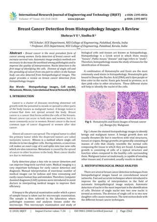
  • 1. International Research Journal of Engineering and Technology (IRJET) e-ISSN: 2395-0056 Volume: 07 Issue: 02 | Feb 2020 www.irjet.net p-ISSN: 2395-0072 © 2020, IRJET | Impact Factor value: 7.34 | ISO 9001:2008 Certified Journal | Page 2562 Breast Cancer Detection from Histopathology Images: A Review Shehras V U 1, Sindhu R2 1PG Scholar, ECE Department, NSS College of Engineering, Palakkad, Kerala, India 2 Professor, ECE Department, NSS College of Engineering, Palakkad, Kerala, India ---------------------------------------------------------------------***--------------------------------------------------------------------- Abstract - Breast cancer is the most prevalent form of cancer among women. Early detection of breast cancer will increase survival rate. Automatic image analysis methods are necessary to decreasetheworkloadamongpathologistsandto improve the quality of interpretation. Nuclei detection is the first stage of identifying the cells. Division of single cell and the spreading of cancer from one part to others in the human body can also detected from histopathological images. This paper provides a review on breast cancer detection from histopathology images. Key Words: Histopathology images, Cell nuclei, Metastasis,Mitosis,Convolutional Neural Network(CNN) 1. INTRODUCTION Cancer is a cluster of diseases involving abnormal cell growth with the potential to invade or spread to other parts of the body known as malignant tumor. A benign tumor is a tumor that does not spread around the body. Breast cancer is a cancer that forms within the cells of the breasts. Breast cancer can occur in both men and women, but it is more commonly occur in women. Breast cancer is the most common type of cancer diagnosed in women after skin cancer. Almost all cancers can spread. The original tumoriscalled the primary tumor while the dispersed tumors are called metastatic tumors. Mitosis is a method where single cell divides in to two daughter cells. During mitosis, a cancerous cell makes an exact copy of it and splits into two new cells which are also cancerous. Metastasis iscausedbythespread of cancer to other locations in the body. Most cancer deaths are due to metastasis. Early detection plays a key role in cancer detection and can improve long-term survival rates. Medical imaging is a very important technique for early cancer detection and diagnosis. Manual interpretation of enormous number of medical images can be tedious and time consuming and easily causes human biasandmistakes.Therefore,Computer Assisted diagnosis (CAD) systems were introduced to assist doctors in interpreting medical images to improve their efficiency. A biopsy is the physical examination under which a piece of sample tissue is taken out for microscopic examination. The sample is then referred to the laboratory where pathologist examines and analyses tissues under the microscope. This microscopic examination and study of biological cells and tissues are known as histopathology. Histopathology is a Greek word in which Histo means “tissues”, Patho means “disease” and logo refers to “study”. Therefore, histopathology means the study of tissues forthe identification of diseases. A combination of Hematoxylin and Eosin are the most commonly used stains in histopathology. Hematoxylin gets bound to Deoxyribo Nucleic Acid(DNA)anditdyespurpleor blue color to the nuclei. Eosin gets bound to proteins, so it dyes pink color to other structures. These different stains will help to identify the nuclei of the cells. Fig-1: Hematoxylin and Eosin Images of breast cancer (a): Benign (b): Malignant Fig-1 shows the stained histopathology images to identify benign and malignant tumor. A benign growth does not usually threaten life but it interferes with vital structures, tissues or organs. Benign growths aregenerally consistingof masses of cells that closely resemble the normal cells composing the tissue in which they are found. A malignant growth is consisting of cells of a typical structure and function when compared to the healthy cells surrounding them. Malignanttumoractmaliciously,iscapableofinvading other tissues and, if untreated, usually results in death. 2. HISTOPATHOLOGICAL IMAGE ANALYSIS There are several breastcancerdetectiontechniquesfrom histopathological images based on convolutional neural networks. Fast and accuratetechniqueswhereintroducedto improve the quality of interpretation in the field of automatic histopathological image analysis. Automatic detection of nuclei is the most importantintheidentification of cells. Division of single nuclei into two new nuclei is termed as mitosis. The division of single cell in to two new daughter cells causes metastasis. Thenextsectiondealswith the different breast cancer techniques.
  • 2. International Research Journal of Engineering and Technology (IRJET) e-ISSN: 2395-0056 Volume: 07 Issue: 02 | Feb 2020 www.irjet.net p-ISSN: 2395-0072 © 2020, IRJET | Impact Factor value: 7.34 | ISO 9001:2008 Certified Journal | Page 2563 2.1 Breast Cancer Detection Techniques M. M. Dundar et.al [1] developed a prototype system for classifying breast microscopic tissues to distinguish between Usual Ductal Hyperplasia (UDH) and actionable subtypes Atypical Ductal Hyperplasia (ADH), and Ductal Carcinoma In Situ (DCIS). The introduced system automatically evaluates digitized slides of tissues forcertain cytological criteria and classifies the tissues based on the features derived. The developed system has great potential in improving diagnostic accuracy and reproducibility. H. Su et.al [2] proposed a method to apply a fast scanning deep convolutional network (fCNN) to pixel wise region segmentation. The aim of fCNN was to removes the redundant computations in the original CNN without affecting its performance. This method is robust, efficient and scalable based on fast scanning deep neural network. A method based on the extractionofimagepatchesfor training CNN and from the combination of these patches final classification was done by F.A. Spanhol et.al [3]. This method aims to allow using high resolution images from BreaKHis as input to existing CNN in order to avoid adaptations of the model that led to a more complex and computationally costly architecture. When compared to previously reported results obtained by other machine learning models trained with handcrafted textural descriptors, a better performed CNN is introduced. T. Wan et.al [4] presented a novel image analysis based method for automatically identifying different breast cancer grades such as low, intermediate, and high in digitized histopathology. To segment cell nuclei in histopathology images, an improved hybrid active contour model based segmentation method was introduced. This method provides an accuracy of 0.92forlowVshigh,0.77 for low Vs intermediate, 0.76 for intermediate Vs high. In 2018, a method for automatic classification of breast cancer using neural network were introduced by S. Kaymak et.al [5]. The proposed methodology representsthe images using discrete Haar Wavelets which provides better image content representation and then in putting them in to neural networks. Classification of the images was achieved using Back Propagation Neural Network (BPNN) and Radial Basis Neural Network (RBFN). B. Gecer et.al [6] proposed a system that classifies whole slide images of breast biopsies in to five diagnostic categories. A saliency detector by using a pipeline of four sequential fully convolutional networks was used for multi scale processing. For diagnosis, patch based multi class convolutional network was used. Finally, a fusionofsaliency detector and the fully convolutionalized classifier network for pixel wise labelling of the whole slide. In 2019, C. Kaushal et.al [7] introduced a Computer Assisted Diagnosis (CAD) system to diagnose and detect breast cancer from histopathology images which will helps pathologists to detect the cancer earlier. CAD system will help to reduce the time taken to diagnose the cancer as well as computational complexity. Since it requires large amount of data for training, data augmentation with the support of various deep learning techniques had led to provide reliable and accurate results. R. Yan et.al [8] proposed a new hybrid model combining both convolutional and recurrent deep neural networks for breast cancer image classification which integrates the advantages of the both convolutional and recurrent deep neural networks. In this model, both the short and long term spatial correlations were preserved between the patches. This new hybrid model provides an average accuracy of 91.3%. When compared to manual detection system, Computer Aideddiagnosissystemwill providebetterresults. In deep learning, this is generally done by using convolutional neural network for extracting features and these features are classified usingfullyconnectednetwork.S. Dabeer et.al [9] trained a convolutional neural network and provided a prediction accuracy of up to 99.86%. X. Li et.al [10] studied a practical, generalizable,and self-interpretable solution to pathology image based cancer diagnosis in 2019. The proposed method learns discriminative patterns in weak-supervised manner from Whole slide imaging (WSI) labels and explains its diagnosis results via inferring locations of abnormalities in a histopathology image. Nuclei detectionistheimportantstageofidentifying the cells from histopathological images. Accurate detection and segmentation of nuclei can be done using convolutional networks and auto encoder from high resolution images of breast cancer. 2.2 Nuclei Detection Y.Al-Kofahi et.al [11] presented a robust and accurate method for the segmentation of nuclei. The image foreground is automatically extracted using a graph-cuts- based binarization. A novel method combining Multiscale Laplacian of Gaussian filtering constrained by distance map based adaptive scale selection were used to detect nuclear seed points. An initial segmentation which is refined using a second graph cuts based algorithm incorporating the method of alpha expansions were performed using these seed points. P.Wang et.al [12] proposed an automatic quantitative image analysis technique. To enhancetheimagequality,top- bottom Hat transform were applied in nuclei segmentation. To obtain the region of interest, Wavelet decompositionand Multi scale region grown (WDMR) were used. To split overlapped cells, a Double Strategy Splitting Model (DSS)
  • 3. International Research Journal of Engineering and Technology (IRJET) e-ISSN: 2395-0056 Volume: 07 Issue: 02 | Feb 2020 www.irjet.net p-ISSN: 2395-0072 © 2020, IRJET | Impact Factor value: 7.34 | ISO 9001:2008 Certified Journal | Page 2564 containing adaptive mathematical morphology and Curvature Scale Space (CSS) corner detection method were applied for better accuracy and robustness. J. Xu et.al [13] in 2015 introduced a Stacked Sparse Auto Encoder (SSAE) basedalgorithmwhichisaninstanceofdeep learning strategy for efficient nuclei detection on high resolution histopathological images of breast cancer. The SSAE learns high level features from pixel intensities in order to identify distinguishing features of cell nuclei. For the efficient and accurate detection of cell nuclei, H. Xu et.al [14]presented an automatic technique based on generalized Laplacian of Gaussian(gLoG)filterin2016. They proposed computationally efficient nuclei seed detection algorithm based on directionalsgLoGkernelsandmean-shift clustering to remove false seeds in the image background. In 2017, L. Hou et.al [15] proposed a sparse Convolutional Auto Encoder (CAE) that uses visual characteristicsofnuclei for simultaneous unsupervised nucleus detection and feature extraction in histopathology tissue images. CAE detects and encodes nuclei in image patches into sparse feature maps that encode both the location and appearance of nuclei. In 2018, X. Li et.al [16] proposed a staged identification framework for cell nuclei in colon cancer histopathology images. A cascade residual fusion block was presented to enhance the detection performance during the decoding process. A multicropping module was designed for effectively capturing contextual feature contentsaroundthe centre of a nucleus for reducing the impact of uncertainty. P. Naylor et.al [17] described a new method to automatically segment nuclei from Haematoxylin and Eosin (H&E) stained histopathology images with fully convolutional networks. They addressed the problem of segmenting touchingnuclei byformulatingthesegmentation problem as a regression task of the distance map. They showed that the fully convolutional networksarewell suited for the task of nuclei segmentation. The spreading of cancer cells is mainly due to the division of single cell to two daughter cells. The cancer causing cells will replace with the normal cells and led to the growth of abnormal cells out of control. 2.3 Mitosis Detection In 2016, A. Albayrak et.al [18] proposed a deep learning based feature extraction method by Convolutional Neural Network (CNN) for automated mitosis detection for cancer diagnosis and grading by histopathological images. After Pre processing, cell structures are found by combined clustering based segmentation and blob analysis. CNN is a prominent deep learning method on image processing tasks which were used for extractingdiscriminativefeatures.They used a robust kernel based classifier, support vector machine (SVM) for final classification of mitotic and non- mitotic cells. H. Chen et.al [19] introduced a fast and accurate method to detect mitosis by designinga novel deepcascaded convolutional neural network composedoftwocomponents. They proposed a coarse retrieval model to identify and locate the candidates of mitosis while preserving a high sensitivity. A fine discrimination model utilizing knowledge transferred from cross-domainisdevelopedtofurthersingle out mitoses from hard mimics. In S. Albarqouni et.al [20] proposed a new concept for learning from crowds that handle data aggregation directly as part of the learning process of CNN via additional crowd sourcing layer AggNet. An aggregationlayerisintroduced to aggregate the prediction results with annotation results from multiple participations. In 2017, M. Saha et.al [21] presented a supervised model to detect mitosis signature for breast histopathology WSI (Whole Slide Images) images. This model was designed using deep learning architecture with handcrafted features. The proposed architecture had an improved 92% precision, 88% recall and 90% F-score. B.K. Sabeena et.al [22] transformed a pre-trained Convolutional Neural Network by coupling random forest classifier with the initial fully connected layers to extract discriminant features from nuclei patches and to precisely prognosticate the class label of cell nuclei. The designed framework gives higher classification accuracy by carefully fine tune the pre trained model and pre processing the extraction features. For weakly supervised breast cancer diagnosis, C. Li et.al [23] proposed a deep learning scheme with novel loss function in 2019. This method utilizes a deep segmentation network to produce segmentation map. Then a filtering operation was applied to produce the detection results. The spread of cancer cells from the place where they first formed to another part of the body after the division of nuclei. In metastasis, cancer cells break away from the original (primary) tumor, travel through the blood orlymph system, and form a new tumor in other organs or tissues of the body. 2.4 Metastasis Detection M. Valkonen et.al [24] described a machine learning approach for detection of cancerous tissue from scanned whole slide images. This method was based on feature engineering and supervisor leaning with random forest model. Several local descriptors related to image texture, spatial structure, and distribution of nuclei was the features extracted from the whole slide images. In 2018, P. Grover et.al [25] proposed an algorithm for automated detection of breast cancer metastasis in whole
  • 4. International Research Journal of Engineering and Technology (IRJET) e-ISSN: 2395-0056 Volume: 07 Issue: 02 | Feb 2020 www.irjet.net p-ISSN: 2395-0072 © 2020, IRJET | Impact Factor value: 7.34 | ISO 9001:2008 Certified Journal | Page 2565 slide images. To improve the detection accuracy and overall time to localize tumorous regions, proposed algorithm leverages the capability of advanced image processing and machine learning. K. Fukuta et.al [26] developed a system for the automatic detection of breast cancer metastasis in whole slide images of histopathological lymph node sections. The system focuses on extracting feature and spatial information. They created tumor probability heat maps and perform post- processing to make patient level predictions. In 2018, H. Lin et.al [27] presented a framework by leveraging fully convolutional networks that overcomes the major speed bottleneck in whole-slide image analysis for efficient inference. For ensuring accurate detection on both micro- and macro-metastases, framework reconstructs dense heat maps. This method achieved superior performance compared to other method on Camelyon 2016 Grand Challenge dataset. In order to increase the survival of patients by early diagnosis of breast cancer, L. Tapak et.al [28] compared the performance of six machine learning techniques, two traditional methods for the prediction of breast cancer and metastasis. Results shows that the average specificity of all techniques was greater than 94% and when compared to other techniques, Support Vector Machine (SVM)andLinear Discriminant Analysis (LDA)havegreatersensitivityof73%. 3. CONCLUSIONS The analysis of histopathological images remains the most widely used method for breast cancer diagnosis. The use of computer-assisted analysis of histopathological images is a promising way to improve the analytical and predictive capabilities. The computer aided diagnosis of breast cancer from Histopathology will reduce the complexity and increase the accuracy. To classify breast cancer efficiently,different neural networks are used. Auto encoder based algorithms are proposed to identify thedistinguishingfeaturesofcell nuclei. Deep learning basedfeature extractionsformitosisdetection are performed using Convolutional Neural Networks. There are various machine learning approach for the detection of cancerous tissue from scanned Whole Slide Images. REFERENCES [1] M. M. Dundar et al., “Computerized classification of intraductal breast lesions using histopathological images,” IEEE Trans. Biomed. Eng., vol. 58, no. 7, pp. 1977–1984, 2011. [2] H. Su, F. Liu, Y. Xie, F. Xing, S. Meyyappan, and L. Yang, “Region segmentation in histopathological breast cancer images using deep convolutional neural network,” Proc. - Int. Symp. Biomed. Imaging, vol. 2015-July, pp. 55–58, 2015. [3] F. A. Spanhol, L. S. Oliveira, C. Petitjean,andL.Heutte, “Breast Cancer Histopathological Image Classification usingConvolutional Neural Networks,” Int. Jt. Conf. Neural Networks (IJCNN 2016), 2016. [4] T. Wan, J. Cao, J. Chen, and Z. Qin, “Automated grading of breast cancer histopathology using cascaded ensemble with combination of multi-level image features,” Neurocomputing, vol. 229, pp. 34– 44, 2017. [5] S. Kaymak, A. Helwan, and D. Uzun, “Breast cancer image classification usingartificial neural networks,” Procedia Comput. Sci., vol. 120, pp. 126–131, 2018. [6] B. Gecer, S. Aksoy, E. Mercan, L. G. Shapiro, D. L. Weaver, and J. G. Elmore, “Detection and Classification of Cancer in Whole Slide Breast Histopathology Images Using Deep Convolutional Networks,” Pattern Recognit., 2018. [7] C. Kaushal, S. Bhat, D. Koundal, and A. Singla, “Recent Trends in Computer Assisted Diagnosis ( CAD ) System for Breast Cancer Diagnosis Using Histopathological Images,” IRBM, vol. 1, pp. 1–17, 2019. [8] R. Yan et al., “Breast cancer histopathological image classification using a hybrid deep neural network,” Methods, no. February, pp. 1–9, 2019. [9] S. Dabeer, M. M. Khan, and S. Islam,“Cancerdiagnosis in histopathological image: CNN based approach,” Informatics Med. Unlocked, p. 100231, 2019. [10] X. Li, M. Radulovic, K. Kanjer, and K. N. Plataniotis, “Discriminative Pattern Mining for Breast Cancer Histopathology Image Classification via Fully Convolutional Autoencoder,” IEEE Access, vol. 7, no. c, pp. 36433–36445, 2019. [11] Y. Al-Kofahi, W. Lassoued, W. Lee, and B. Roysam, “Improved automatic detection and segmentationof cell nuclei in histopathology images,” IEEE Trans. Biomed. Eng., vol. 57, no. 4, pp. 841–852, 2010. [12] P. Wang, X. Hu, Y. Li, Q. Liu, and X. Zhu, “Automatic cell nuclei segmentation and classification of breast cancer histopathology images,” Signal Processing, vol. 122, pp. 1–13, 2016.
  • 5. International Research Journal of Engineering and Technology (IRJET) e-ISSN: 2395-0056 Volume: 07 Issue: 02 | Feb 2020 www.irjet.net p-ISSN: 2395-0072 © 2020, IRJET | Impact Factor value: 7.34 | ISO 9001:2008 Certified Journal | Page 2566 [13] J. Xu et al., “Stacked sparse autoencoder (SSAE) for nuclei detection on breast cancer histopathology images,” IEEE Trans. Med. Imaging, vol. 35, no. 1, pp. 119–130, 2016. [14] H. Xu, C. Lu, R. Berendt, N. Jha, and M. Mandal, “Automatic Nuclei Detection Based on Generalized Laplacian of Gaussian Filters,” IEEE J. Biomed. Heal. Informatics, vol. 21, no. 3, pp. 826–837, 2017. [15] L. Hou et al., “Sparse autoencoder for unsupervised nucleus detection and representation in histopathology images,” Pattern Recognit., vol. 86, pp. 188–200, 2019. [16] X. Li, W. Li, S. Member, R. Tao, and S. Member, “Staged Detection – Identification Framework for Cell Nuclei in Histopathology Images,” IEEE Trans. Instrum. Meas., vol. PP, pp. 1–11, 2019. [17] P. Naylor, M. Laé, F. Reyal, and T. Walter, “Segmentation of NucleiinHistopathologyImages by Deep Regression of the Distance Map,” IEEE Trans. Med. Imaging, vol. 38, no. 2, pp. 448–459, 2019. [18] A. Albayrak and G. Bilgin, “Mitosis detection using convolutional neural network basedfeatures,” CINTI 2016 - 17th IEEE Int. Symp. Comput. Intell. Informatics Proc., pp. 335–340, 2017. [19] H. Chen, Q. Dou, X. Wang, J. Qin, and P. A. Heng, “Mitosis detection in breast cancer histology images via deep cascaded networks,” 30th AAAI Conf. Artif. Intell. AAAI 2016, pp. 1160–1166, 2016. [20] S. Albarqouni, C. Baur, F. Achilles, V. Belagiannis, S. Demirci, and N. Navab,“AggNet:DeepLearningFrom Crowds for Mitosis Detection in Breast Cancer Histology Images,” IEEE Trans. Med.Imaging,vol.35, no. 5, pp. 1313–1321, 2016. [21] M. Saha, C. Chakraborty, and D. Racoceanu, “Efficient deep learning model for mitosis detection using breast histopathology images,” Comput. Med. Imaging Graph., vol. 64, pp. 29–40, 2018. [22] K. Sabeena Beevi, M. S. Nair, and G. R. Bindu, “Automatic mitosis detection in breast histopathology images using Convolutional Neural Network based deep transfer learning,” Biocybern. Biomed. Eng., vol. 39, no. 1, pp. 214–223, 2019. [23] C. Li, X. Wang, W. Liu, L. J. Latecki, B. Wang, and J. Huang, “Weakly supervised mitosis detection in breast histopathology images using concentricloss,” Med. Image Anal., vol. 53, pp. 165–178, 2019. [24] M. Valkonen, K. Kartasalo, K. Liimatainen, M. Nykter, L. Latonen, and P. Ruusuvuori, “Metastasis detection from whole slide images using local features and random forests,” Cytom. Part A, vol. 91, no. 6, pp. 555–565, 2017. [25] P. Grover, “Automated Detection of Breast Cancer Metastases in Whole Slide Images,” 2018 First Int. Conf. Secur. Cyber Comput. Commun., pp. 1–6, 2018. [26] K. Fukuta, “Identifyingmetastaticbreastcancerusing deep texture representation,” The Univercity of Tokyo , Tokyo Medical and Dental University Camelyon17.Grand-Challenge.Org. [27] H. Lin, H. Chen, S. Graham, Q. Dou, N. Rajpoot, and P. A. Heng, “Fast ScanNet: Fast and Dense Analysis of Multi-Gigapixel Whole-Slide Images for Cancer Metastasis Detection,” IEEETrans.Med.Imaging,vol. 38, no. 8, pp. 1948–1958, 2019. [28] L. Tapak, N. Shirmohammadi-Khorram, P. Amini, B. Alafchi, O. Hamidi, and J. Poorolajal, “Prediction of survival and metastasis in breast cancer patients using machine learning classifiers,” Clin. Epidemiol. Glob. Heal., vol. 7, no. 3, pp. 293–299, 2019. BIOGRAPHIES Shehras V U, Currently pursuing M.Tech in Communication Engineering from NSS College of Engineering, Palakkad, Kerala, India Dr. Sindhu R, Professor, ECE Dept, NSS College of Engineering, Palakkad, Kerala, India