CVS handouts
•
2 gostaram•1,081 visualizações

... lecture handouts, summamry of cardiovascular system

Recomendados
Mais conteúdo relacionado
Mais procurados
Mais procurados (18)
Destaque
Destaque (20)
Semelhante a CVS handouts
Semelhante a CVS handouts (20)
Mais de Ronald Magbitang
Mais de Ronald Magbitang (10)
Último
Último (20)
CVS handouts
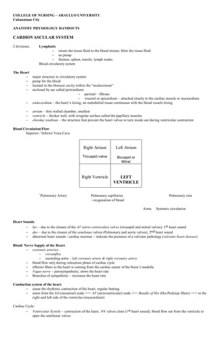
- 1. COLLEGE OF NURSING – ARAULLO UNIVERSITY Cabanatuan City ANATOMY PHYSIOLOGY HANDOUTS CARDIOVASCULAR SYSTEM 2 divisions: Lymphatic return the tissue fluid to the blood stream; filter the tissue fluid no pump thymus, spleen, tonsils, lymph nodes Blood circulatory system The Heart major structure in circulatory system pump for the blood located in the thoracic cavity within the “mediastinum” enclosed by sac called pericardium: parietal – fibrous visceral or epicardium – attached closely to the cardiac muscle or myocardium endocardium – the heart’s lining; an endothelial tissue continuous with the blood vessels lining atrium – thin walled chamber, smallest ventricle – thicker wall, with irregular surface called the papillary muscles chordae tendinae – the structure that prevent the heart valves to turn inside out during ventricular contraction Blood Circulation/Flow: Superior / Inferior Vena Cava Right Atrium Left Atrium Tricuspid valve Bicuspid or Mitral Right Ventricle LEFT VENTRICLE `Pulmonary Artery Pulmonary capillaries Pulmonary vein - oxygenation of blood Aorta Systemic circulation Heart Sounds: luv – due to the closure of the AV (atrio-ventricular) valves (tricuspid and mitral valves); 1st heart sound duv – due to the closure of the semilunar valves (Pulmonary and aortic valves); 2nd heart sound abnormal heart sounds / cardiac murmur – indicate the presence of a valvular pathology (valvular heart disease) Blood/ Nerve Supply of the Heart: coronary arteries: circumflex ascending aorta – left coronary artery & right coronary artery blood flow only during relaxation phase of cardiac cycle efferent fibers to the heart is coming from the cardiac center of the brain’s medulla Vagus nerve – parasympathetic; slows the heart rate Branches of sympathetic – increases the heart rate Conduction system of the heart: cause the rhythmic contraction of the heart, regular beating starts from the SA (sinoatrial) node >>> AV (atrioventricular) node >>> Bundle of His (His-Purkinje fibers) >>> to the right and left side of the ventricles (myocardium) Cardiac Cycle: Ventricular Systole – contraction of the heart, AV valves close (1st heart sound); blood flow out from the ventricle to open the semilunar valves
- 2. Ventricular Diastole – relaxation; AV valves open and the ventricles will be filled with blood; blood in the pulmonary artery flow back to the ventricle; semilunar valves close (2nd heart sound) Electrical Changes during Cardiac Cycle: Electrocardiogram (ECG/ or EKG) records the electrical activity of the heart as P-QRS-T waves P wave – cause by atrial depolarization QRS wave – due to ventricular depolarization/ contraction T wave – vemtricular repolarization/ relaxation U wave – not commonly recorded in ECG, due to atrial repolarization Arterial Circulation: carries blood from the heart to the capillaries capillaries are the microscopic connections between arterioles and venules veins – thin walled and will collapse when cut and severe supplies blood to the different areas/ structures of the body pulmonary circulation – for blood oxygenation; starts from the pulmonary artery to the capillary network around the alveoli (gas exchange of oxygen and carbon dioxide) to the pulmonary vein and brought the oxygenated blood to the left atrium systemic circulation – from the aorta gives the 3 main branches: left common carotid left subclavian a. innominate or brachiocephalic – gives the right common carotid and right subclavian a. ligamentum arteriosum (ductus arteriosus) – blood flow from the pulmonary artery into the aorta and bypassing the pulmonary circulation during the fetal life DISEASES OF THE CVS Diagnostics: ECG/EKG, phonocardiogram, echocardiography, doppler, arteriography, cardiac catheterization, radionucleide History and physical examination Diseases: Atherosclerosis or Arteriosclerosis thickening and hardening of the arteries caused by the deposition of the fatty substance on the walls of the arteries Hypertension (HTN/HPN) increase in the BP above 139 mmHg systolic BP and 89 mmHg diastolic BP cause by the increase in the peripheral resistance can be essential (i.e. no known cause) can be secondary (cardiovascular, renal or endocrine pathology) Ischemic Heart Disease (IHD) or Coronary Artery Disease (CAD) caused by the decrease in the blood supply to the heart maybe due to a decrease blood flow or decrease supply due to either increased peripheral resistance or narrowing or blockage of the coronary artery Dysrythmias or Cardiac arrhythmia irregular cardiac rhythm due to either pathology in the conduction system of the heart or contractility of the heart Valvular Heart Disease (VHD) problem on the either of the different valves (AV or semilunar valves), resulting to either narrowing or tightening of the different valves may cause stenosis (tight) or regurgitation (looses) of the valves maybe caused by either an inborn defect or due to a connective tissue disease (i.e. Rheumatic Heart Disease/RHD) Congenital Heart Disease (CHD) an inborn defect on either of the different structures of the heart maybe a defect on the septum or partition between the atrium (Atrial septal defect/ ASD) or between the ventricle (Ventricular septal defect/ VSD); may also present as valvular pathology; conduction problem; or pathology on the areas of the great blood vessels, the aorta or pulmonary vessels (i.e. Tetralogy of Fallot) Congestive Heart Failure (CHF) the failing heart maybe caused by either an increase in the peripheral resistance, contraction and/or arrhythmia,
- 3. Peripheral Arterial disease Arterial aneurysm Varicose veins Blood Cell Diseases: Anemia; Polycythemia vera; Hemorrhagic diseases RONALD S. MAGBITANG, M.D