SlideShare uma empresa Scribd logo
1 de 42
Smear Layer In Endodontics
The presence or absence of the smear layer is of interest not only to
restorative dentists, but to endodontists as well. Whenever dentin is filed, a smear
is produced on its surface. This has been demonstrated in many studies with SEM
(Baker & others, 1975; McComb, Smith &Beagrie, 1976; Lester & Boyde,
1977; Goldman & others, 1982).
Fig. 32
Dentin of a root canal treated with 5% NaOCl and 17% EDTA to remove pulpal tissue and the
smear layer. A no. 8 file was drawn over the clean surface in the middle of the scanning electron
micrograph, creating a smear layer. Scanning electron micrograph X2000.
The morphology of the canal wall is of interest in this context. In adult teeth
the walls may be partly covered with atubular, irregular dentin and thus the
tubules are blocked in the same way as under erosion and abrasion. Infection may
not be seen in the tubules in such an area. However, in many adult teeth and
especially in young teeth we may have large areas with primary dentin facing the
root canal. From a necrotic and infected canal, bacteria enter the dentin and can be
found rather deep in the tubules. Infected tubules with fluid communication to the
exterior may cause pathological complications such as external resorption of roots
and periapical pathosis.
In the treatment of infected roots there is a good reason to remove smear
plugs from the apertures of the tubules by using, for instance, EDTA. In this same
way, the bacteria within the tubules at some distance can be more easily destroyed
71
Smear Layer In Endodontics
by an intracanal dressing. On the other hand, if the asepsis or the sealing is poor,
we may run the risk of reinfecting dentinal tubules opened and widened by
treatment with EDTA. The situation is similar to that for cavities.
Another important consideration is that the smear layer is a separate structure
from the underlying dentin.
Fig. 31: fig 4,pg 18,op dent 1984 is a fortuitous scanning electron micrograph of
the smear layer lining a root canal that pulled away from the underlying dentin
during processing. The absence of superficial smear layer may facilitate good
contact between the sealing material and the wall of cut dentin.
Fig 33
SEM of a region in the root canal where the smear layer (SL) cracked open
and pulled away from the underlying dentinal tubules (DT). X 1700
The removal of the smear layer from the dentin lining the pulp chamber and
root canals has been the subject of numerous investigations. Lester & Boyde
(1977) found that treatment for 3 days with 5% sodium hypochlorite did not
remove smear plugs from apertures of tubules and may not diffuse into the
dentinal tubules sufficiently to take care of microbes that have penetrated deeper
into the dentinal tubules. However, Cameron (1983), using an ultrasonic
technique, found that 3% sodium hypochlorite combined with ultrasound for 3 to
5 minutes removed not only the superficial smear layer but also the smear plugs;
72
Smear Layer In Endodontics
but one minute of ultrasound removed only the superficial smear layer. In this
method we may have a selective technique that can modify the effect of an
irrigating solution. As suggested by Yamada & others (1983), an alternative
would be a combination of irrigants. They found the combined use of 10 ml of
17% EDTA, followed by 10 ml of sodium hypochlorite effective. Goldman &
others (1982) recommend alternate use of sodium hypochlorite and EDTA to
remove smeared dentin. The sodium hypochlorite removes the organic material,
including the collagenous matrix of dentin, and EDTA removes the mineralized
dentin, thereby exposing more collagen. There are no reasons to believe that a
short application of these solutions would have any deleterious effects on the
periapical tissues already replaced by granulation tissue. Such preparative
treatment of root canals presumably permits a better adaptation of obturating
materials and sealers to the dentin.
To reduce the risk of reinfection, but also to avoid the development of
secondary caries, in permanent coronal restorations of root-filled teeth the cavity
should be treated in the same way as cavities in vital teeth, that is, a proper
cleansing and lining of all cavity walls.
Smear layer in Endodontics can be discussed under the following headings:
(I) Effect of instrumentation on endodontic smear layer
(II) Intracanal medicaments and smear layer
(III) Obturation and smear layer
(IV) Post cementation and smear layer
(V) Root-end filing and smear layer
73
Smear Layer In Endodontics
(I) EFFECT OF INSTRUMENTATION ON ENDODONTIC
SMEAR LAYER
Thorough biomechanical preparation of the root canal is unanimously
considered to be one of the major requirements for successful endodontic
treatment. The prime objectives of this phase are to remove completely the
organic substance that may be infected, or become so, and to shape the root canal
in conformity with the principles of obturation. Over the years a variety of
instruments and techniques have been proposed to reach this goal. Manual
preparation techniques vary both in the type of instruments used and in the
sequence of using them. Hand preparation techniques are time consuming,
technically demanding and may lead to iatrogenic errors (ledging, zipping, canal
transportation and apical blockage). Since optimal shaping and cleaning of root
canals with manual files is one of the most difficult aspects of root canal
treatment, many alternative techniques have been suggested. Since the 1950s a
number of different endodontic handpieces have been developed for root canal
preparation with the aim to mainly decrease instrumentation time and to simplify
root canal preparation. Automated techniques utilize devices (Canal Finder,
Excalibur, Giromatic, Endolift, Cavi-Endo etc.) that rotate files, displace them
vertically, or that do both movements. Numerous investigations have
demonstrated the limitation of manual and automated root canal instrumentation
regarding the overall quality of preparation (Weine et al, 1976; Lehman &
Girstein, 1982; Turek & Langeland, 1982; Bolanos et al, 1988; Hulsmann &
Stryga, 1993, Bertrand et al, 1999). These problems have resulted in a wide
search for innovative materials, instruments and techniques to obtain a clean,
disinfected debris-free canal for obturation (Yang et al, 1996).
During the last decade several new nickel-titanium instruments for manual
root canal preparation as well as for use in rotary endodontic handpiece, have
been developed in order to facilitate the difficult and time-consuming process of
cleaning and shaping the root canal system and to improve the final quality of root
canal preparation. Numerous studies have reported they could efficiently create a
74
Smear Layer In Endodontics
smooth, predetermined funnel-form shape, with minimal risk of ledging and
transporting the canals (Esposito & Cunningham, 1995; Glosson et al, 1995,
Short et al, 1997; Thompson & Dummer, 1997b). Shaping procedures can be
completed more easily, quickly and predictably.
Cleansing efficiency has been one of the issues discussed with regard to
preparation techniques. It has been studied extensively, mainly by means of
observation of the root canal walls and contents after preparation. The pulpal
debris and smear layer produced by instrumentation of the canal walls must be
removed (Cergneux, 1987; Gettleman et al, 1991). During preparation,
insufficient removal of debris and smear layer material can induce stresses on the
cutting segment of endodontic instruments. Their removal depends not only on the
irrigation method but also on the endodontic instrument, i.e., the way the
instrument is used and the method of preparation.
Numerous studies have been reported on the relative effectiveness of
different instrumentation techniques, based on a variety of ways of evaluating
canal debridement. Outcomes of instrumentation differ according to the method of
canal preparation and evaluation, each method showing advantages and
disadvantages (Heard & Walton, 1997). Introduction of the scanning electron
microscope (SEM) has proved to be a valuable method for assessment of the
ability of the endodontic procedures to remove debris from the root canals, thus
enabling comparison of instruments and techniques. Numerous authors (Bolanos
& Jensen, 1980; Haikel & Allemann, 1988, Hulsmann et al, 1997) have
indicated that scanning electron microscopes are indeed useful in examining
instrumented root canal walls within the context of evaluating the efficiency of
different instrumentation systems. SEM studies focus on observing the smear
layer, the patency of dentinal tubules, instrumentation striations and
calcospherites. These studies have conflicting results. In most investigations
manual preparation was found to be more effective and superior to automated
preparation and ultrasonic preparation. In other studies ultrasonic preparation
resulted in cleaner canals. Several researchers did not find significant differences
75
Smear Layer In Endodontics
between preparation techniques. Similarly, the cleaning effectiveness of the rotary
nickel-titanium systems is controversial. As both manual and mechanical shaping
produce smear layer and debris (Hulsmann & Stryga, 1993; Heard & Walton,
1997), it is important to develop an instrumentation technique for endodontic
treatment that produces a minimal amount of smear layer and debris in order to
obtain the optimal seal of the root canals.
Studies evaluating the cleansing efficiency of preparation techniques are as
follows:
(A) THOSE THAT INDICATE BETTER CLEANSING EFFICIENCY
WITH MANUAL INSTRUMENTATION:
Velvart (1987) found hand instrumentation to be superior to ultrasonic
cleaning in the apical third of the canal. Complete cleanliness could be achieved
by none of the techniques and devices investigated.
Baker et al (1988) compared the canal cleanliness following ultrasonic
preparation and manual preparation. They found significantly cleaner root canal
walls in the middle part of the root canal following hand instrumentation; in the
apical and coronal parts no significant differences were detected.
Ahlquist et al (2001) compared the cleanliness of the root canal walls
following either a manual technique using stainless steel S-files (Sjodings,
Sendoline, Sweden) or a rotary technique (ProFile® Rotary nickel-titanium files
in a handpiece) of canal instrumentation. It was found that the manual technique
employed produced cleaner canal walls than the rotary technique.
76
Smear Layer In Endodontics
(B) THOSE THAT INDICATE BETTER CLEANSING EFFICIENCY
WITH ULTRASONIC AND SONIC DEVICES:
Bolanos et al (1988) compared sonic instrumentation with hand
instrumentation and the Giromatic system. The sonic preparation resulted in the
best cleanliness in all parts of the root canal in straight or curved canals. Hand
instrumentation was slightly superior to Giromatic preparation.
Schadle et al (1990) compared the efficacy of various root canal cleaning
instruments, i.e. hand-instruments, sonic instruments (Sonic Air 3000 and
Endostar 5), mechanical instruments (Canalfinder System), and ultrasonic
instruments (Cavi-Endo with or without integrated rinsing). The smear layer was
somewhat reduced by the Cavi-Endo without the integrated spray.
Prati et al (1994) evaluated the morphology of the smear layer and the
amount of debris and pulpal residues in the apical third of human teeth by
comparing four manual endodontic instruments, an ultrasonic/endosonic system
and an automated system (Excalibur). The scoring system was as described by
Ahmed et al (1988), i.e. a three point scoring system:
0: smear layer was absent and more than 50% of tubules were visible and partially
open.
1: less than 50% of dentinal tubules were visible; smear layer was present in some
areas.
2: smear layer covered the canal wall; dentinal tubules were partially visible in
limited area.
3: smear layer covered the dentinal tubules.
Ultrasonic technique showed the complete removal of the smear layer
(scores 0 to 2), leaving small amounts of pulp debris at the apical third.
Cameron (1995) reported a superior cleaning ability of the ultrasonic
systems. Even most of the smear layer could be removed.
77
Smear Layer In Endodontics
Hulsmann et al (1997) evaluated the root canal cleanliness after preparation
with eight different automated devices and hand instruments. The teeth were
investigated under the SEM using the five category scoring system which is as
follows (Fig. 34) :
Score 1: No smear layer, dentinal tubuli open.
Score 2: Small amount of smear layer, some dentinal tubuli open.
Score 3: Homogenous smear layer covering the root canal wall, only few dentinal
tubuli open.
Score 4: Complete root canal wall covered by a homogenous smear layer, no open
dentinal tubuli.
Score 5: Heavy, nonhomogenous smear layer covering the complete root canal
wall.
No preparation system or technique resulted in complete removal of smear
layer and debris. The ultrasonic unit performed best followed by the Canal
Leader 2000 and hand instrumentation.
78
Smear Layer In Endodontics
Fig 34
Score 1 Score 2
Score 3 Score 4
Score 5
79
Smear Layer In Endodontics
(C) THOSE THAT INDICATE BETTER CLEANSING EFFICACY WITH
AUTOMATED HANDPIECES:
Bartha & Rocca (1993) assessed the cleansing-effect of two endodontic
handpieces (Excalibur, W & H; MM 1400 Micro Mega). It was found that
Excalibur was superior in eliminating the SL.
Schafer et al (2000) investigated the cleaning effectiveness of automated
(KaVo-Endo Flash device and ProFile system) and manual root canal
instrumentation with the aid of SEM. Evaluation for smear layer was done using
the 5-step scale as given by Hulsmann et al (1997). Comparison of manual
instrumentation with the automated KaVo-Endo Flash resulted in an
equivalent degree of canal cleaning. Furthermore, in comparison with the
canals that were instrumented with the ProFile instruments, the KaVo-Endo
Flash resulted in significantly better root canal cleanliness in curved canals.
These results corroborate the reports from other authors, in that most cases of
manual instrumentation proved to be superior to mechanical instrumentation as far
as cleaning efficiency is concerned (Hulsmann et al, 1997; Mizrahi et al, 1975;
Schwarze & Geurtsen, 1996) and that automated devices in most cases create a
thicker smear layer than manual instrumentation (Schwarze & Geurtsen, 1996).
(D) THOSE THAT INDICATE BETTER CLEANSING EFFICIENCY
WITH ROTARY TECHNIQUES:
Valli et al (1996) compared the debridement ability of Canal Master and K-
Files using scanning electron microscope. The results showed that the Canal
Master produced cleaner showing lesser debris than that produced by K-Files.
Bertrand et al (1999) determined the ability of the Quantec Series 2000
rotary nickel-titanium endodontic system to remove the dentinal debris and smear
layer produced during canal preparation. The absence or presence of a smear layer
80
Smear Layer In Endodontics
was rated and scored on three appearances using the scale described by Ciucchi et
al (1989) as follows:
Score 0: No visible tubule orifices-surface is totally smeared.
Score 5: Scattered open tubules orifices-surface is partially free of smear layer.
Score 10: Regularly distributed open tubule orifices-surface is free of smear layer.
The conclusion was that Quantec rotary system produced cleaner canal
walls than conventional manual instrumentation, particularly in the middle and
apical third.
Bechelli et al (1999) compared the efficacy of root canal wall debridement
following hand versus Lightspeed ™ instrumentation. Less smear layer was
present in the apical region following Lightspeed™ instrumentation than
stainless steel hand files, but this difference was statistically not significant.
Roggendorf et al (1999) found ProFile superior to Lightspeed and
Quantec instruments (no specification on the type of Quantec files) or hand
instrumentation.
Medioni et al (1999) reported the superior cleaning ability of Quantec
Series 2000 when compared to HERO 642, ProFile, and hand instrumentation.
Peters & Barbakow (2000) studied the effect of irrigation on debris and
smear layer on the canal walls prepared by two rotary techniques, i.e.: Lightspeed
(LS) and Profiles (PS) systems. The larger canal preparations obtained in this
study with Lightspeed instruments enabled a more effective removal of the
smear layer than the PF group.
Hulsmann, Schade & Schafers (2001) compared cleaning ability using two
different rotary nickel-titanium instruments: HERO 642 (MicroMega, France) and
Quantec SC (Tycom, CA, USA). Results indicated that cleaner root canal walls
were found after preparation with HERO 642 (53% scores 1 and 2), followed by
Quantec SC (41%).
81
Smear Layer In Endodontics
Gambarini & Laszkiewicz (2002) assessed the debris and smear layer
remaining following canal preparation with GT rotary instruments. It was
concluded that the GT™ rotary instruments removed debris effectively, but left
root canal walls covered with smear layer particularly n the apical third.
Baumann et al (2003) found that RCT with RaCe™ had better smear layer
removal than hand instrumentation.
Almannai et al (2003) compared 4 different rotary nickel-titanium
instruments (ProFile®, ProTaper®, RaCe®, and NiTee®) with regards to canal
cleanliness. Significantly less smear layer was observed in the apical third of the
ProTaper group when compared with the ProFile group.
In-Soo Jeon et al (2003) compared the quality and amount of smear layer
generated in the apical third of straight root canals by 2 rotary nickel-titanium
reamers and I rotary steel reamer with different cutting blade designs. Automated
preparation was performed with ProFile and HERO 642 reamers using the crown-
down technique and with a stainless steel engine reamer (Mani) by using a
reaming motion. A 4 – category scoring system for smear layer was used which is
as follows:
0: no smear layer/all tubules clean and open.
1: no superficial smear layer/tubule openings visible, but some contain debris plug
or soft tissue remnants.
2: moderate smear layer/some tubules open and others closed.
3: heavy smear layer and most/all tubule openings obscured.
The results indicated that the least smear layer remained in the HERO 642
group. In the ProFile group, the smear layer had a shiny and burnished
appearance with few openings of dentinal tubules. The depth of the packed smear
layer into the tubules was deeper than in the Hero group. The smear layer
generated by the steel reamers appeared thinner and less compressed than in the
ProFile group.
82
Smear Layer In Endodontics
Fig 35
A B
A scanning electron micrograph of a canal wall
prepared with Hero 642 rotary reamers has a
snowy appearance with many open dentinal
tubules (Score 1: Original magnification
X1500).
A scanning electron micrograph of a canal wall
prepared with ProFile rotary reamers. The
canal wall was to be covered by a thick,
nonhomogenous layer, with a shiny burnished
appearance and almost no open dentinal tubule
(Score 3; original magnification x1500).
C D
Cut-view of a canal wall prepared with Pro-
File rotary reamers at the apical 2mm level.
The smear layer had a muddy appearance
(X5000).
Cut-view of a canal wall prepared with the
Hero rotary reamers at the apical 2mm level.
Generally, the tubular packing is less frequent
than that obtained in the ProFile group. Thin
smear layer with many open dentinal tubules
covered on the root canal wall (X3000).
83
Smear Layer In Endodontics
(E) THOSE THAT HAVE SHOWN INCONCLUSIVE RESULTS OR
SIMILAR RESULTS WITH DIFFERENT TECHNIQUES:
Klimm et al (1990) evaluated the efficacy of irrigated, hand root canal
preparation by SEM. In all groups a homogeneous or inhomogeneous smear
layer of different thickness and extent dominated.
Mandel et al (1990) evaluated the cleansing efficiency of manual serial
preparation, endosonic (Cavi-Endo) preparation and automated preparation with
the Canal Finder System. No preparation technique was found to result in any
characteristic microscopic features that distinguished it from the other
techniques.
Lumley et al (1992) investigated the effect of precurving Endosonic files on
the amount of debris and smear layer remaining in curved root canals. The result
of this study indicated that precurving of files decreased the amount of debris
but did not affect smear layer removal.
Sydney et al (1996) analyzed the smear layer removal after root canal
preparation by a manual technique and by an automated handpiece, the Canal
Finder System (CFS).Both the techniques showed root canal walls with dense
smear layer.
Heard & Walton (1997) compared four root canal preparation techniques in
small curved canals. The four methods were: (1) step-back without initial coronal
flaring, (2) step-back with coronal flaring, (3) step-back with initial coronal
flaring and finished by ultrasonic irrigation and, (4) ultrasonics only. The results
indicated that there were no statistically significant differences between the
techniques.
Kochi et al (1998) could find no difference between Quantec (No
information on type of file) and manual preparation using K-files.
84
Smear Layer In Endodontics
Mayer et al (2002) evaluated smear layer scores after two types of
instruments to ultrasonically activate irrigants during 2 types of canal preparation
(ProFile .04 and Lightspeed). It was found that ultrasonically activated irrigants
did not reduce smear layer scores and this finding was not influenced by the
material or by the design of the instrument used to transmit ultrasonic activation.
There were no differences in amount of smear layer on canal wall prepared
with PF or LS instruments.
Lichota et al (2003) assessed the cleanliness of root canal walls following
ProFile rotary technique of instrumentation. Thus though they removed debris
effectively, ProFiles left the root canal walls covered with smear layer in the
apical and middle thirds.
Thus, from the above studies we can conclude that there is still no consensus
on the best instrumentation technique which may result in cleanest possible canal
without the presence of a smear layer. Further research is required to resolve the
current controversy on the best instrumentation technique.
85
Smear Layer In Endodontics
(II) INTRACANAL MEDICAMENTS AND SMEAR LAYER
The goal of clinical treatment is to completely disrupt and destroy the
bacteria involved in the endodontic infection. Endodontic infection will persist
until the source of irritation is removed. Previous studies have shown that bacteria
in infected root canals and periradicular tissues are capable of invading and
residing deeply within dentin and in cementum around the periapex. (Ando &
Hoshino, 1990; Kiryu et al, 1994; Peters et al, 2001). Further more, it has been
demonstrated that although bacteria in artificial smear layers and prepared
reservoir channels in deeper layers of root dentin could be eliminated by
procedures such as ultrasonic irrigation with NaOCl (Huque et al, 1998),
microorganisms within fins and isthmuses could still remain viable (Sato et al,
1996). Even after chemomechanical instrumentation of the canal, some bacteria
still remain in the canal and dentinal tubules (Bystrom & Sundqvist, 1981, 1983,
1985). Total debridement is impeded because of accessory canals, fins, cul-de-
sacs and communications between the main canals. Such microorganisms may
cause root canal treatment to fail.
The rationale behind intracanal medication is to destroy residual
microorganisms and their toxins and to remove organic tissue. The medicament
should inhibit microbial recolonization of the cleaned parts of root canal system
by preventing residual microorganisms from growing and new organisms
invading through lateral communications and coronal access. For this reason,
chemomechanical cleansing is often supported by the use of disinfectants.
Various medicaments have been proposed for disinfection of root canals.
The traditional phenolic or fixative agents include camphorated
monochlorophenol (CMCP), formocresol, and cresatin. Iodine potassium iodide
and calcium hydroxide are the main nonphenolic intracanal medications. These
medicaments are potent antibacterial agents under laboratory test conditions;
however their effectiveness in clinical use is unpredictable (Messer & Chen,
1984). According to some researchers, they also neutralize and render canal tissue
86
Smear Layer In Endodontics
remnants inert. Some medications contain aldehyde derivatives that can be used to
fix fresh tissues for histological examination; however, they may not effectively
fix necrotic or decomposed tissues (Walton & Rivera, 2002). According to
Wesselink et al (1977), fixed tissues are not inert and may become more toxic
and antigenic after fixation. Intracanal medications have also been used clinically
to prevent posttreatment pain. Studies have shown, however, that routine use of
these materials as intracanal medication has no significant effect on the prevention
of pain. According to Oguntebi (1994), most currently used intracanal
medicaments have a limited antibacterial spectrum and antigenic potential. In
addition, some of them have a limited ability to diffuse into dentinal tubules.
Effect of intracanal medications on bacteria in dentinal tubules:
After removal of the smear layer, Haapasalo & Orstavik (1987) inoculated
the bovine incisors with E. faecalis and found penetration of these bacteria into
the dentinal tubules upto 1 mm. They demonstrate that liquid CMCP completely
disinfected the dentinal tubules but that Ca(OH)2 was ineffective. Behnen et al
(2001) had better success killing E. faecalis with either Pulpdent or a 10%
solution of Ca(OH)2 than with the traditional thick mixes. The difference might
have been attributable to the different viscosity of various types of Ca(OH)2 used
in this experiment. Heling & Chandler (1998) also inoculated dentinal tubules of
bovine teeth with E. faecalis and then examined the disinfecting effect of various
irrigants. They found that none of the test irrigants (chlorhexidine, hydrogen
peroxide, sodium hypochlorite, EDTA or their combinations) were totally
effective. Siqueira & De Uzeda (1996) inoculated bovine dentinal cylinders with
1 facultative and 2 obligate anaerobic bacteria and examined the disinfecting
effect of Ca(OH)2 mixed with saline solution or CMCP for 1 hour, 1 day, and 1
week. Their results showed that Ca(OH)2 and saline solution were ineffective in
disinfecting the dentinal tubules after 1 week of application. In contrast, a mixture
of Ca(OH)2 and CMCP resulted in complete disinfection of the dentinal tubules in
1 day. Jeansonne & White (1994) and Kuruvilla & Kamath (1998) examined
the antibacterial effect of chlorhexidine gluconate and sodium hypochlorite on
87
Smear Layer In Endodontics
inoculated human teeth in vitro. They reported a reduction in bacterial counts but
not total disinfection of the root canals.
In an in vivo experiment, Katebzadeh et al (1999) showed that infected dog
teeth that were filled experienced treatment failure more frequently than those
medicated with Ca(OH)2 before obturation. Sjogren et al (1997) examined the
presence and the influence of bacteria on the long-term success of root canal
therapy. Their results show that 40% of root canals remain infected after
instrumentation. In addition, they reported that teeth filled in 1 visit without the
use of Ca(OH)2 experienced treatment failure significantly more frequently than
those that were medicated for 1 week with Ca(OH)2 (68% vs. 94%). The results of
this study corroborate the findings of Bystrom et al (1987), who showed
improved clinical success rates after effective disinfection of root canals.
Gutierrez et al (1991) evaluated the diffusion of medicaments within the root
canal dentin after mechanical preparation and flushing with alternate NaOCl and
hydrogen peroxide followed by placement of the medicaments (CMCP, CMCP
with formocresol and methylcresylacetate) by paper points. It was found that all 3
medicaments crystallized both on dentin walls and inside the dentinal tubules.
Bacteria remaining in dentinal tubules in close relationship with CMCP crystals
appeared shrunken.
Smear layer- a physical barrier for disinfectants:
According to some authors (Goldberg & Abramovich, 1977; Wayman et
al, 1979; Yamada et al, 1983; Berg et al, 1986; Baumgartner & Mader, 1987)
the presence of the smear layer may block the antimicrobial effect of intracanal
disinfectants into the tubules by preventing their penetration into the tubules.
In an in vitro study, Orstavik & Haapasalo (1990) showed the importance
of removal of the smear layer and the presence of patent dentinal tubules for
decreasing the time necessary to achieve the disinfecting effect of intracanal
medications. They found that camphorated p-monochlorophenol was generally
more efficient than Calasept, and of the irrigants tested, iodine potassium iodide
88
Smear Layer In Endodontics
appeared more potent than sodium hypochlorite or chlorhexidine. They also
concluded that the smear layer did delay, but not abolish the action of
disinfectants. However, following the removal of the smear layer, bacteria in
dentinal tubules can be easily destroyed (Brannstrom, 1984) and in this way, it
may be beneficial to use lower concentrations and/or amounts of antibacterial
agents since all of these agents show some degree of toxicity to viable host cells.
Bystrom & Sundqvist (1985) have also shown that the presence of a smear
layer can inhibit or significantly delay the penetration of antimicrobial agents
such as intracanal irrigants and medications into the dentinal tubules.
Diffusion through dentin is directly proportional to the surface area of
dentinal tubules and inversely proportional to the dentin thickness. Also the
presence of the smear layer acts as a diffusion barrier that can reduce diffusion by
25-30% (Pashley, 1988). The presence of a smear layer has previously been
demonstrated to decrease the diffusion of triamcinolone and
demethylchlortetracycline, two active components of the root canal medicament,
Ledermix, through radicular dentin (Abbott et al, 1989).
Foster et al (1993) evaluated the effect of smear layer removal on the
diffusion of calcium hydroxide through radicular dentin. Pulp extirpation and the
placement of Ca(OH)2 into the root canal 7 to 14 days after a traumatic episode
are recommended to decrease the incidence of inflammatory root resorption.
Tronstad (1981) suggested that Ca(OH)2 placed in the root canal elevates the pH
in areas of resorption on the surface of the root by diffusion of OH-
through the
dentin, thereby decreasing osteoclastic activity and activating alkaline
phosphatases. The result of this study demonstrated that Ca(OH)2 diffuses from
the root canal to the exterior surface of the root and that the removal of the
smear layer may facilitate this diffusion. Thus the root canal space may be
considered a route for medication delivery to the dentinal tubules themselves, if
disinfection is required, and/or to the external root surface.
89
Smear Layer In Endodontics
Cruz et al (2002) evaluated the penetration of propylene glycol into root
dentin. Propylene glycol is a vehicle that has the potential for use in root canal
medicaments. It has been reported to be a widely used vehicle for various
pharmaceutical and commercial products such as drugs, cosmetics and foods
(Morshed et al, 1988) and it is also used as a constituent of Caries Detector®
(Fusayama, 1988). In endodontics, it has been used as a vehicle for calcium
hydroxide (Saiijo, 1957; Laws, 1962; Laws, 1971; Simon et al, 1995). In this
study, Safranin O in propylene glycol and in distilled water was introduced into
root canals with and without smear layer. It was found that the area and depth of
penetration with propylene glycol was significantly greater than with distilled
water. Smear layer significantly delayed the penetration of dye. Thus it is seen
that propylene glycol delivered dye through root canal system rapidly and more
effectively indicating its potential use in delivering intracanal medicaments. Also,
propylene glycol is less cytotoxic than other vehicles, possesses antibacterial
properties and is hygroscopic, thus allowing absorption of water which results in
sustained release of intracanal medicaments for prolonged periods (Fava &
Saunders, 1999).
Thus, it is seen that generally, removal of smear layer helps in diffusion of
the intracanal medicaments and thereby improves their efficacy.
90
Smear Layer In Endodontics
(III) OBTURATION AND SMEAR LAYER
Complete obturation of the root canal with an inert filling material and
creation of a hermetic, apical seal have been proposed as goals for successful
endodontic treatment (Nguyen, 1984). To achieve this, the root canal filling must
seal the canal space both apically and coronally to prevent the ingress of
microorganisms or tissue fluids into the canal space. Apical leakage is considered
a common reason for the clinical failure of endodontic therapy. Likewise, coronal
leakage is also reported to be an important reason for failure (Madison et al,
1987; Saunders & Saunders, 1994). Gutta-percha is considered an impermeable
core material; therefore, leakage through an obturated canal is expected to take
place at the interface between sealer and dentin or sealer and gutta-percha, or
through voids within the sealer. Hence, the sealing quality of a root canal filling
depends much on the sealing ability of the sealer.
A review of a large number of published leakage studies points to general
agreement that leakage occurs between the root filling and the root canal wall.
Therefore, anything that may influence the adaptation of the root filling to the
canal wall is of great significance in determining the degree and the extent of
leakage, and ultimately the prognosis of the endodontic therapy. Because the
smear layer remaining on the root canal wall has been characterized, there has
been much interest in the possible effect of the smear layer on the leakage of the
root canal sealer. There is a controversy over whether or not to remove this layer
before obturation and there is still no consensus on the influence of the smear
layer on the development of an effective seal of the root canal system.
Some consider that it is desirable to remove this layer as it covers prepared
areas and prevents medicaments and filling material from penetrating the dentinal
tubules or even contacting the canal wall. As of today, almost no material has
been shown to enter dentinal tubules with the smear layer present. Removal of the
smear layer is also considered to be desirable by others because, in addition to
inorganic particles, it may contain some organic material, as well as viable
91
Smear Layer In Endodontics
bacteria. Biologically, the presence of the smear layer has been postulated to be an
avenue for leakage and a source of substrate for bacterial growth and ingress
(Pashley, 1984). The frequency of bacterial penetration in the presence of a smear
layer, when canals were obturated with thermoplasticized gutta-percha and sealer,
has been shown to be significantly higher than with smear layer removal and
obturation (Behrend et al, 1996). A further concern is the presence of viable
bacteria that may remain in the dentinal tubules and use the smear layer for
sustained growth and activity (Brannstrom, 1984; Olgart et al, 1974).
Because of the bacterial content of the smear layer, any apical extrusion of
the smear layer during instrumentation or obturation can defeat one of the goals of
endodontic therapy: the return to and maintenance of an inflammation-free state in
the periapical area. To be considered an ideal sealer, a material should not itself
cause or further irritate the periapical tissue. Some root canal filling materials,
especially N2 paste and silver points, are not biocompatible. To risk further tissue
trauma with a technique that may induce or potentiate periapical inflammation is
unthinkable. Smear layer induced inflammation of the periapical area can be
caused by over instrumentation or by the careless measurement and filling of a
master cone. It has been recommended that master cones be foreshortened to fit
1mm short of the apex as an effective countermeasure to creating pre- and post
obturation periapical inflammation.
Some doubt exists if the formation of apical plugs from a combination of
dentin chips, soft-tissue fragments and fibers from instrumentation may enhance
the seal of a root canal filling. Dentin filings occur during instrumentation, but the
formation of an apical plug, from them is often an inadvertent or accidental
occurrence. Well-formed apical plugs can often reduce the inflammatory reactions
following filling by limiting the apical extension of filling materials thereby
encouraging healing. If more cementum is going to form to create a better apical
seal, dentin chips at the apex of a root can act as a nidus for formation of hard
tissue. Bacterial contamination by the presence of the smear layer can prevent
this.
92
Smear Layer In Endodontics
Endodontic sealers act as a glue to ensure a good adaptation of gutta percha
to the canal walls. If the smear layer is not removed, the gutta percha may
occasionally be glued to the dentin in the smear layer as well as to exposed parts
of the canal wall. The smear layer may interfere with the penetration of the gutta-
percha into the tubules and the adhesion and penetration of the sealers into the
dentinal tubules. Not being firmly attached to the dentin, the smear layer may
laminate off the canal wall and create a false seal, voids in the fill and an expected
environment for microleakage. Thus the retention or removal of the smear layer
before obturation may influence the quality of the obturation.
In contrast, other investigators (Pashley et al, 1981) consider that the smear
layer should be left intact, as it may actually form a protective barrier. The smear
layer may be responsible for excluding bacteria from dentinal tubules as well as
restricting the surface area available for the diffusion of many molecules. One
study (Michelich et al, 1980) has shown that the smear layer will prevent
bacterial penetration (Streptococcus mutans), but will permit fluid filtration.
Another study (Williams & Goldman, 1985) has revealed that the smear layer
simply delays the penetration of some bacteria (Proteus vulgaris) rather than
preventing it.
Many articles have been written on the physical properties of root canal
sealers, including the their adhesive strength to dentin (Orstavik et al, 1983;
McComb & Smith, 1976) and to gutta-percha (Orstavik et al, 1983). Adhesive
strength measurements may be important to clinical usage, because higher
adhesive strengths may reduce leakage in clinical situations. Opening of the tubuli
by removal of the smear layer prior to filling the root canal system makes great
sense. If chemical adhesion between the dentin and sealers, pastes, cements or
plastics cannot be easily achieved, then why not a mechanical lock? The material
will flow or be forced back into the empty dentinal tubules, gripping like tentacles
and forever resisting displacement! This may actually increase the adhesive
strength of sealers to dentin and improve the sealing ability of the filling.
93
Smear Layer In Endodontics
The type of sealer used has different implications once the smear layer is
removed. A powder-liquid combination, the most common of which is
Grossman's sealer, contains small particles in the powder that could enter the
orifices of the dentinal tubules and help create a secure interface between sealer
and canal wall. But studies have shown that the adhesion of Grossman type root
canal sealers to dentin is established by electrostatic bonding and not by its
penetration into the tubules. So the action of EDTA to remove the smear layer and
open the dentinal tubule orifices does not significantly increase the adhesion value
of Grossman type root canal sealers.
Calcium hydroxide based sealers have the advantage of promoting the
apposition of cementum and sealing it off against microleakage. Although
calcium hydroxide has dentin regenerating properties, the formation of secondary
dentin along the canal wall is prevented by the absence of vital pulp tissue. The
calcium ion is used in the formation of osteoid or dentoid type material.
Circulation of blood (which is absent in filled canals) is needed for the calcium
ion to promote new tissue; thus the calcium hydroxide sealers are effective for
sealing only at the root apex.
It is also seen that the adhesion values of Sealapex, a calcium hydroxide
based sealer is quite low even with the removal of the smear layer, which allows
greater penetration into the tubules. This can be explained by the high setting
time, solubility and disintegration of the sealer. Also the sealer is easily displaced
from the tubules due to low cohesive structure.
Epoxy based resin sealers showed increased adhesion values with dentin
treated with EDTA when compared to the dentin without any treatment. This is
due to the fact that EDTA removes the smear layer and opens the tubule orifices,
which are then partially filled with the sealer with the resultant formation of resin
tags, causing a higher mechanical bonding between the sealer and the root canal
wall. Since the epoxy resin based sealers are strongly cross- and end to end linked
with ether bonds and had improved physical integrity, they appeared more likely
94
Smear Layer In Endodontics
to remain intact in the tubules after penetration (White, Goldman & Peck Sun
Lin, 1987).
A glass-ionomer based sealer Ketac-Endo has been shown to bond
chemically with the hydroxyapatite of the root dentin. Several studies have shown
that that the removal of the smear layer from the root canals decreased
microleakage. Conditioning of the tooth surfaces with acids can remove the smear
layer from the instrumented root canals and alter the surface energy, allowing the
glass ionomer to flow and adapt more easily, enhancing its adhesion. In addition,
acid conditioning removes surface contaminants before glass ionomer sealer
placement, possibly permitting greater ionic exchange and better bonding between
the cement and tooth structure.
There is no practical advantage to the use of an autocure unfilled resin as a
seal over the tubules before gutta percha obturation. The resin would be
susceptible to moisture through the lateral canals and through the apex. Upon
polymerization, the resin would shrink creating a gap between the fill and the
canal wall.
The use of some dentin-bonding agents to harden the smear layer to the canal
wall and to harden the apical plug is a subject of research. It is doubtful that the
bonding agent would be antimicrobial to the bacteria in the smear layer. It is
probable that the insertion of a tight-fitting post into the canal wall would help to
prevent lamination of the smear layer off of the canal wall if the layer wad
retained and treated with a dentin-bonding agent. Possibly, it would be better to
remove the smear layer. Once packed down, the apical debris is hard; whether
further hardening with a dentin-bonding agent would improve the seal
permanently and enhance retention is problematical.
Another important factor to be considered is the obturation technique which
is used. Since the most common cause of endodontic failure has been attributed to
incomplete obturation (Ingle showed 60% of endodontic failures to be caused by
95
Smear Layer In Endodontics
incomplete obturation), many different obturation techniques have been
developed in order to increase the success of root canal treatment. Gutta-percha
has been the material of choice for obturation since 1867 and many different
gutta-percha techniques have been introduced in order to increase the quality of
the apical seal of the root canal. Lateral condensation has proven to be a very
popular gutta-percha technique. However, Schilder (DCNA, 1967) and Brayton
et al (1973) noted that with lateral condensation of gutta-percha at no time did a
homogenous mass form nor did the gutta-percha adapt to the root canal walls. The
voids in this nonhomogenous mass may remain empty or be filled with sealer
which may resorb in time, decreasing the effectiveness of root canal obturation
(Peters, 1986).
The concept of root canal obturation with thermoplasticized gutta-percha
was introduced by Yee et al (1977). A major impetus for this development was
the need to ensure better adaptation of the root canal filling material to the
prepared canal walls. They reported injected gutta-percha to be superior to lateral
condensation and capable of filling multiple foramina and other ramification.
Further studies by Torabinejad et al (1978), Marlin et al (1981) and Budd et al
(1991) have supported this achievement in that the thermoplasticized gutta-percha
was shown to replicate the intricacies of the root canal system and achieve a seal
equal to, if not superior to, that produced by other obturation methods
(Michanowicz & Czonstkowsky, 1984; Czonstkowsky et al, 1985; ElDeeb,
1985; Evans &Simon, 1986; Mann & McWalter, 1987). Favourable results for
the sealing ability of the low-temperature thermoplasticized gutta-percha
technique (Ultrafil) and close adaptation to the canal walls have been reported
(Michanowicz & Czonstkowsky, 1984; Michanowicz et al, 1986). Another
thermoplasticized gutta-percha technique, Thermafil which consists of a metal
carried with α-phase gutta-percha was introduced by Johnson (1978, 1988).
Beatty et al (1989) found that Thermafil produced a better apical seal than lateral
condensation.
96
Smear Layer In Endodontics
Current methods of canal obturation using thermoplasticized gutta-percha all
extol the virtues of the adaptation of the softened materials to the canal
irregularities. One technique has been shown to have dentinal tubule penetration
with the softened gutta-percha even without the use of sealer (Michanowicz et al,
1986). When sealer was used no penetration was evident. However, they still
advised using sealer to produce better apical seal. Other plastic-type filling
materials (pHEMA and silicone) have also been shown to penetrate the dentinal
tubules in the absence of root canal sealer (White et al, 1984). However, this
situation poses a dilemma, as the use of root canal sealer has been advocated as
essential with thermoplasticized gutta-percha to achieve the best possible seal
(ElDeeb, 1985; Evans & Simon, 1986; Skinner & Himel, 1987; Bradshaw et
al, 1989).
An additional dilemma in this issue is the ability of the gutta-percha to be
adapted intimately to the root canal wall when the smear layer is present
(Moodnik et al, 1976; White et al, 1984). The effect of removal of smear layer
on the overall success rate of endodontic therapy is unclear. Although Biesterfeld
& Taintor (1980), Madison & Krell (1984), Goldberg et al (1985) and Evans
& Simon (1986) did not find any improvement in sealing of smear-free canals,
experiments by Kennedy et al (1986), Abramovich & Goldberg (1976),
Tidmarsh (1978) and White et al (1984) showed better adhesion/sealing of
smear-free canals. Removing of the smear layer prior to filling may actually
increase the adhesive strength of the filling to dentin and improve the sealing
ability of the filling.
However, most of the studies carried out with a variety of sealers and by
different obturating techniques do indicate that removal of smear layer does
indeed improve the adaptation and the seal of the filling material. Given below are
some of the several studies that support this, though different sealers and
obturating techniques do vary in their sealing ability/penetration/adhesion when
smear layer has been removed.
97
Smear Layer In Endodontics
(A) Sealers and smear layer:
Yamada et al (1983) stated that a case could be made for the removal of
smear layer: “For instance, it may interfere with the adaptation of filling
materials to the canal wall by imposing an additional interface”. They also went
on to state, “in addition, opening all the tubules can perhaps provide a better seal
by allowing sealer or filling material to penetrate the dentin”.
Cergneux et al (1987) compared the sealing ability of obturated root canals
which had previously been cleaned chemically by EDTA or mechanically by
ultrasound. The results indicated that when the smear layer is not eliminated
(control group), there is a tendency for greater dye infiltration. Also that the
EDTA-treated canals showed the least infiltration, while those treated with
ultrasound showed less sealing ability that the EDTA treated group but
significantly less leakage than the control group.
White, Goldman & Peck Sun Lin (1987) evaluated the influence of the
smeared layer upon dentinal tubule penetration by different endodontic filling
materials, i.e., pHEMA (Hydron), silicone (Endo-fill) and laterally condensed
gutta-percha with 2 different sealers (Roth 801 and AH 26). It was found that
pHEMA, silicone and both sealers consistently entered the dentinal tubules
when the smear layer was removed with EDTA prior to filling. When the
smeared layer was present during filling, tubular penetration was unpredictable
and infrequent.
Gettleman et al (1991) assessed the influence of smear layer on the
adhesion of sealer cements to dentin. The results showed significant differences
among AH26, Sultan, and Sealapex, with AH26 being the strongest and
Sealapex being the weakest and that AH26 had a stronger bond when smear
layer was removed.
98
Smear Layer In Endodontics
Oskan et al (1993) evaluated the effect of smear layer on the penetration of
4 root canal sealers into dentinal tubules. It was observed that the smear layer
obstructed the penetration of the tubules by the sealers. However, the
penetration into the tubules was better with Diaket, N2 and SPAD, than with
Forfenan when the smear layer was removed.
Kouvas et al (1998) examined the effect of the smear layer on the
penetration depth of Sealapex, Roth 811, and CRCS root canal sealers into the
dentinal tubules. The removal of the smear layer allowed penetration of all 3
sealers into the dentinal tubules to a depth of between 35 µm and 80 µm, with
penetration depth of Roth 811 sealer being 10-60 µm, that of Sealapex being 30-
80 µm and that of CRCS being 1-35 µm. The Sealapex and Roth 811 sealers
penetrated deeper than the CRCS sealer since their particle size was smaller than
that of CRCS. The degree of obstruction of the dentinal tubules differed
depending on the sealer, with CRCS providing a better, tighter seal. This
suggested that the microstructure of the sealer paste in the dentinal tubules and the
degree of their closure, rather than their depth of penetration, might be an
important factor for a tight obturation of a smear-free root canal. The presence of
smear layer at the root canal walls obstructed the penetration of all sealers into
the dentinal tubules.
Fig 36
Picture of CRCS in smear layer-free dentinal
tubule the stratified appearance of the sealer.
The presence of smear layer inhibited Sealapex
from striating the dentinal tubules
99
Smear Layer In Endodontics
Economides et al (1999) evaluated the influence of the smear layer removal
on the apical sealing ability of different sealers (Roth 811 and AH26) The results
indicated that the smear layer removal resulted in statistically significant
reduction of microleakage values in the groups obturated with AH26.
Stevens (2003) investigated the sealer penetration of Roth’s 801 sealer into
smear-free dentinal tubules following a final rinse of 95% ethyl alcohol and also
ascertained the whether this final rinse affects leakage. It was found that following
smear layer removal with 17% EDTA/5.25% NaOCl, flushing the canal with
95% ethyl alcohol prior to obturation will significantly improve sealer
penetration and significantly decrease leakage as compared to the group in
which the final rinse was carried out with NaOCl after smear layer removal.
Cobankara et al (2004) determined the effect of the smear layer on apical
and coronal leakage in root canals obturated with AH26 or RoekoSeal (a
polydimethylsiloxane based sealer) sealers. According to the results of this study,
the smear (+) groups displayed higher apical and coronal leakage than those
smear (-) groups for both root canal sealers. There was no statistical significant
difference in either apical or coronal leakage between RoekoSeal and AH26,
regardless of the presence or absence of the smear layer.
Economides et al (2004) compared the microleakage of 2 root canal sealers
with and without the smear layer being present. In this study, a resin-based sealer
Fibrefill and the Calcibiotic root canal sealer (CRCS), which is a calcium
hydroxide-based sealer, were assessed. The results indicated that the canals
treated with Fibrefill leaked less than those treated with CRCS, regardless of
whether the smear layer had been removed. Less microleakage was found with
both sealers when the smear layer was removed, but the value did not differ
statistically from the results with an intact smear layer.
Kokkas et al (2004) examined the influence of the smear layer on dentinal
tubule penetration depth by three different root canal sealers (AH Plus, Apexit,
100
Smear Layer In Endodontics
and Roth 811). Examination in SEM revealed that the smear layer obstructed all
the sealers from penetrating dentinal tubules. In contrast, smear layer removal
allowed the penetration of all sealers to occur to a varying depth.
These findings suggest that smear layer plays an important role in sealer
penetration into the dentinal tubules, as well as in the potential clinical
implications.
(B) Obturation techniques and smear layer:
Gutmann (1993) examined with SEM the adaptation of thermoplasticized,
injected gutta-percha (Obtura,) to prepared dentin devoid of smear layer. It was
found in this study that the gutta-percha penetrated into the patent dentinal
tubules with or without the root canal sealer. This was in direct contrast to the
findings of Michanowicz et al (1986), in which gutta-percha did not penetrate the
dentinal tubules in the presence of sealer. However, here a different
thermoplasticized injection system (Ultrafil) was used and condensation was not
performed (Fig. 37).
101
Smear Layer In Endodontics
Fig 37
Discrete penetration of softened gutta-percha into
the patern tubules in the form of plugs. Note that
seperation of the plugs suggesting a decreased
number of dentinal tubules in the apical position
of the canal. No sealer used X1500
Guttapercha matted together with sealer, SEM
X940.
Guttapercha plugs showing discrete penetration
into the tubules without the use of sealer, SEM
original magnification X940.
Entanglement of gutta-percha plugs and sealer,
SEM X720
102
Smear Layer In Endodontics
Gencoglu et al (1993) evaluated the dentinal adaptation of injected
thermoplasticized gutta-percha and thermoplasticized gutta-percha resulting from
Ultrafil and Thermafil systems respectively, by SEM, and compared it with
adaptation obtained with lateral condensation technique. Each method was
evaluated with and without the smear layer removed. The results showed that
thermoplasticized gutta-percha resulting from either system had better dentinal
wall adaptation than lateral condensation in either the absence or presence of the
smear layer. In the absence of smear layer, the adaptation of gutta-percha was
improved in all groups.
In another study by Gencoglu et al (1993) these 2 thermoplasticized gutta-
percha techniques showed significantly less apical leakage than the lateral
condensation technique with and without the smear layer. Removing the smear
layer reduced the leakage significantly in all groups. It has been postulated that
heated gutta-percha forced into the patent tubules may establish a mechanical lock
or bond between the filling material and the canal wall in the absence of the smear
layer (Evans & Simon, 1986).
Lloyd et al (1995) assessed the sealability of another thermoplasticized
gutta-percha technique, i.e. is the Trifecta™ technique, in the presence or absence
of smear layer. The results also showed that the prevalence of sealer and gutta-
percha penetration into the dentinal tubules was significantly more common in
the absence of smear layer. Removal of the smear layer did not enhance the
sealability despite increasing the proportion of specimens with dentinal
penetration of sealer and gutta-percha.
Pallares et al (1995) compared the adaptation of mechanically softened
gutta-percha (J.S. Quick-fill™ and AH-26 sealer) to the root canal in the presence
and absence of smear layer. It was found that the sealer penetrated into the
dentinal tubules along with projections of gutta-percha only in those teeth
without the smear layer.
103
Smear Layer In Endodontics
Kytridou et al (1999) evaluated the adaptation and the short- and long-term
sealability of two different thermoplastic techniques (Thermafil and System B), in
the absence of smear layer. It was found that both obturation techniques were not
significantly different in the apical third adaptation. But it was seen that
Thermafil demonstrated significantly more long-term apical leakage than
System B.
Following are the few studies which indicated that the smear layer removal
did not help to improve the adhesion /penetration/sealing ability of sealers and
various obturation techniques:
Saleh et al (2002) studied the effects of dentine pretreatment on the adhesion
of root-canal sealers (Grossman's sealer, Apexit, Ketac-Endo, AH Plus,
RoekoSeal Automix or RoekoSeal Automix with an experimental primer). It was
concluded that removal of the smear layer might impair sealer adhesion to
dentine. Different sealer types require different dentine pretreatments for optimal
adhesion. In another study, Saleh et al (2003) found that penetration of the
endodontic sealers into the dentinal tubules when the smear layer was removed
was not associated with higher bond strength.
Cobankara et al (2002) tested the effect of two different root canal sealers
(i.e. Ketac-Endo and AH26) on resistance to root fracture and also evaluated the
effect of smear layer. The use of adhesive sealers in the root canal system may
offer an opportunity to reinforce the endodontically treated tooth. The results of
this study indicated that the canals obturated with either sealer were
significantly stronger than roots whose canals were instruments but not obturated.
The presence of absence of the smear layer did not cause any significant effect
on the root fracture resistance of the teeth.
Karadag et al (2004) evaluated the influence of passive ultrasonic activation
of the irrigants on the penetration depth of different sealers. Under the conditions
of this study, it can be said that ultrasonically activated irrigation did not reduce
104
Smear Layer In Endodontics
the smear layer effectively at 1 min and 0.5 min time intervals. According to this
result, no difference was observed between the penetrations of sealers.
Even though most of these in vitro findings showed that removing the smear
layer reduced the apical leakage and improved the adhesion and penetration of the
sealers, the effects of the smear layer on in vivo leakage cannot be easily
determined. Further work should therefore include more clinical studies
correlating with these in vitro studies. In addition, studies are needed which
further evaluates which sealers seal better in the presence and as well as the
absence of the smear layer. Also, evaluation on which sealers work best utilizing
specific obturating techniques should be completed. Clearly, further research is
necessary into the properties of different sealer cements, in order to establish the
factors that affect and determine their clinical usefulness.
105
Smear Layer In Endodontics
(IV) POST CEMENTATION AND SMEAR LAYER
Endodontically treated teeth with loss of coronal tooth structure generally
require a radicular post for restoration of the tooth function. In the last 10 years,
several experimental and clinical studies established that pre-fabricated posts offer
a better prognosis than cast post and cores for endodontically treated teeth (Isidor
& Brondum, 1992 and Mannocci et al, 1998).
Retention of all types of posts is affected by the cement selected for luting it
to the post space. Removal of smear layer increases the cementation bond and the
tensile strength of the cementing medium by allowing penetration of these luting
cements into the dentinal tubules. Glass ionomer cements are effective in post
cementation after smear layer removal because the glass ionomer has a better
union with tooth structure.
The need for removal of smear layer is controversial while using glass
ionomer luting cements. Few reports suggest that the smear layer could assist
adhesion because its high calcium and phosphate content provide potential sites
for strong adhesive bonding. However, in clinical situations, contamination of the
smear layer is unavoidable and therefore removal of the whole contaminated layer
may become necessary.
Heretofore, there has been no significant difference between cements when
the final rinse was 2cc of 5.25% NaOCl (Goldman, De Vitre & Tenca, 1984).
When an unfilled Bis-GMA resin was used after NaOCl rinse, the strength of the
resin bond was better than that of polycarboxylate cement. When the smear layer
was removed by flushing with EDTA and NaOCl rinse, the Bis-GMA resin
followed into the exposed dentinal tubules and into the serrations on the post,
vastly improving retention. Post length only made minimal difference when the
canals were flushed with either rinse and the post was cemented with one of the
conventional cements. But with removal of smear layer and an unfilled resin
bonding agent, shorter posts can be used (Goldman, De Vitre & Tenca, 1984).
Scanning electron microscopic studies have shown that with the unfilled resins,
106
Smear Layer In Endodontics
serrated posts are recommended over smooth posts, and parallel posts show more
retentive ability than tapered posts (Goldman, De Vitre & Tenca, 1984). The use
of a dentin bonding agent prior to cementing a post with a composite cement or a
glass ionomer cement may or may not dictate removal of the smear layer,
depending upon which bonding agent is used or whether a glass ionomer is used.
Goldman, De Vitre & Pier (1984a) and Goldman, De Vitre, White &
Nathanson (1984b) demonstrated that removing the smear layer with EDTA
from the root canal permits increased tensile strength of plastic posts. The
increased retention was associated with penetration of the resin into the open
dentinal tubules.
Fig 38
Unfilled resin was applied to cleaned dentin inside root canal just prior to insertion of a plastic
post covered with more unfilled resin. After polymerization, the tooth substance was
demineralized and the organic matrix digested away to leave a plastic cast of the thousands of
resin tags extending into open dentinal tubules. Root canals covered with a smear layer did not
permit resin to penetrate the tubules. X89.
Since 1990, the use of resin cement has increased in order to reduce the
stress created by post cementation. Adhesive resin cements are also more
desirable than other luting cements as they are insoluble in oral fluids and produce
much higher enamel and dentin bond strengths. Several factors, including post
retention and apical and marginal seal, are influenced by the methods of gutta-
percha removal and by post space preparation. When a fiber post is used to restore
endodontically treated teeth, the bonding mechanism of adhesive systems to root
107
Smear Layer In Endodontics
dental walls is essentially micromechanical in nature, based on hybridization of
the demineralized surface and on resin tags and adhesive lateral branch formation.
It has been assumed that to achieve the hybrid layer it is necessary to remove the
smear layer and debris from the dentin canal walls and the initial part of dentinal
tubules to increase retention when resin cement is used (Morris et al, 2001). Post
space preparation is commonly performed using rotary instruments, and the
mechanical removal of the sealer impregnated dentin from the canal walls during
post space preparations the critical step in achieving optimum retention (Boone et
al, 2001). Wu et al (2000) suggested that the percentage of sealer-coated
perimeter was influenced by different condensation procedures: higher after
lateral condensation than after vertical condensation. Another factor influencing
the characteristics of the canal dentin surface and the percentage of sealers and
gutta-percha into dentinal tubules is the irrigation regimen. Commonly used
agents are NaOCl and EDTA that are used alternately with a final flush of NaOCl
to stop the chelating effect (Dogan & Calt, 2001).
Thus, Serafino et al (2004) evaluated the surface cleanliness of root canal
walls along post space after endodontic treatment using 2 different irrigant
regimens (5.25% NaOCl or alternate use of 5.25% NaOCl and 10% EDTA),
obturation techniques (cold lateral condensation and warm vertical condensation)
and post space preparation for adhesive bonding (use of drills followed by 35%
phosphoric acid gel). The amount of openings of the dentinal tubules compared to
smear layer, debris, and sealer/gutta-percha remnants were graded between 0 and
2 (Fig. 39):
Score of 0 was assigned when all dentinal tubules were open and no smear layer,
debris, or sealer/gutta-percha was present or no instrumented calcospherites were
noted;
Score of 1 was recorded when some dentinal tubules were open and a thin smear
layer, debris, or sealer/gutta-percha remnants covered the openings of the cut
dentinal tubules;
Score of 2 was recorded when all dentinal tubules were covered by smear layer,
debris, or sealer/gutta-percha remnants.
108
Smear Layer In Endodontics
It was found that canal walls along post space shows large areas (covered by
smear layer, debris, or sealer/gutta-percha remnants) not available for adhesive
bonding and resin cementation of fiber posts. Thus, the results of this study
underscore the difficulty in obtaining a dentin surface cleaned and suitable for
resin adhesion in endodontically treated tooth after a routine post space
preparation, despite acid etching and irrigation. It is therefore suggested that a
pretreatment with chelating agent and sodium hypochlorite be carried out before
post cementation (Standlee & Caputo, 1992).
Fig 39
Score-0 Score-1
Score-2
Li & Liao (1999) evaluated the effect of EDTA with different density on
adhesive power of casting post and its scanning electromicroscopy (SEM)
observation. Four different densities of EDTA (5%, 10%, 15%, 17%) were used to
rinse the smear layer of root canal before they were cemented with ZP. It was
found that EDTA with different densities has different effect in the rinse of smear
109
Smear Layer In Endodontics
layer of root canal; it was proved by both SEM observation and the records of
tensile strength of casting post. EDTA with density 10% is more effective in both
the rinse of smear layer of root canal and the enhancement of adhesive power
of casting post.
On the other hand, Burns et al (1993) found that EDTA did not
significantly affect retention, when they compared the retention of endodontic
posts after preparation with ETDA. This study indicated that EDTA treatment for
the removal of smear layer within a root canal space before endodontic post
cementation can be harmful. EDTA removed the inorganic smear layer, but the
adjacent dentin of the canal wall may also be appreciably weakened by it because
of the deterioration to the inorganic matrix of dentin. Thus, though the resin did
form mechanical interlocks with the dentin, the diminished dentinal strength due
to EDTA, caused a degradation of the interface, resulting in less retention.
Therefore, use of a milder dentinal smear layer solvent may be less detrimental to
the adjacent dentinal wall.
110
Smear Layer In Endodontics
(V) ROOT-END FILLING AND SMEAR LAYER
Instrumentation of dentin results in accumulation of a smear layer covering
the dentinal surface and occluding the dentinal tubules. It has been shown that
bacteria may colonize in the smear layer and penetrate the dentinal tubules
(Michelich et al, 1980). Removal of this smear layer seems desirable in the
situation of root-end fillings that are placed in bacterially contaminated root apex.
Irrigation with tetracycline has been shown to remove the smear layer
(Barkhordar et al, 1997). Smear layer removal from resected root ends and
dentin demineralization by citric acid has been shown to be associated with more
rapid healing and deposition of cementum o the resected root ends (Craig &
Harrison, 1993).
Tetracyclines have a number of properties of interest to the endodontists;
they are antimicrobial agents, effective against periodontal pathogens; they bind
strongly to dentin; and when released they are still biologically active (Rifkin et
al, 1993). Root surfaces exposed to anaerobic bacteria accumulate endotoxin and
exhibit collagen loss, which may suppress fibroblast migration and proliferation,
thus interfering with healing (Aleo et al, 1975). Root surface conditioning with
acidic agents such as tetracycline, not only removes the smear layer, it also
removes endotoxin from contaminated root surfaces (Minabe et al, 1994).
Barkhordar & Russel (1998) reported on an in vitro studied that examined
the effect of irrigation with doxycycline hydrochloride, a hydroxy derivative of
tetracycline, on the sealing ability of IRM and amalgam when used as root end
fillings. Their results indicated significantly less microleakage following irrigation
with doxycycline involving both IRM and amalgam, compared with the control
irrigation with saline. They also suggested that because of the long lasting
sustentative of doxycycline on root surfaces and its slow release in a biologically
active state, their results support its use for dentin conditioning before placement
of a root-end filling in a periradicular surgery.
111
Smear Layer In Endodontics
It has also been suggested by Pitt Ford & Roberts (1990) that the failure of
glass-ionomer retrograde fillings after apical surgery may result from the
degradation of the smear layer.
In a latest study done by Gouw-Soares S et al (2004) it was shown that the
Er:YAG 2.94 micro m and the 9.6 micro m CO(2) laser used for root canal
resection and dentine surface treatment, prior to retrofilling during apicoectomy
resulted in a reduction of permeability to methylene blue dye.
112

Mais conteúdo relacionado

Mais procurados

Irrigation in endo ppt
 Irrigation in endo ppt Irrigation in endo ppt
Irrigation in endo pptDaniel Baiyee
 
Double seal in endodontics and conservative dentistry
Double seal in endodontics and conservative dentistryDouble seal in endodontics and conservative dentistry
Double seal in endodontics and conservative dentistrydrepsitaghosh
 
Irrigation in endodontics
Irrigation in endodonticsIrrigation in endodontics
Irrigation in endodonticsMohamed Zeglam
 
Reciprocation in Endodontics
Reciprocation in EndodonticsReciprocation in Endodontics
Reciprocation in EndodonticsHateem Sharief
 
Recent advances in dental caries diagnosis
Recent advances in dental caries diagnosisRecent advances in dental caries diagnosis
Recent advances in dental caries diagnosisGousalyaVelu
 
LASERS IN ENDODONTICS AND CONSERVATIVE DENTISTRY
LASERS IN ENDODONTICS AND CONSERVATIVE DENTISTRYLASERS IN ENDODONTICS AND CONSERVATIVE DENTISTRY
LASERS IN ENDODONTICS AND CONSERVATIVE DENTISTRYBlagoja Lazovski
 
Current concept of endodontic irrigation
Current concept of endodontic irrigationCurrent concept of endodontic irrigation
Current concept of endodontic irrigationDenny Rinto Alam DDS
 
Working length determination
Working length determinationWorking length determination
Working length determinationliya thomas
 
Root Canal Irrigants or Endodontic irrigants
Root Canal Irrigants or Endodontic irrigants Root Canal Irrigants or Endodontic irrigants
Root Canal Irrigants or Endodontic irrigants surabhisoumya1
 
Sodium hypochlorite and its use as an endodontic irrigant Australian Dental J...
Sodium hypochlorite and its use as an endodonticirrigantAustralian Dental J...Sodium hypochlorite and its use as an endodonticirrigantAustralian Dental J...
Sodium hypochlorite and its use as an endodontic irrigant Australian Dental J...Anubhuti Singh
 
Removal of broken endodontic instruments
Removal of broken endodontic instrumentsRemoval of broken endodontic instruments
Removal of broken endodontic instrumentsPalaniselvi Kamaraj
 
Bioceramic in dentistry ( Endodontics)
Bioceramic in dentistry ( Endodontics)Bioceramic in dentistry ( Endodontics)
Bioceramic in dentistry ( Endodontics)Nakul Patidar
 
Minimally invasive endodontics by Dr. JAGADEESH KODITYALA
Minimally invasive endodontics by Dr. JAGADEESH KODITYALAMinimally invasive endodontics by Dr. JAGADEESH KODITYALA
Minimally invasive endodontics by Dr. JAGADEESH KODITYALAJagadeesh Kodityala
 
root canal sealers
root canal sealersroot canal sealers
root canal sealersSai D
 
014.irrigation and intracanal medicaments
014.irrigation and intracanal  medicaments014.irrigation and intracanal  medicaments
014.irrigation and intracanal medicamentsDr.Jaffar Raza BDS
 

Mais procurados (20)

Hot tooth
Hot toothHot tooth
Hot tooth
 
Irrigation in endo ppt
 Irrigation in endo ppt Irrigation in endo ppt
Irrigation in endo ppt
 
Double seal in endodontics and conservative dentistry
Double seal in endodontics and conservative dentistryDouble seal in endodontics and conservative dentistry
Double seal in endodontics and conservative dentistry
 
Irrigation in endodontics
Irrigation in endodonticsIrrigation in endodontics
Irrigation in endodontics
 
Reciprocation in Endodontics
Reciprocation in EndodonticsReciprocation in Endodontics
Reciprocation in Endodontics
 
Recent advances in dental caries diagnosis
Recent advances in dental caries diagnosisRecent advances in dental caries diagnosis
Recent advances in dental caries diagnosis
 
Root Canal Irrigants
Root Canal IrrigantsRoot Canal Irrigants
Root Canal Irrigants
 
Endo Perio Lesions
Endo Perio LesionsEndo Perio Lesions
Endo Perio Lesions
 
LASERS IN ENDODONTICS AND CONSERVATIVE DENTISTRY
LASERS IN ENDODONTICS AND CONSERVATIVE DENTISTRYLASERS IN ENDODONTICS AND CONSERVATIVE DENTISTRY
LASERS IN ENDODONTICS AND CONSERVATIVE DENTISTRY
 
Current concept of endodontic irrigation
Current concept of endodontic irrigationCurrent concept of endodontic irrigation
Current concept of endodontic irrigation
 
Working length determination
Working length determinationWorking length determination
Working length determination
 
Root Canal Irrigants or Endodontic irrigants
Root Canal Irrigants or Endodontic irrigants Root Canal Irrigants or Endodontic irrigants
Root Canal Irrigants or Endodontic irrigants
 
Sodium hypochlorite and its use as an endodontic irrigant Australian Dental J...
Sodium hypochlorite and its use as an endodonticirrigantAustralian Dental J...Sodium hypochlorite and its use as an endodonticirrigantAustralian Dental J...
Sodium hypochlorite and its use as an endodontic irrigant Australian Dental J...
 
Removal of broken endodontic instruments
Removal of broken endodontic instrumentsRemoval of broken endodontic instruments
Removal of broken endodontic instruments
 
NiTi files - RECENT ADVANCES
NiTi files - RECENT ADVANCESNiTi files - RECENT ADVANCES
NiTi files - RECENT ADVANCES
 
Root canal sealers
Root canal sealersRoot canal sealers
Root canal sealers
 
Bioceramic in dentistry ( Endodontics)
Bioceramic in dentistry ( Endodontics)Bioceramic in dentistry ( Endodontics)
Bioceramic in dentistry ( Endodontics)
 
Minimally invasive endodontics by Dr. JAGADEESH KODITYALA
Minimally invasive endodontics by Dr. JAGADEESH KODITYALAMinimally invasive endodontics by Dr. JAGADEESH KODITYALA
Minimally invasive endodontics by Dr. JAGADEESH KODITYALA
 
root canal sealers
root canal sealersroot canal sealers
root canal sealers
 
014.irrigation and intracanal medicaments
014.irrigation and intracanal  medicaments014.irrigation and intracanal  medicaments
014.irrigation and intracanal medicaments
 

Destaque

The Smear layer in endodontics
The Smear layer in endodonticsThe Smear layer in endodontics
The Smear layer in endodonticsDr. Arpit Viradiya
 
Sodium hypochlorite
Sodium hypochloriteSodium hypochlorite
Sodium hypochloriteAnoop Nair
 
DENTIN BONDING AGENTS
DENTIN BONDING AGENTSDENTIN BONDING AGENTS
DENTIN BONDING AGENTSTaduri Vivek
 
smear layer /certified fixed orthodontic courses by Indian dental academy
smear layer /certified fixed orthodontic courses by Indian dental academy smear layer /certified fixed orthodontic courses by Indian dental academy
smear layer /certified fixed orthodontic courses by Indian dental academy Indian dental academy
 
removal of the smear layer /rotary endodontic courses by indian dental academy
removal of the smear layer /rotary endodontic courses by indian dental academyremoval of the smear layer /rotary endodontic courses by indian dental academy
removal of the smear layer /rotary endodontic courses by indian dental academyIndian dental academy
 
what is smear layer/rotary endodontic courses by indian dental academy
what is smear layer/rotary endodontic courses by indian dental academywhat is smear layer/rotary endodontic courses by indian dental academy
what is smear layer/rotary endodontic courses by indian dental academyIndian dental academy
 
Endodontic irrigating solutions
Endodontic irrigating solutionsEndodontic irrigating solutions
Endodontic irrigating solutionsMarwa Ahmed
 

Destaque (11)

Bond
BondBond
Bond
 
The Smear layer in endodontics
The Smear layer in endodonticsThe Smear layer in endodontics
The Smear layer in endodontics
 
Smear layer / dental courses
Smear layer / dental coursesSmear layer / dental courses
Smear layer / dental courses
 
Sodium hypochlorite
Sodium hypochloriteSodium hypochlorite
Sodium hypochlorite
 
DENTIN BONDING AGENTS
DENTIN BONDING AGENTSDENTIN BONDING AGENTS
DENTIN BONDING AGENTS
 
smear layer /certified fixed orthodontic courses by Indian dental academy
smear layer /certified fixed orthodontic courses by Indian dental academy smear layer /certified fixed orthodontic courses by Indian dental academy
smear layer /certified fixed orthodontic courses by Indian dental academy
 
removal of the smear layer /rotary endodontic courses by indian dental academy
removal of the smear layer /rotary endodontic courses by indian dental academyremoval of the smear layer /rotary endodontic courses by indian dental academy
removal of the smear layer /rotary endodontic courses by indian dental academy
 
what is smear layer/rotary endodontic courses by indian dental academy
what is smear layer/rotary endodontic courses by indian dental academywhat is smear layer/rotary endodontic courses by indian dental academy
what is smear layer/rotary endodontic courses by indian dental academy
 
Dentistry mcqs
Dentistry mcqsDentistry mcqs
Dentistry mcqs
 
Endodontic irrigating solutions
Endodontic irrigating solutionsEndodontic irrigating solutions
Endodontic irrigating solutions
 
Irrigation in endodontics
Irrigation in endodonticsIrrigation in endodontics
Irrigation in endodontics
 

Semelhante a smear layer in endodontics/ rotary endodontic courses by indian dental academy

microleakage in restorative dentistry/rotary endodontic courses by indian den...
microleakage in restorative dentistry/rotary endodontic courses by indian den...microleakage in restorative dentistry/rotary endodontic courses by indian den...
microleakage in restorative dentistry/rotary endodontic courses by indian den...Indian dental academy
 
structure and components of the smear layer/ rotary endodontic courses by ind...
structure and components of the smear layer/ rotary endodontic courses by ind...structure and components of the smear layer/ rotary endodontic courses by ind...
structure and components of the smear layer/ rotary endodontic courses by ind...Indian dental academy
 
Restoration of endodontically treated teeth review & treatment recomendations
Restoration of endodontically treated teeth review & treatment recomendationsRestoration of endodontically treated teeth review & treatment recomendations
Restoration of endodontically treated teeth review & treatment recomendationsJinny Shaw
 
Applications of ultrasonics in endodontics
Applications of ultrasonics in endodonticsApplications of ultrasonics in endodontics
Applications of ultrasonics in endodonticsMettinaAngela
 
A clinical guide to endodontics
A clinical guide to endodonticsA clinical guide to endodontics
A clinical guide to endodonticsNay Aung
 
Comparison of Different Dentin Pretreatment Protocols on the Bond Strength of...
Comparison of Different Dentin Pretreatment Protocols on the Bond Strength of...Comparison of Different Dentin Pretreatment Protocols on the Bond Strength of...
Comparison of Different Dentin Pretreatment Protocols on the Bond Strength of...Indian dental academy
 
Effect Of Solvent Type On Microtensile Bond Strength Of TotalEtch One-Bottle ...
Effect Of Solvent Type On Microtensile Bond Strength Of TotalEtch One-Bottle ...Effect Of Solvent Type On Microtensile Bond Strength Of TotalEtch One-Bottle ...
Effect Of Solvent Type On Microtensile Bond Strength Of TotalEtch One-Bottle ...iosrjce
 
,Dana info=www.blackwell synergy.com+j.1601-1546.2005.00129
,Dana info=www.blackwell synergy.com+j.1601-1546.2005.00129,Dana info=www.blackwell synergy.com+j.1601-1546.2005.00129
,Dana info=www.blackwell synergy.com+j.1601-1546.2005.00129ibugesic1234
 
Micro-computed tomographic assessment of dentinal microcrack formation in str...
Micro-computed tomographic assessment of dentinal microcrack formation in str...Micro-computed tomographic assessment of dentinal microcrack formation in str...
Micro-computed tomographic assessment of dentinal microcrack formation in str...DR.AJAY BABU GUTTI M.D.S
 
final prof lecture on pit and fissure sealant.pptx
final prof lecture on pit and fissure sealant.pptxfinal prof lecture on pit and fissure sealant.pptx
final prof lecture on pit and fissure sealant.pptxpedodonticsdepartmen1
 
Strip Crowns Technique for Restoration of Primary Anterior Teeth: Case Report
Strip Crowns Technique for Restoration of Primary Anterior Teeth: Case ReportStrip Crowns Technique for Restoration of Primary Anterior Teeth: Case Report
Strip Crowns Technique for Restoration of Primary Anterior Teeth: Case ReportAbu-Hussein Muhamad
 
06-.pptx oval shaped root canals in endodontics
06-.pptx oval shaped root canals in endodontics06-.pptx oval shaped root canals in endodontics
06-.pptx oval shaped root canals in endodonticsSiddheshKokitkar
 
JOURNAL CLUB: Impact of Access Cavity Design and Root Canal Taper on Fracture...
JOURNAL CLUB: Impact of Access Cavity Design and Root Canal Taper on Fracture...JOURNAL CLUB: Impact of Access Cavity Design and Root Canal Taper on Fracture...
JOURNAL CLUB: Impact of Access Cavity Design and Root Canal Taper on Fracture...Urvashi Sodvadiya
 
Obturation of root canal system
Obturation of root canal systemObturation of root canal system
Obturation of root canal systemAniqua Sami Khan
 
Obturation of root canal system
Obturation of root canal systemObturation of root canal system
Obturation of root canal systemAniqua Sami Khan
 

Semelhante a smear layer in endodontics/ rotary endodontic courses by indian dental academy (20)

microleakage in restorative dentistry/rotary endodontic courses by indian den...
microleakage in restorative dentistry/rotary endodontic courses by indian den...microleakage in restorative dentistry/rotary endodontic courses by indian den...
microleakage in restorative dentistry/rotary endodontic courses by indian den...
 
structure and components of the smear layer/ rotary endodontic courses by ind...
structure and components of the smear layer/ rotary endodontic courses by ind...structure and components of the smear layer/ rotary endodontic courses by ind...
structure and components of the smear layer/ rotary endodontic courses by ind...
 
Restoration of endodontically treated teeth review & treatment recomendations
Restoration of endodontically treated teeth review & treatment recomendationsRestoration of endodontically treated teeth review & treatment recomendations
Restoration of endodontically treated teeth review & treatment recomendations
 
Micro
MicroMicro
Micro
 
Applications of ultrasonics in endodontics
Applications of ultrasonics in endodonticsApplications of ultrasonics in endodontics
Applications of ultrasonics in endodontics
 
A clinical guide to endodontics
A clinical guide to endodonticsA clinical guide to endodontics
A clinical guide to endodontics
 
Comparison of Different Dentin Pretreatment Protocols on the Bond Strength of...
Comparison of Different Dentin Pretreatment Protocols on the Bond Strength of...Comparison of Different Dentin Pretreatment Protocols on the Bond Strength of...
Comparison of Different Dentin Pretreatment Protocols on the Bond Strength of...
 
Smear layer
Smear layerSmear layer
Smear layer
 
Effect Of Solvent Type On Microtensile Bond Strength Of TotalEtch One-Bottle ...
Effect Of Solvent Type On Microtensile Bond Strength Of TotalEtch One-Bottle ...Effect Of Solvent Type On Microtensile Bond Strength Of TotalEtch One-Bottle ...
Effect Of Solvent Type On Microtensile Bond Strength Of TotalEtch One-Bottle ...
 
,Dana info=www.blackwell synergy.com+j.1601-1546.2005.00129
,Dana info=www.blackwell synergy.com+j.1601-1546.2005.00129,Dana info=www.blackwell synergy.com+j.1601-1546.2005.00129
,Dana info=www.blackwell synergy.com+j.1601-1546.2005.00129
 
Micro-computed tomographic assessment of dentinal microcrack formation in str...
Micro-computed tomographic assessment of dentinal microcrack formation in str...Micro-computed tomographic assessment of dentinal microcrack formation in str...
Micro-computed tomographic assessment of dentinal microcrack formation in str...
 
final prof lecture on pit and fissure sealant.pptx
final prof lecture on pit and fissure sealant.pptxfinal prof lecture on pit and fissure sealant.pptx
final prof lecture on pit and fissure sealant.pptx
 
Strip Crowns Technique for Restoration of Primary Anterior Teeth: Case Report
Strip Crowns Technique for Restoration of Primary Anterior Teeth: Case ReportStrip Crowns Technique for Restoration of Primary Anterior Teeth: Case Report
Strip Crowns Technique for Restoration of Primary Anterior Teeth: Case Report
 
articol ISI
articol ISI articol ISI
articol ISI
 
06-.pptx oval shaped root canals in endodontics
06-.pptx oval shaped root canals in endodontics06-.pptx oval shaped root canals in endodontics
06-.pptx oval shaped root canals in endodontics
 
JOURNAL CLUB: Impact of Access Cavity Design and Root Canal Taper on Fracture...
JOURNAL CLUB: Impact of Access Cavity Design and Root Canal Taper on Fracture...JOURNAL CLUB: Impact of Access Cavity Design and Root Canal Taper on Fracture...
JOURNAL CLUB: Impact of Access Cavity Design and Root Canal Taper on Fracture...
 
Obturation of root canal system
Obturation of root canal systemObturation of root canal system
Obturation of root canal system
 
55041413951400
5504141395140055041413951400
55041413951400
 
Interproximal enamel reduction
Interproximal enamel reductionInterproximal enamel reduction
Interproximal enamel reduction
 
Obturation of root canal system
Obturation of root canal systemObturation of root canal system
Obturation of root canal system
 

Mais de Indian dental academy

Indian Dentist - relocate to united kingdom
Indian Dentist - relocate to united kingdomIndian Dentist - relocate to united kingdom
Indian Dentist - relocate to united kingdomIndian dental academy
 
1ST, 2ND AND 3RD ORDER BENDS IN STANDARD EDGEWISE APPLIANCE SYSTEM /Fixed ort...
1ST, 2ND AND 3RD ORDER BENDS IN STANDARD EDGEWISE APPLIANCE SYSTEM /Fixed ort...1ST, 2ND AND 3RD ORDER BENDS IN STANDARD EDGEWISE APPLIANCE SYSTEM /Fixed ort...
1ST, 2ND AND 3RD ORDER BENDS IN STANDARD EDGEWISE APPLIANCE SYSTEM /Fixed ort...Indian dental academy
 
Invisalign -invisible aligners course in india
Invisalign -invisible aligners course in india Invisalign -invisible aligners course in india
Invisalign -invisible aligners course in india Indian dental academy
 
Invisible aligners for your orthodontics pratice
Invisible aligners for your orthodontics praticeInvisible aligners for your orthodontics pratice
Invisible aligners for your orthodontics praticeIndian dental academy
 
Development of muscles of mastication / dental implant courses
Development of muscles of mastication / dental implant coursesDevelopment of muscles of mastication / dental implant courses
Development of muscles of mastication / dental implant coursesIndian dental academy
 
Corticosteriods uses in dentistry/ oral surgery courses  
Corticosteriods uses in dentistry/ oral surgery courses  Corticosteriods uses in dentistry/ oral surgery courses  
Corticosteriods uses in dentistry/ oral surgery courses  Indian dental academy
 
Cytotoxicity of silicone materials used in maxillofacial prosthesis / dental ...
Cytotoxicity of silicone materials used in maxillofacial prosthesis / dental ...Cytotoxicity of silicone materials used in maxillofacial prosthesis / dental ...
Cytotoxicity of silicone materials used in maxillofacial prosthesis / dental ...Indian dental academy
 
Diagnosis and treatment planning in completely endntulous arches/dental courses
Diagnosis and treatment planning in completely endntulous arches/dental coursesDiagnosis and treatment planning in completely endntulous arches/dental courses
Diagnosis and treatment planning in completely endntulous arches/dental coursesIndian dental academy
 
Properties of Denture base materials /rotary endodontic courses
Properties of Denture base materials /rotary endodontic coursesProperties of Denture base materials /rotary endodontic courses
Properties of Denture base materials /rotary endodontic coursesIndian dental academy
 
Use of modified tooth forms in complete denture occlusion / dental implant...
Use of modified  tooth forms  in  complete denture occlusion / dental implant...Use of modified  tooth forms  in  complete denture occlusion / dental implant...
Use of modified tooth forms in complete denture occlusion / dental implant...Indian dental academy
 
Dental luting cements / oral surgery courses  
Dental   luting cements / oral surgery courses  Dental   luting cements / oral surgery courses  
Dental luting cements / oral surgery courses  Indian dental academy
 
Dental casting alloys/ oral surgery courses  
Dental casting alloys/ oral surgery courses  Dental casting alloys/ oral surgery courses  
Dental casting alloys/ oral surgery courses  Indian dental academy
 
Dental casting investment materials/endodontic courses
Dental casting investment materials/endodontic coursesDental casting investment materials/endodontic courses
Dental casting investment materials/endodontic coursesIndian dental academy
 
Dental casting waxes/ oral surgery courses  
Dental casting waxes/ oral surgery courses  Dental casting waxes/ oral surgery courses  
Dental casting waxes/ oral surgery courses  Indian dental academy
 
Dental ceramics/prosthodontic courses
Dental ceramics/prosthodontic coursesDental ceramics/prosthodontic courses
Dental ceramics/prosthodontic coursesIndian dental academy
 
Dental implant/ oral surgery courses  
Dental implant/ oral surgery courses  Dental implant/ oral surgery courses  
Dental implant/ oral surgery courses  Indian dental academy
 
Dental perspective/cosmetic dentistry courses
Dental perspective/cosmetic dentistry coursesDental perspective/cosmetic dentistry courses
Dental perspective/cosmetic dentistry coursesIndian dental academy
 
Dental tissues and their replacements/ oral surgery courses  
Dental tissues and their replacements/ oral surgery courses  Dental tissues and their replacements/ oral surgery courses  
Dental tissues and their replacements/ oral surgery courses  Indian dental academy
 

Mais de Indian dental academy (20)

Indian Dentist - relocate to united kingdom
Indian Dentist - relocate to united kingdomIndian Dentist - relocate to united kingdom
Indian Dentist - relocate to united kingdom
 
1ST, 2ND AND 3RD ORDER BENDS IN STANDARD EDGEWISE APPLIANCE SYSTEM /Fixed ort...
1ST, 2ND AND 3RD ORDER BENDS IN STANDARD EDGEWISE APPLIANCE SYSTEM /Fixed ort...1ST, 2ND AND 3RD ORDER BENDS IN STANDARD EDGEWISE APPLIANCE SYSTEM /Fixed ort...
1ST, 2ND AND 3RD ORDER BENDS IN STANDARD EDGEWISE APPLIANCE SYSTEM /Fixed ort...
 
Invisalign -invisible aligners course in india
Invisalign -invisible aligners course in india Invisalign -invisible aligners course in india
Invisalign -invisible aligners course in india
 
Invisible aligners for your orthodontics pratice
Invisible aligners for your orthodontics praticeInvisible aligners for your orthodontics pratice
Invisible aligners for your orthodontics pratice
 
online fixed orthodontics course
online fixed orthodontics courseonline fixed orthodontics course
online fixed orthodontics course
 
online orthodontics course
online orthodontics courseonline orthodontics course
online orthodontics course
 
Development of muscles of mastication / dental implant courses
Development of muscles of mastication / dental implant coursesDevelopment of muscles of mastication / dental implant courses
Development of muscles of mastication / dental implant courses
 
Corticosteriods uses in dentistry/ oral surgery courses  
Corticosteriods uses in dentistry/ oral surgery courses  Corticosteriods uses in dentistry/ oral surgery courses  
Corticosteriods uses in dentistry/ oral surgery courses  
 
Cytotoxicity of silicone materials used in maxillofacial prosthesis / dental ...
Cytotoxicity of silicone materials used in maxillofacial prosthesis / dental ...Cytotoxicity of silicone materials used in maxillofacial prosthesis / dental ...
Cytotoxicity of silicone materials used in maxillofacial prosthesis / dental ...
 
Diagnosis and treatment planning in completely endntulous arches/dental courses
Diagnosis and treatment planning in completely endntulous arches/dental coursesDiagnosis and treatment planning in completely endntulous arches/dental courses
Diagnosis and treatment planning in completely endntulous arches/dental courses
 
Properties of Denture base materials /rotary endodontic courses
Properties of Denture base materials /rotary endodontic coursesProperties of Denture base materials /rotary endodontic courses
Properties of Denture base materials /rotary endodontic courses
 
Use of modified tooth forms in complete denture occlusion / dental implant...
Use of modified  tooth forms  in  complete denture occlusion / dental implant...Use of modified  tooth forms  in  complete denture occlusion / dental implant...
Use of modified tooth forms in complete denture occlusion / dental implant...
 
Dental luting cements / oral surgery courses  
Dental   luting cements / oral surgery courses  Dental   luting cements / oral surgery courses  
Dental luting cements / oral surgery courses  
 
Dental casting alloys/ oral surgery courses  
Dental casting alloys/ oral surgery courses  Dental casting alloys/ oral surgery courses  
Dental casting alloys/ oral surgery courses  
 
Dental casting investment materials/endodontic courses
Dental casting investment materials/endodontic coursesDental casting investment materials/endodontic courses
Dental casting investment materials/endodontic courses
 
Dental casting waxes/ oral surgery courses  
Dental casting waxes/ oral surgery courses  Dental casting waxes/ oral surgery courses  
Dental casting waxes/ oral surgery courses  
 
Dental ceramics/prosthodontic courses
Dental ceramics/prosthodontic coursesDental ceramics/prosthodontic courses
Dental ceramics/prosthodontic courses
 
Dental implant/ oral surgery courses  
Dental implant/ oral surgery courses  Dental implant/ oral surgery courses  
Dental implant/ oral surgery courses  
 
Dental perspective/cosmetic dentistry courses
Dental perspective/cosmetic dentistry coursesDental perspective/cosmetic dentistry courses
Dental perspective/cosmetic dentistry courses
 
Dental tissues and their replacements/ oral surgery courses  
Dental tissues and their replacements/ oral surgery courses  Dental tissues and their replacements/ oral surgery courses  
Dental tissues and their replacements/ oral surgery courses  
 

Último

Influencing policy (training slides from Fast Track Impact)
Influencing policy (training slides from Fast Track Impact)Influencing policy (training slides from Fast Track Impact)
Influencing policy (training slides from Fast Track Impact)Mark Reed
 
INTRODUCTION TO CATHOLIC CHRISTOLOGY.pptx
INTRODUCTION TO CATHOLIC CHRISTOLOGY.pptxINTRODUCTION TO CATHOLIC CHRISTOLOGY.pptx
INTRODUCTION TO CATHOLIC CHRISTOLOGY.pptxHumphrey A Beña
 
Earth Day Presentation wow hello nice great
Earth Day Presentation wow hello nice greatEarth Day Presentation wow hello nice great
Earth Day Presentation wow hello nice greatYousafMalik24
 
USPS® Forced Meter Migration - How to Know if Your Postage Meter Will Soon be...
USPS® Forced Meter Migration - How to Know if Your Postage Meter Will Soon be...USPS® Forced Meter Migration - How to Know if Your Postage Meter Will Soon be...
USPS® Forced Meter Migration - How to Know if Your Postage Meter Will Soon be...Postal Advocate Inc.
 
ACC 2024 Chronicles. Cardiology. Exam.pdf
ACC 2024 Chronicles. Cardiology. Exam.pdfACC 2024 Chronicles. Cardiology. Exam.pdf
ACC 2024 Chronicles. Cardiology. Exam.pdfSpandanaRallapalli
 
Judging the Relevance and worth of ideas part 2.pptx
Judging the Relevance  and worth of ideas part 2.pptxJudging the Relevance  and worth of ideas part 2.pptx
Judging the Relevance and worth of ideas part 2.pptxSherlyMaeNeri
 
ISYU TUNGKOL SA SEKSWLADIDA (ISSUE ABOUT SEXUALITY
ISYU TUNGKOL SA SEKSWLADIDA (ISSUE ABOUT SEXUALITYISYU TUNGKOL SA SEKSWLADIDA (ISSUE ABOUT SEXUALITY
ISYU TUNGKOL SA SEKSWLADIDA (ISSUE ABOUT SEXUALITYKayeClaireEstoconing
 
Keynote by Prof. Wurzer at Nordex about IP-design
Keynote by Prof. Wurzer at Nordex about IP-designKeynote by Prof. Wurzer at Nordex about IP-design
Keynote by Prof. Wurzer at Nordex about IP-designMIPLM
 
Full Stack Web Development Course for Beginners
Full Stack Web Development Course  for BeginnersFull Stack Web Development Course  for Beginners
Full Stack Web Development Course for BeginnersSabitha Banu
 
Difference Between Search & Browse Methods in Odoo 17
Difference Between Search & Browse Methods in Odoo 17Difference Between Search & Browse Methods in Odoo 17
Difference Between Search & Browse Methods in Odoo 17Celine George
 
Barangay Council for the Protection of Children (BCPC) Orientation.pptx
Barangay Council for the Protection of Children (BCPC) Orientation.pptxBarangay Council for the Protection of Children (BCPC) Orientation.pptx
Barangay Council for the Protection of Children (BCPC) Orientation.pptxCarlos105
 
Grade 9 Q4-MELC1-Active and Passive Voice.pptx
Grade 9 Q4-MELC1-Active and Passive Voice.pptxGrade 9 Q4-MELC1-Active and Passive Voice.pptx
Grade 9 Q4-MELC1-Active and Passive Voice.pptxChelloAnnAsuncion2
 
Inclusivity Essentials_ Creating Accessible Websites for Nonprofits .pdf
Inclusivity Essentials_ Creating Accessible Websites for Nonprofits .pdfInclusivity Essentials_ Creating Accessible Websites for Nonprofits .pdf
Inclusivity Essentials_ Creating Accessible Websites for Nonprofits .pdfTechSoup
 
ECONOMIC CONTEXT - PAPER 1 Q3: NEWSPAPERS.pptx
ECONOMIC CONTEXT - PAPER 1 Q3: NEWSPAPERS.pptxECONOMIC CONTEXT - PAPER 1 Q3: NEWSPAPERS.pptx
ECONOMIC CONTEXT - PAPER 1 Q3: NEWSPAPERS.pptxiammrhaywood
 
DATA STRUCTURE AND ALGORITHM for beginners
DATA STRUCTURE AND ALGORITHM for beginnersDATA STRUCTURE AND ALGORITHM for beginners
DATA STRUCTURE AND ALGORITHM for beginnersSabitha Banu
 
Computed Fields and api Depends in the Odoo 17
Computed Fields and api Depends in the Odoo 17Computed Fields and api Depends in the Odoo 17
Computed Fields and api Depends in the Odoo 17Celine George
 

Último (20)

Influencing policy (training slides from Fast Track Impact)
Influencing policy (training slides from Fast Track Impact)Influencing policy (training slides from Fast Track Impact)
Influencing policy (training slides from Fast Track Impact)
 
INTRODUCTION TO CATHOLIC CHRISTOLOGY.pptx
INTRODUCTION TO CATHOLIC CHRISTOLOGY.pptxINTRODUCTION TO CATHOLIC CHRISTOLOGY.pptx
INTRODUCTION TO CATHOLIC CHRISTOLOGY.pptx
 
Raw materials used in Herbal Cosmetics.pptx
Raw materials used in Herbal Cosmetics.pptxRaw materials used in Herbal Cosmetics.pptx
Raw materials used in Herbal Cosmetics.pptx
 
Earth Day Presentation wow hello nice great
Earth Day Presentation wow hello nice greatEarth Day Presentation wow hello nice great
Earth Day Presentation wow hello nice great
 
USPS® Forced Meter Migration - How to Know if Your Postage Meter Will Soon be...
USPS® Forced Meter Migration - How to Know if Your Postage Meter Will Soon be...USPS® Forced Meter Migration - How to Know if Your Postage Meter Will Soon be...
USPS® Forced Meter Migration - How to Know if Your Postage Meter Will Soon be...
 
ACC 2024 Chronicles. Cardiology. Exam.pdf
ACC 2024 Chronicles. Cardiology. Exam.pdfACC 2024 Chronicles. Cardiology. Exam.pdf
ACC 2024 Chronicles. Cardiology. Exam.pdf
 
Judging the Relevance and worth of ideas part 2.pptx
Judging the Relevance  and worth of ideas part 2.pptxJudging the Relevance  and worth of ideas part 2.pptx
Judging the Relevance and worth of ideas part 2.pptx
 
ISYU TUNGKOL SA SEKSWLADIDA (ISSUE ABOUT SEXUALITY
ISYU TUNGKOL SA SEKSWLADIDA (ISSUE ABOUT SEXUALITYISYU TUNGKOL SA SEKSWLADIDA (ISSUE ABOUT SEXUALITY
ISYU TUNGKOL SA SEKSWLADIDA (ISSUE ABOUT SEXUALITY
 
Keynote by Prof. Wurzer at Nordex about IP-design
Keynote by Prof. Wurzer at Nordex about IP-designKeynote by Prof. Wurzer at Nordex about IP-design
Keynote by Prof. Wurzer at Nordex about IP-design
 
Full Stack Web Development Course for Beginners
Full Stack Web Development Course  for BeginnersFull Stack Web Development Course  for Beginners
Full Stack Web Development Course for Beginners
 
OS-operating systems- ch04 (Threads) ...
OS-operating systems- ch04 (Threads) ...OS-operating systems- ch04 (Threads) ...
OS-operating systems- ch04 (Threads) ...
 
Difference Between Search & Browse Methods in Odoo 17
Difference Between Search & Browse Methods in Odoo 17Difference Between Search & Browse Methods in Odoo 17
Difference Between Search & Browse Methods in Odoo 17
 
Barangay Council for the Protection of Children (BCPC) Orientation.pptx
Barangay Council for the Protection of Children (BCPC) Orientation.pptxBarangay Council for the Protection of Children (BCPC) Orientation.pptx
Barangay Council for the Protection of Children (BCPC) Orientation.pptx
 
Grade 9 Q4-MELC1-Active and Passive Voice.pptx
Grade 9 Q4-MELC1-Active and Passive Voice.pptxGrade 9 Q4-MELC1-Active and Passive Voice.pptx
Grade 9 Q4-MELC1-Active and Passive Voice.pptx
 
LEFT_ON_C'N_ PRELIMS_EL_DORADO_2024.pptx
LEFT_ON_C'N_ PRELIMS_EL_DORADO_2024.pptxLEFT_ON_C'N_ PRELIMS_EL_DORADO_2024.pptx
LEFT_ON_C'N_ PRELIMS_EL_DORADO_2024.pptx
 
Inclusivity Essentials_ Creating Accessible Websites for Nonprofits .pdf
Inclusivity Essentials_ Creating Accessible Websites for Nonprofits .pdfInclusivity Essentials_ Creating Accessible Websites for Nonprofits .pdf
Inclusivity Essentials_ Creating Accessible Websites for Nonprofits .pdf
 
ECONOMIC CONTEXT - PAPER 1 Q3: NEWSPAPERS.pptx
ECONOMIC CONTEXT - PAPER 1 Q3: NEWSPAPERS.pptxECONOMIC CONTEXT - PAPER 1 Q3: NEWSPAPERS.pptx
ECONOMIC CONTEXT - PAPER 1 Q3: NEWSPAPERS.pptx
 
FINALS_OF_LEFT_ON_C'N_EL_DORADO_2024.pptx
FINALS_OF_LEFT_ON_C'N_EL_DORADO_2024.pptxFINALS_OF_LEFT_ON_C'N_EL_DORADO_2024.pptx
FINALS_OF_LEFT_ON_C'N_EL_DORADO_2024.pptx
 
DATA STRUCTURE AND ALGORITHM for beginners
DATA STRUCTURE AND ALGORITHM for beginnersDATA STRUCTURE AND ALGORITHM for beginners
DATA STRUCTURE AND ALGORITHM for beginners
 
Computed Fields and api Depends in the Odoo 17
Computed Fields and api Depends in the Odoo 17Computed Fields and api Depends in the Odoo 17
Computed Fields and api Depends in the Odoo 17
 

smear layer in endodontics/ rotary endodontic courses by indian dental academy

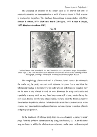
  • 1. Smear Layer In Endodontics The presence or absence of the smear layer is of interest not only to restorative dentists, but to endodontists as well. Whenever dentin is filed, a smear is produced on its surface. This has been demonstrated in many studies with SEM (Baker & others, 1975; McComb, Smith &Beagrie, 1976; Lester & Boyde, 1977; Goldman & others, 1982). Fig. 32 Dentin of a root canal treated with 5% NaOCl and 17% EDTA to remove pulpal tissue and the smear layer. A no. 8 file was drawn over the clean surface in the middle of the scanning electron micrograph, creating a smear layer. Scanning electron micrograph X2000. The morphology of the canal wall is of interest in this context. In adult teeth the walls may be partly covered with atubular, irregular dentin and thus the tubules are blocked in the same way as under erosion and abrasion. Infection may not be seen in the tubules in such an area. However, in many adult teeth and especially in young teeth we may have large areas with primary dentin facing the root canal. From a necrotic and infected canal, bacteria enter the dentin and can be found rather deep in the tubules. Infected tubules with fluid communication to the exterior may cause pathological complications such as external resorption of roots and periapical pathosis. In the treatment of infected roots there is a good reason to remove smear plugs from the apertures of the tubules by using, for instance, EDTA. In this same way, the bacteria within the tubules at some distance can be more easily destroyed 71
  • 2. Smear Layer In Endodontics by an intracanal dressing. On the other hand, if the asepsis or the sealing is poor, we may run the risk of reinfecting dentinal tubules opened and widened by treatment with EDTA. The situation is similar to that for cavities. Another important consideration is that the smear layer is a separate structure from the underlying dentin. Fig. 31: fig 4,pg 18,op dent 1984 is a fortuitous scanning electron micrograph of the smear layer lining a root canal that pulled away from the underlying dentin during processing. The absence of superficial smear layer may facilitate good contact between the sealing material and the wall of cut dentin. Fig 33 SEM of a region in the root canal where the smear layer (SL) cracked open and pulled away from the underlying dentinal tubules (DT). X 1700 The removal of the smear layer from the dentin lining the pulp chamber and root canals has been the subject of numerous investigations. Lester & Boyde (1977) found that treatment for 3 days with 5% sodium hypochlorite did not remove smear plugs from apertures of tubules and may not diffuse into the dentinal tubules sufficiently to take care of microbes that have penetrated deeper into the dentinal tubules. However, Cameron (1983), using an ultrasonic technique, found that 3% sodium hypochlorite combined with ultrasound for 3 to 5 minutes removed not only the superficial smear layer but also the smear plugs; 72
  • 3. Smear Layer In Endodontics but one minute of ultrasound removed only the superficial smear layer. In this method we may have a selective technique that can modify the effect of an irrigating solution. As suggested by Yamada & others (1983), an alternative would be a combination of irrigants. They found the combined use of 10 ml of 17% EDTA, followed by 10 ml of sodium hypochlorite effective. Goldman & others (1982) recommend alternate use of sodium hypochlorite and EDTA to remove smeared dentin. The sodium hypochlorite removes the organic material, including the collagenous matrix of dentin, and EDTA removes the mineralized dentin, thereby exposing more collagen. There are no reasons to believe that a short application of these solutions would have any deleterious effects on the periapical tissues already replaced by granulation tissue. Such preparative treatment of root canals presumably permits a better adaptation of obturating materials and sealers to the dentin. To reduce the risk of reinfection, but also to avoid the development of secondary caries, in permanent coronal restorations of root-filled teeth the cavity should be treated in the same way as cavities in vital teeth, that is, a proper cleansing and lining of all cavity walls. Smear layer in Endodontics can be discussed under the following headings: (I) Effect of instrumentation on endodontic smear layer (II) Intracanal medicaments and smear layer (III) Obturation and smear layer (IV) Post cementation and smear layer (V) Root-end filing and smear layer 73
  • 4. Smear Layer In Endodontics (I) EFFECT OF INSTRUMENTATION ON ENDODONTIC SMEAR LAYER Thorough biomechanical preparation of the root canal is unanimously considered to be one of the major requirements for successful endodontic treatment. The prime objectives of this phase are to remove completely the organic substance that may be infected, or become so, and to shape the root canal in conformity with the principles of obturation. Over the years a variety of instruments and techniques have been proposed to reach this goal. Manual preparation techniques vary both in the type of instruments used and in the sequence of using them. Hand preparation techniques are time consuming, technically demanding and may lead to iatrogenic errors (ledging, zipping, canal transportation and apical blockage). Since optimal shaping and cleaning of root canals with manual files is one of the most difficult aspects of root canal treatment, many alternative techniques have been suggested. Since the 1950s a number of different endodontic handpieces have been developed for root canal preparation with the aim to mainly decrease instrumentation time and to simplify root canal preparation. Automated techniques utilize devices (Canal Finder, Excalibur, Giromatic, Endolift, Cavi-Endo etc.) that rotate files, displace them vertically, or that do both movements. Numerous investigations have demonstrated the limitation of manual and automated root canal instrumentation regarding the overall quality of preparation (Weine et al, 1976; Lehman & Girstein, 1982; Turek & Langeland, 1982; Bolanos et al, 1988; Hulsmann & Stryga, 1993, Bertrand et al, 1999). These problems have resulted in a wide search for innovative materials, instruments and techniques to obtain a clean, disinfected debris-free canal for obturation (Yang et al, 1996). During the last decade several new nickel-titanium instruments for manual root canal preparation as well as for use in rotary endodontic handpiece, have been developed in order to facilitate the difficult and time-consuming process of cleaning and shaping the root canal system and to improve the final quality of root canal preparation. Numerous studies have reported they could efficiently create a 74
  • 5. Smear Layer In Endodontics smooth, predetermined funnel-form shape, with minimal risk of ledging and transporting the canals (Esposito & Cunningham, 1995; Glosson et al, 1995, Short et al, 1997; Thompson & Dummer, 1997b). Shaping procedures can be completed more easily, quickly and predictably. Cleansing efficiency has been one of the issues discussed with regard to preparation techniques. It has been studied extensively, mainly by means of observation of the root canal walls and contents after preparation. The pulpal debris and smear layer produced by instrumentation of the canal walls must be removed (Cergneux, 1987; Gettleman et al, 1991). During preparation, insufficient removal of debris and smear layer material can induce stresses on the cutting segment of endodontic instruments. Their removal depends not only on the irrigation method but also on the endodontic instrument, i.e., the way the instrument is used and the method of preparation. Numerous studies have been reported on the relative effectiveness of different instrumentation techniques, based on a variety of ways of evaluating canal debridement. Outcomes of instrumentation differ according to the method of canal preparation and evaluation, each method showing advantages and disadvantages (Heard & Walton, 1997). Introduction of the scanning electron microscope (SEM) has proved to be a valuable method for assessment of the ability of the endodontic procedures to remove debris from the root canals, thus enabling comparison of instruments and techniques. Numerous authors (Bolanos & Jensen, 1980; Haikel & Allemann, 1988, Hulsmann et al, 1997) have indicated that scanning electron microscopes are indeed useful in examining instrumented root canal walls within the context of evaluating the efficiency of different instrumentation systems. SEM studies focus on observing the smear layer, the patency of dentinal tubules, instrumentation striations and calcospherites. These studies have conflicting results. In most investigations manual preparation was found to be more effective and superior to automated preparation and ultrasonic preparation. In other studies ultrasonic preparation resulted in cleaner canals. Several researchers did not find significant differences 75
  • 6. Smear Layer In Endodontics between preparation techniques. Similarly, the cleaning effectiveness of the rotary nickel-titanium systems is controversial. As both manual and mechanical shaping produce smear layer and debris (Hulsmann & Stryga, 1993; Heard & Walton, 1997), it is important to develop an instrumentation technique for endodontic treatment that produces a minimal amount of smear layer and debris in order to obtain the optimal seal of the root canals. Studies evaluating the cleansing efficiency of preparation techniques are as follows: (A) THOSE THAT INDICATE BETTER CLEANSING EFFICIENCY WITH MANUAL INSTRUMENTATION: Velvart (1987) found hand instrumentation to be superior to ultrasonic cleaning in the apical third of the canal. Complete cleanliness could be achieved by none of the techniques and devices investigated. Baker et al (1988) compared the canal cleanliness following ultrasonic preparation and manual preparation. They found significantly cleaner root canal walls in the middle part of the root canal following hand instrumentation; in the apical and coronal parts no significant differences were detected. Ahlquist et al (2001) compared the cleanliness of the root canal walls following either a manual technique using stainless steel S-files (Sjodings, Sendoline, Sweden) or a rotary technique (ProFile® Rotary nickel-titanium files in a handpiece) of canal instrumentation. It was found that the manual technique employed produced cleaner canal walls than the rotary technique. 76
  • 7. Smear Layer In Endodontics (B) THOSE THAT INDICATE BETTER CLEANSING EFFICIENCY WITH ULTRASONIC AND SONIC DEVICES: Bolanos et al (1988) compared sonic instrumentation with hand instrumentation and the Giromatic system. The sonic preparation resulted in the best cleanliness in all parts of the root canal in straight or curved canals. Hand instrumentation was slightly superior to Giromatic preparation. Schadle et al (1990) compared the efficacy of various root canal cleaning instruments, i.e. hand-instruments, sonic instruments (Sonic Air 3000 and Endostar 5), mechanical instruments (Canalfinder System), and ultrasonic instruments (Cavi-Endo with or without integrated rinsing). The smear layer was somewhat reduced by the Cavi-Endo without the integrated spray. Prati et al (1994) evaluated the morphology of the smear layer and the amount of debris and pulpal residues in the apical third of human teeth by comparing four manual endodontic instruments, an ultrasonic/endosonic system and an automated system (Excalibur). The scoring system was as described by Ahmed et al (1988), i.e. a three point scoring system: 0: smear layer was absent and more than 50% of tubules were visible and partially open. 1: less than 50% of dentinal tubules were visible; smear layer was present in some areas. 2: smear layer covered the canal wall; dentinal tubules were partially visible in limited area. 3: smear layer covered the dentinal tubules. Ultrasonic technique showed the complete removal of the smear layer (scores 0 to 2), leaving small amounts of pulp debris at the apical third. Cameron (1995) reported a superior cleaning ability of the ultrasonic systems. Even most of the smear layer could be removed. 77
  • 8. Smear Layer In Endodontics Hulsmann et al (1997) evaluated the root canal cleanliness after preparation with eight different automated devices and hand instruments. The teeth were investigated under the SEM using the five category scoring system which is as follows (Fig. 34) : Score 1: No smear layer, dentinal tubuli open. Score 2: Small amount of smear layer, some dentinal tubuli open. Score 3: Homogenous smear layer covering the root canal wall, only few dentinal tubuli open. Score 4: Complete root canal wall covered by a homogenous smear layer, no open dentinal tubuli. Score 5: Heavy, nonhomogenous smear layer covering the complete root canal wall. No preparation system or technique resulted in complete removal of smear layer and debris. The ultrasonic unit performed best followed by the Canal Leader 2000 and hand instrumentation. 78
  • 9. Smear Layer In Endodontics Fig 34 Score 1 Score 2 Score 3 Score 4 Score 5 79
  • 10. Smear Layer In Endodontics (C) THOSE THAT INDICATE BETTER CLEANSING EFFICACY WITH AUTOMATED HANDPIECES: Bartha & Rocca (1993) assessed the cleansing-effect of two endodontic handpieces (Excalibur, W & H; MM 1400 Micro Mega). It was found that Excalibur was superior in eliminating the SL. Schafer et al (2000) investigated the cleaning effectiveness of automated (KaVo-Endo Flash device and ProFile system) and manual root canal instrumentation with the aid of SEM. Evaluation for smear layer was done using the 5-step scale as given by Hulsmann et al (1997). Comparison of manual instrumentation with the automated KaVo-Endo Flash resulted in an equivalent degree of canal cleaning. Furthermore, in comparison with the canals that were instrumented with the ProFile instruments, the KaVo-Endo Flash resulted in significantly better root canal cleanliness in curved canals. These results corroborate the reports from other authors, in that most cases of manual instrumentation proved to be superior to mechanical instrumentation as far as cleaning efficiency is concerned (Hulsmann et al, 1997; Mizrahi et al, 1975; Schwarze & Geurtsen, 1996) and that automated devices in most cases create a thicker smear layer than manual instrumentation (Schwarze & Geurtsen, 1996). (D) THOSE THAT INDICATE BETTER CLEANSING EFFICIENCY WITH ROTARY TECHNIQUES: Valli et al (1996) compared the debridement ability of Canal Master and K- Files using scanning electron microscope. The results showed that the Canal Master produced cleaner showing lesser debris than that produced by K-Files. Bertrand et al (1999) determined the ability of the Quantec Series 2000 rotary nickel-titanium endodontic system to remove the dentinal debris and smear layer produced during canal preparation. The absence or presence of a smear layer 80
  • 11. Smear Layer In Endodontics was rated and scored on three appearances using the scale described by Ciucchi et al (1989) as follows: Score 0: No visible tubule orifices-surface is totally smeared. Score 5: Scattered open tubules orifices-surface is partially free of smear layer. Score 10: Regularly distributed open tubule orifices-surface is free of smear layer. The conclusion was that Quantec rotary system produced cleaner canal walls than conventional manual instrumentation, particularly in the middle and apical third. Bechelli et al (1999) compared the efficacy of root canal wall debridement following hand versus Lightspeed ™ instrumentation. Less smear layer was present in the apical region following Lightspeed™ instrumentation than stainless steel hand files, but this difference was statistically not significant. Roggendorf et al (1999) found ProFile superior to Lightspeed and Quantec instruments (no specification on the type of Quantec files) or hand instrumentation. Medioni et al (1999) reported the superior cleaning ability of Quantec Series 2000 when compared to HERO 642, ProFile, and hand instrumentation. Peters & Barbakow (2000) studied the effect of irrigation on debris and smear layer on the canal walls prepared by two rotary techniques, i.e.: Lightspeed (LS) and Profiles (PS) systems. The larger canal preparations obtained in this study with Lightspeed instruments enabled a more effective removal of the smear layer than the PF group. Hulsmann, Schade & Schafers (2001) compared cleaning ability using two different rotary nickel-titanium instruments: HERO 642 (MicroMega, France) and Quantec SC (Tycom, CA, USA). Results indicated that cleaner root canal walls were found after preparation with HERO 642 (53% scores 1 and 2), followed by Quantec SC (41%). 81
  • 12. Smear Layer In Endodontics Gambarini & Laszkiewicz (2002) assessed the debris and smear layer remaining following canal preparation with GT rotary instruments. It was concluded that the GT™ rotary instruments removed debris effectively, but left root canal walls covered with smear layer particularly n the apical third. Baumann et al (2003) found that RCT with RaCe™ had better smear layer removal than hand instrumentation. Almannai et al (2003) compared 4 different rotary nickel-titanium instruments (ProFile®, ProTaper®, RaCe®, and NiTee®) with regards to canal cleanliness. Significantly less smear layer was observed in the apical third of the ProTaper group when compared with the ProFile group. In-Soo Jeon et al (2003) compared the quality and amount of smear layer generated in the apical third of straight root canals by 2 rotary nickel-titanium reamers and I rotary steel reamer with different cutting blade designs. Automated preparation was performed with ProFile and HERO 642 reamers using the crown- down technique and with a stainless steel engine reamer (Mani) by using a reaming motion. A 4 – category scoring system for smear layer was used which is as follows: 0: no smear layer/all tubules clean and open. 1: no superficial smear layer/tubule openings visible, but some contain debris plug or soft tissue remnants. 2: moderate smear layer/some tubules open and others closed. 3: heavy smear layer and most/all tubule openings obscured. The results indicated that the least smear layer remained in the HERO 642 group. In the ProFile group, the smear layer had a shiny and burnished appearance with few openings of dentinal tubules. The depth of the packed smear layer into the tubules was deeper than in the Hero group. The smear layer generated by the steel reamers appeared thinner and less compressed than in the ProFile group. 82
  • 13. Smear Layer In Endodontics Fig 35 A B A scanning electron micrograph of a canal wall prepared with Hero 642 rotary reamers has a snowy appearance with many open dentinal tubules (Score 1: Original magnification X1500). A scanning electron micrograph of a canal wall prepared with ProFile rotary reamers. The canal wall was to be covered by a thick, nonhomogenous layer, with a shiny burnished appearance and almost no open dentinal tubule (Score 3; original magnification x1500). C D Cut-view of a canal wall prepared with Pro- File rotary reamers at the apical 2mm level. The smear layer had a muddy appearance (X5000). Cut-view of a canal wall prepared with the Hero rotary reamers at the apical 2mm level. Generally, the tubular packing is less frequent than that obtained in the ProFile group. Thin smear layer with many open dentinal tubules covered on the root canal wall (X3000). 83
  • 14. Smear Layer In Endodontics (E) THOSE THAT HAVE SHOWN INCONCLUSIVE RESULTS OR SIMILAR RESULTS WITH DIFFERENT TECHNIQUES: Klimm et al (1990) evaluated the efficacy of irrigated, hand root canal preparation by SEM. In all groups a homogeneous or inhomogeneous smear layer of different thickness and extent dominated. Mandel et al (1990) evaluated the cleansing efficiency of manual serial preparation, endosonic (Cavi-Endo) preparation and automated preparation with the Canal Finder System. No preparation technique was found to result in any characteristic microscopic features that distinguished it from the other techniques. Lumley et al (1992) investigated the effect of precurving Endosonic files on the amount of debris and smear layer remaining in curved root canals. The result of this study indicated that precurving of files decreased the amount of debris but did not affect smear layer removal. Sydney et al (1996) analyzed the smear layer removal after root canal preparation by a manual technique and by an automated handpiece, the Canal Finder System (CFS).Both the techniques showed root canal walls with dense smear layer. Heard & Walton (1997) compared four root canal preparation techniques in small curved canals. The four methods were: (1) step-back without initial coronal flaring, (2) step-back with coronal flaring, (3) step-back with initial coronal flaring and finished by ultrasonic irrigation and, (4) ultrasonics only. The results indicated that there were no statistically significant differences between the techniques. Kochi et al (1998) could find no difference between Quantec (No information on type of file) and manual preparation using K-files. 84
  • 15. Smear Layer In Endodontics Mayer et al (2002) evaluated smear layer scores after two types of instruments to ultrasonically activate irrigants during 2 types of canal preparation (ProFile .04 and Lightspeed). It was found that ultrasonically activated irrigants did not reduce smear layer scores and this finding was not influenced by the material or by the design of the instrument used to transmit ultrasonic activation. There were no differences in amount of smear layer on canal wall prepared with PF or LS instruments. Lichota et al (2003) assessed the cleanliness of root canal walls following ProFile rotary technique of instrumentation. Thus though they removed debris effectively, ProFiles left the root canal walls covered with smear layer in the apical and middle thirds. Thus, from the above studies we can conclude that there is still no consensus on the best instrumentation technique which may result in cleanest possible canal without the presence of a smear layer. Further research is required to resolve the current controversy on the best instrumentation technique. 85
  • 16. Smear Layer In Endodontics (II) INTRACANAL MEDICAMENTS AND SMEAR LAYER The goal of clinical treatment is to completely disrupt and destroy the bacteria involved in the endodontic infection. Endodontic infection will persist until the source of irritation is removed. Previous studies have shown that bacteria in infected root canals and periradicular tissues are capable of invading and residing deeply within dentin and in cementum around the periapex. (Ando & Hoshino, 1990; Kiryu et al, 1994; Peters et al, 2001). Further more, it has been demonstrated that although bacteria in artificial smear layers and prepared reservoir channels in deeper layers of root dentin could be eliminated by procedures such as ultrasonic irrigation with NaOCl (Huque et al, 1998), microorganisms within fins and isthmuses could still remain viable (Sato et al, 1996). Even after chemomechanical instrumentation of the canal, some bacteria still remain in the canal and dentinal tubules (Bystrom & Sundqvist, 1981, 1983, 1985). Total debridement is impeded because of accessory canals, fins, cul-de- sacs and communications between the main canals. Such microorganisms may cause root canal treatment to fail. The rationale behind intracanal medication is to destroy residual microorganisms and their toxins and to remove organic tissue. The medicament should inhibit microbial recolonization of the cleaned parts of root canal system by preventing residual microorganisms from growing and new organisms invading through lateral communications and coronal access. For this reason, chemomechanical cleansing is often supported by the use of disinfectants. Various medicaments have been proposed for disinfection of root canals. The traditional phenolic or fixative agents include camphorated monochlorophenol (CMCP), formocresol, and cresatin. Iodine potassium iodide and calcium hydroxide are the main nonphenolic intracanal medications. These medicaments are potent antibacterial agents under laboratory test conditions; however their effectiveness in clinical use is unpredictable (Messer & Chen, 1984). According to some researchers, they also neutralize and render canal tissue 86
  • 17. Smear Layer In Endodontics remnants inert. Some medications contain aldehyde derivatives that can be used to fix fresh tissues for histological examination; however, they may not effectively fix necrotic or decomposed tissues (Walton & Rivera, 2002). According to Wesselink et al (1977), fixed tissues are not inert and may become more toxic and antigenic after fixation. Intracanal medications have also been used clinically to prevent posttreatment pain. Studies have shown, however, that routine use of these materials as intracanal medication has no significant effect on the prevention of pain. According to Oguntebi (1994), most currently used intracanal medicaments have a limited antibacterial spectrum and antigenic potential. In addition, some of them have a limited ability to diffuse into dentinal tubules. Effect of intracanal medications on bacteria in dentinal tubules: After removal of the smear layer, Haapasalo & Orstavik (1987) inoculated the bovine incisors with E. faecalis and found penetration of these bacteria into the dentinal tubules upto 1 mm. They demonstrate that liquid CMCP completely disinfected the dentinal tubules but that Ca(OH)2 was ineffective. Behnen et al (2001) had better success killing E. faecalis with either Pulpdent or a 10% solution of Ca(OH)2 than with the traditional thick mixes. The difference might have been attributable to the different viscosity of various types of Ca(OH)2 used in this experiment. Heling & Chandler (1998) also inoculated dentinal tubules of bovine teeth with E. faecalis and then examined the disinfecting effect of various irrigants. They found that none of the test irrigants (chlorhexidine, hydrogen peroxide, sodium hypochlorite, EDTA or their combinations) were totally effective. Siqueira & De Uzeda (1996) inoculated bovine dentinal cylinders with 1 facultative and 2 obligate anaerobic bacteria and examined the disinfecting effect of Ca(OH)2 mixed with saline solution or CMCP for 1 hour, 1 day, and 1 week. Their results showed that Ca(OH)2 and saline solution were ineffective in disinfecting the dentinal tubules after 1 week of application. In contrast, a mixture of Ca(OH)2 and CMCP resulted in complete disinfection of the dentinal tubules in 1 day. Jeansonne & White (1994) and Kuruvilla & Kamath (1998) examined the antibacterial effect of chlorhexidine gluconate and sodium hypochlorite on 87
  • 18. Smear Layer In Endodontics inoculated human teeth in vitro. They reported a reduction in bacterial counts but not total disinfection of the root canals. In an in vivo experiment, Katebzadeh et al (1999) showed that infected dog teeth that were filled experienced treatment failure more frequently than those medicated with Ca(OH)2 before obturation. Sjogren et al (1997) examined the presence and the influence of bacteria on the long-term success of root canal therapy. Their results show that 40% of root canals remain infected after instrumentation. In addition, they reported that teeth filled in 1 visit without the use of Ca(OH)2 experienced treatment failure significantly more frequently than those that were medicated for 1 week with Ca(OH)2 (68% vs. 94%). The results of this study corroborate the findings of Bystrom et al (1987), who showed improved clinical success rates after effective disinfection of root canals. Gutierrez et al (1991) evaluated the diffusion of medicaments within the root canal dentin after mechanical preparation and flushing with alternate NaOCl and hydrogen peroxide followed by placement of the medicaments (CMCP, CMCP with formocresol and methylcresylacetate) by paper points. It was found that all 3 medicaments crystallized both on dentin walls and inside the dentinal tubules. Bacteria remaining in dentinal tubules in close relationship with CMCP crystals appeared shrunken. Smear layer- a physical barrier for disinfectants: According to some authors (Goldberg & Abramovich, 1977; Wayman et al, 1979; Yamada et al, 1983; Berg et al, 1986; Baumgartner & Mader, 1987) the presence of the smear layer may block the antimicrobial effect of intracanal disinfectants into the tubules by preventing their penetration into the tubules. In an in vitro study, Orstavik & Haapasalo (1990) showed the importance of removal of the smear layer and the presence of patent dentinal tubules for decreasing the time necessary to achieve the disinfecting effect of intracanal medications. They found that camphorated p-monochlorophenol was generally more efficient than Calasept, and of the irrigants tested, iodine potassium iodide 88
  • 19. Smear Layer In Endodontics appeared more potent than sodium hypochlorite or chlorhexidine. They also concluded that the smear layer did delay, but not abolish the action of disinfectants. However, following the removal of the smear layer, bacteria in dentinal tubules can be easily destroyed (Brannstrom, 1984) and in this way, it may be beneficial to use lower concentrations and/or amounts of antibacterial agents since all of these agents show some degree of toxicity to viable host cells. Bystrom & Sundqvist (1985) have also shown that the presence of a smear layer can inhibit or significantly delay the penetration of antimicrobial agents such as intracanal irrigants and medications into the dentinal tubules. Diffusion through dentin is directly proportional to the surface area of dentinal tubules and inversely proportional to the dentin thickness. Also the presence of the smear layer acts as a diffusion barrier that can reduce diffusion by 25-30% (Pashley, 1988). The presence of a smear layer has previously been demonstrated to decrease the diffusion of triamcinolone and demethylchlortetracycline, two active components of the root canal medicament, Ledermix, through radicular dentin (Abbott et al, 1989). Foster et al (1993) evaluated the effect of smear layer removal on the diffusion of calcium hydroxide through radicular dentin. Pulp extirpation and the placement of Ca(OH)2 into the root canal 7 to 14 days after a traumatic episode are recommended to decrease the incidence of inflammatory root resorption. Tronstad (1981) suggested that Ca(OH)2 placed in the root canal elevates the pH in areas of resorption on the surface of the root by diffusion of OH- through the dentin, thereby decreasing osteoclastic activity and activating alkaline phosphatases. The result of this study demonstrated that Ca(OH)2 diffuses from the root canal to the exterior surface of the root and that the removal of the smear layer may facilitate this diffusion. Thus the root canal space may be considered a route for medication delivery to the dentinal tubules themselves, if disinfection is required, and/or to the external root surface. 89
  • 20. Smear Layer In Endodontics Cruz et al (2002) evaluated the penetration of propylene glycol into root dentin. Propylene glycol is a vehicle that has the potential for use in root canal medicaments. It has been reported to be a widely used vehicle for various pharmaceutical and commercial products such as drugs, cosmetics and foods (Morshed et al, 1988) and it is also used as a constituent of Caries Detector® (Fusayama, 1988). In endodontics, it has been used as a vehicle for calcium hydroxide (Saiijo, 1957; Laws, 1962; Laws, 1971; Simon et al, 1995). In this study, Safranin O in propylene glycol and in distilled water was introduced into root canals with and without smear layer. It was found that the area and depth of penetration with propylene glycol was significantly greater than with distilled water. Smear layer significantly delayed the penetration of dye. Thus it is seen that propylene glycol delivered dye through root canal system rapidly and more effectively indicating its potential use in delivering intracanal medicaments. Also, propylene glycol is less cytotoxic than other vehicles, possesses antibacterial properties and is hygroscopic, thus allowing absorption of water which results in sustained release of intracanal medicaments for prolonged periods (Fava & Saunders, 1999). Thus, it is seen that generally, removal of smear layer helps in diffusion of the intracanal medicaments and thereby improves their efficacy. 90
  • 21. Smear Layer In Endodontics (III) OBTURATION AND SMEAR LAYER Complete obturation of the root canal with an inert filling material and creation of a hermetic, apical seal have been proposed as goals for successful endodontic treatment (Nguyen, 1984). To achieve this, the root canal filling must seal the canal space both apically and coronally to prevent the ingress of microorganisms or tissue fluids into the canal space. Apical leakage is considered a common reason for the clinical failure of endodontic therapy. Likewise, coronal leakage is also reported to be an important reason for failure (Madison et al, 1987; Saunders & Saunders, 1994). Gutta-percha is considered an impermeable core material; therefore, leakage through an obturated canal is expected to take place at the interface between sealer and dentin or sealer and gutta-percha, or through voids within the sealer. Hence, the sealing quality of a root canal filling depends much on the sealing ability of the sealer. A review of a large number of published leakage studies points to general agreement that leakage occurs between the root filling and the root canal wall. Therefore, anything that may influence the adaptation of the root filling to the canal wall is of great significance in determining the degree and the extent of leakage, and ultimately the prognosis of the endodontic therapy. Because the smear layer remaining on the root canal wall has been characterized, there has been much interest in the possible effect of the smear layer on the leakage of the root canal sealer. There is a controversy over whether or not to remove this layer before obturation and there is still no consensus on the influence of the smear layer on the development of an effective seal of the root canal system. Some consider that it is desirable to remove this layer as it covers prepared areas and prevents medicaments and filling material from penetrating the dentinal tubules or even contacting the canal wall. As of today, almost no material has been shown to enter dentinal tubules with the smear layer present. Removal of the smear layer is also considered to be desirable by others because, in addition to inorganic particles, it may contain some organic material, as well as viable 91
  • 22. Smear Layer In Endodontics bacteria. Biologically, the presence of the smear layer has been postulated to be an avenue for leakage and a source of substrate for bacterial growth and ingress (Pashley, 1984). The frequency of bacterial penetration in the presence of a smear layer, when canals were obturated with thermoplasticized gutta-percha and sealer, has been shown to be significantly higher than with smear layer removal and obturation (Behrend et al, 1996). A further concern is the presence of viable bacteria that may remain in the dentinal tubules and use the smear layer for sustained growth and activity (Brannstrom, 1984; Olgart et al, 1974). Because of the bacterial content of the smear layer, any apical extrusion of the smear layer during instrumentation or obturation can defeat one of the goals of endodontic therapy: the return to and maintenance of an inflammation-free state in the periapical area. To be considered an ideal sealer, a material should not itself cause or further irritate the periapical tissue. Some root canal filling materials, especially N2 paste and silver points, are not biocompatible. To risk further tissue trauma with a technique that may induce or potentiate periapical inflammation is unthinkable. Smear layer induced inflammation of the periapical area can be caused by over instrumentation or by the careless measurement and filling of a master cone. It has been recommended that master cones be foreshortened to fit 1mm short of the apex as an effective countermeasure to creating pre- and post obturation periapical inflammation. Some doubt exists if the formation of apical plugs from a combination of dentin chips, soft-tissue fragments and fibers from instrumentation may enhance the seal of a root canal filling. Dentin filings occur during instrumentation, but the formation of an apical plug, from them is often an inadvertent or accidental occurrence. Well-formed apical plugs can often reduce the inflammatory reactions following filling by limiting the apical extension of filling materials thereby encouraging healing. If more cementum is going to form to create a better apical seal, dentin chips at the apex of a root can act as a nidus for formation of hard tissue. Bacterial contamination by the presence of the smear layer can prevent this. 92
  • 23. Smear Layer In Endodontics Endodontic sealers act as a glue to ensure a good adaptation of gutta percha to the canal walls. If the smear layer is not removed, the gutta percha may occasionally be glued to the dentin in the smear layer as well as to exposed parts of the canal wall. The smear layer may interfere with the penetration of the gutta- percha into the tubules and the adhesion and penetration of the sealers into the dentinal tubules. Not being firmly attached to the dentin, the smear layer may laminate off the canal wall and create a false seal, voids in the fill and an expected environment for microleakage. Thus the retention or removal of the smear layer before obturation may influence the quality of the obturation. In contrast, other investigators (Pashley et al, 1981) consider that the smear layer should be left intact, as it may actually form a protective barrier. The smear layer may be responsible for excluding bacteria from dentinal tubules as well as restricting the surface area available for the diffusion of many molecules. One study (Michelich et al, 1980) has shown that the smear layer will prevent bacterial penetration (Streptococcus mutans), but will permit fluid filtration. Another study (Williams & Goldman, 1985) has revealed that the smear layer simply delays the penetration of some bacteria (Proteus vulgaris) rather than preventing it. Many articles have been written on the physical properties of root canal sealers, including the their adhesive strength to dentin (Orstavik et al, 1983; McComb & Smith, 1976) and to gutta-percha (Orstavik et al, 1983). Adhesive strength measurements may be important to clinical usage, because higher adhesive strengths may reduce leakage in clinical situations. Opening of the tubuli by removal of the smear layer prior to filling the root canal system makes great sense. If chemical adhesion between the dentin and sealers, pastes, cements or plastics cannot be easily achieved, then why not a mechanical lock? The material will flow or be forced back into the empty dentinal tubules, gripping like tentacles and forever resisting displacement! This may actually increase the adhesive strength of sealers to dentin and improve the sealing ability of the filling. 93
  • 24. Smear Layer In Endodontics The type of sealer used has different implications once the smear layer is removed. A powder-liquid combination, the most common of which is Grossman's sealer, contains small particles in the powder that could enter the orifices of the dentinal tubules and help create a secure interface between sealer and canal wall. But studies have shown that the adhesion of Grossman type root canal sealers to dentin is established by electrostatic bonding and not by its penetration into the tubules. So the action of EDTA to remove the smear layer and open the dentinal tubule orifices does not significantly increase the adhesion value of Grossman type root canal sealers. Calcium hydroxide based sealers have the advantage of promoting the apposition of cementum and sealing it off against microleakage. Although calcium hydroxide has dentin regenerating properties, the formation of secondary dentin along the canal wall is prevented by the absence of vital pulp tissue. The calcium ion is used in the formation of osteoid or dentoid type material. Circulation of blood (which is absent in filled canals) is needed for the calcium ion to promote new tissue; thus the calcium hydroxide sealers are effective for sealing only at the root apex. It is also seen that the adhesion values of Sealapex, a calcium hydroxide based sealer is quite low even with the removal of the smear layer, which allows greater penetration into the tubules. This can be explained by the high setting time, solubility and disintegration of the sealer. Also the sealer is easily displaced from the tubules due to low cohesive structure. Epoxy based resin sealers showed increased adhesion values with dentin treated with EDTA when compared to the dentin without any treatment. This is due to the fact that EDTA removes the smear layer and opens the tubule orifices, which are then partially filled with the sealer with the resultant formation of resin tags, causing a higher mechanical bonding between the sealer and the root canal wall. Since the epoxy resin based sealers are strongly cross- and end to end linked with ether bonds and had improved physical integrity, they appeared more likely 94
  • 25. Smear Layer In Endodontics to remain intact in the tubules after penetration (White, Goldman & Peck Sun Lin, 1987). A glass-ionomer based sealer Ketac-Endo has been shown to bond chemically with the hydroxyapatite of the root dentin. Several studies have shown that that the removal of the smear layer from the root canals decreased microleakage. Conditioning of the tooth surfaces with acids can remove the smear layer from the instrumented root canals and alter the surface energy, allowing the glass ionomer to flow and adapt more easily, enhancing its adhesion. In addition, acid conditioning removes surface contaminants before glass ionomer sealer placement, possibly permitting greater ionic exchange and better bonding between the cement and tooth structure. There is no practical advantage to the use of an autocure unfilled resin as a seal over the tubules before gutta percha obturation. The resin would be susceptible to moisture through the lateral canals and through the apex. Upon polymerization, the resin would shrink creating a gap between the fill and the canal wall. The use of some dentin-bonding agents to harden the smear layer to the canal wall and to harden the apical plug is a subject of research. It is doubtful that the bonding agent would be antimicrobial to the bacteria in the smear layer. It is probable that the insertion of a tight-fitting post into the canal wall would help to prevent lamination of the smear layer off of the canal wall if the layer wad retained and treated with a dentin-bonding agent. Possibly, it would be better to remove the smear layer. Once packed down, the apical debris is hard; whether further hardening with a dentin-bonding agent would improve the seal permanently and enhance retention is problematical. Another important factor to be considered is the obturation technique which is used. Since the most common cause of endodontic failure has been attributed to incomplete obturation (Ingle showed 60% of endodontic failures to be caused by 95
  • 26. Smear Layer In Endodontics incomplete obturation), many different obturation techniques have been developed in order to increase the success of root canal treatment. Gutta-percha has been the material of choice for obturation since 1867 and many different gutta-percha techniques have been introduced in order to increase the quality of the apical seal of the root canal. Lateral condensation has proven to be a very popular gutta-percha technique. However, Schilder (DCNA, 1967) and Brayton et al (1973) noted that with lateral condensation of gutta-percha at no time did a homogenous mass form nor did the gutta-percha adapt to the root canal walls. The voids in this nonhomogenous mass may remain empty or be filled with sealer which may resorb in time, decreasing the effectiveness of root canal obturation (Peters, 1986). The concept of root canal obturation with thermoplasticized gutta-percha was introduced by Yee et al (1977). A major impetus for this development was the need to ensure better adaptation of the root canal filling material to the prepared canal walls. They reported injected gutta-percha to be superior to lateral condensation and capable of filling multiple foramina and other ramification. Further studies by Torabinejad et al (1978), Marlin et al (1981) and Budd et al (1991) have supported this achievement in that the thermoplasticized gutta-percha was shown to replicate the intricacies of the root canal system and achieve a seal equal to, if not superior to, that produced by other obturation methods (Michanowicz & Czonstkowsky, 1984; Czonstkowsky et al, 1985; ElDeeb, 1985; Evans &Simon, 1986; Mann & McWalter, 1987). Favourable results for the sealing ability of the low-temperature thermoplasticized gutta-percha technique (Ultrafil) and close adaptation to the canal walls have been reported (Michanowicz & Czonstkowsky, 1984; Michanowicz et al, 1986). Another thermoplasticized gutta-percha technique, Thermafil which consists of a metal carried with α-phase gutta-percha was introduced by Johnson (1978, 1988). Beatty et al (1989) found that Thermafil produced a better apical seal than lateral condensation. 96
  • 27. Smear Layer In Endodontics Current methods of canal obturation using thermoplasticized gutta-percha all extol the virtues of the adaptation of the softened materials to the canal irregularities. One technique has been shown to have dentinal tubule penetration with the softened gutta-percha even without the use of sealer (Michanowicz et al, 1986). When sealer was used no penetration was evident. However, they still advised using sealer to produce better apical seal. Other plastic-type filling materials (pHEMA and silicone) have also been shown to penetrate the dentinal tubules in the absence of root canal sealer (White et al, 1984). However, this situation poses a dilemma, as the use of root canal sealer has been advocated as essential with thermoplasticized gutta-percha to achieve the best possible seal (ElDeeb, 1985; Evans & Simon, 1986; Skinner & Himel, 1987; Bradshaw et al, 1989). An additional dilemma in this issue is the ability of the gutta-percha to be adapted intimately to the root canal wall when the smear layer is present (Moodnik et al, 1976; White et al, 1984). The effect of removal of smear layer on the overall success rate of endodontic therapy is unclear. Although Biesterfeld & Taintor (1980), Madison & Krell (1984), Goldberg et al (1985) and Evans & Simon (1986) did not find any improvement in sealing of smear-free canals, experiments by Kennedy et al (1986), Abramovich & Goldberg (1976), Tidmarsh (1978) and White et al (1984) showed better adhesion/sealing of smear-free canals. Removing of the smear layer prior to filling may actually increase the adhesive strength of the filling to dentin and improve the sealing ability of the filling. However, most of the studies carried out with a variety of sealers and by different obturating techniques do indicate that removal of smear layer does indeed improve the adaptation and the seal of the filling material. Given below are some of the several studies that support this, though different sealers and obturating techniques do vary in their sealing ability/penetration/adhesion when smear layer has been removed. 97
  • 28. Smear Layer In Endodontics (A) Sealers and smear layer: Yamada et al (1983) stated that a case could be made for the removal of smear layer: “For instance, it may interfere with the adaptation of filling materials to the canal wall by imposing an additional interface”. They also went on to state, “in addition, opening all the tubules can perhaps provide a better seal by allowing sealer or filling material to penetrate the dentin”. Cergneux et al (1987) compared the sealing ability of obturated root canals which had previously been cleaned chemically by EDTA or mechanically by ultrasound. The results indicated that when the smear layer is not eliminated (control group), there is a tendency for greater dye infiltration. Also that the EDTA-treated canals showed the least infiltration, while those treated with ultrasound showed less sealing ability that the EDTA treated group but significantly less leakage than the control group. White, Goldman & Peck Sun Lin (1987) evaluated the influence of the smeared layer upon dentinal tubule penetration by different endodontic filling materials, i.e., pHEMA (Hydron), silicone (Endo-fill) and laterally condensed gutta-percha with 2 different sealers (Roth 801 and AH 26). It was found that pHEMA, silicone and both sealers consistently entered the dentinal tubules when the smear layer was removed with EDTA prior to filling. When the smeared layer was present during filling, tubular penetration was unpredictable and infrequent. Gettleman et al (1991) assessed the influence of smear layer on the adhesion of sealer cements to dentin. The results showed significant differences among AH26, Sultan, and Sealapex, with AH26 being the strongest and Sealapex being the weakest and that AH26 had a stronger bond when smear layer was removed. 98
  • 29. Smear Layer In Endodontics Oskan et al (1993) evaluated the effect of smear layer on the penetration of 4 root canal sealers into dentinal tubules. It was observed that the smear layer obstructed the penetration of the tubules by the sealers. However, the penetration into the tubules was better with Diaket, N2 and SPAD, than with Forfenan when the smear layer was removed. Kouvas et al (1998) examined the effect of the smear layer on the penetration depth of Sealapex, Roth 811, and CRCS root canal sealers into the dentinal tubules. The removal of the smear layer allowed penetration of all 3 sealers into the dentinal tubules to a depth of between 35 µm and 80 µm, with penetration depth of Roth 811 sealer being 10-60 µm, that of Sealapex being 30- 80 µm and that of CRCS being 1-35 µm. The Sealapex and Roth 811 sealers penetrated deeper than the CRCS sealer since their particle size was smaller than that of CRCS. The degree of obstruction of the dentinal tubules differed depending on the sealer, with CRCS providing a better, tighter seal. This suggested that the microstructure of the sealer paste in the dentinal tubules and the degree of their closure, rather than their depth of penetration, might be an important factor for a tight obturation of a smear-free root canal. The presence of smear layer at the root canal walls obstructed the penetration of all sealers into the dentinal tubules. Fig 36 Picture of CRCS in smear layer-free dentinal tubule the stratified appearance of the sealer. The presence of smear layer inhibited Sealapex from striating the dentinal tubules 99
  • 30. Smear Layer In Endodontics Economides et al (1999) evaluated the influence of the smear layer removal on the apical sealing ability of different sealers (Roth 811 and AH26) The results indicated that the smear layer removal resulted in statistically significant reduction of microleakage values in the groups obturated with AH26. Stevens (2003) investigated the sealer penetration of Roth’s 801 sealer into smear-free dentinal tubules following a final rinse of 95% ethyl alcohol and also ascertained the whether this final rinse affects leakage. It was found that following smear layer removal with 17% EDTA/5.25% NaOCl, flushing the canal with 95% ethyl alcohol prior to obturation will significantly improve sealer penetration and significantly decrease leakage as compared to the group in which the final rinse was carried out with NaOCl after smear layer removal. Cobankara et al (2004) determined the effect of the smear layer on apical and coronal leakage in root canals obturated with AH26 or RoekoSeal (a polydimethylsiloxane based sealer) sealers. According to the results of this study, the smear (+) groups displayed higher apical and coronal leakage than those smear (-) groups for both root canal sealers. There was no statistical significant difference in either apical or coronal leakage between RoekoSeal and AH26, regardless of the presence or absence of the smear layer. Economides et al (2004) compared the microleakage of 2 root canal sealers with and without the smear layer being present. In this study, a resin-based sealer Fibrefill and the Calcibiotic root canal sealer (CRCS), which is a calcium hydroxide-based sealer, were assessed. The results indicated that the canals treated with Fibrefill leaked less than those treated with CRCS, regardless of whether the smear layer had been removed. Less microleakage was found with both sealers when the smear layer was removed, but the value did not differ statistically from the results with an intact smear layer. Kokkas et al (2004) examined the influence of the smear layer on dentinal tubule penetration depth by three different root canal sealers (AH Plus, Apexit, 100
  • 31. Smear Layer In Endodontics and Roth 811). Examination in SEM revealed that the smear layer obstructed all the sealers from penetrating dentinal tubules. In contrast, smear layer removal allowed the penetration of all sealers to occur to a varying depth. These findings suggest that smear layer plays an important role in sealer penetration into the dentinal tubules, as well as in the potential clinical implications. (B) Obturation techniques and smear layer: Gutmann (1993) examined with SEM the adaptation of thermoplasticized, injected gutta-percha (Obtura,) to prepared dentin devoid of smear layer. It was found in this study that the gutta-percha penetrated into the patent dentinal tubules with or without the root canal sealer. This was in direct contrast to the findings of Michanowicz et al (1986), in which gutta-percha did not penetrate the dentinal tubules in the presence of sealer. However, here a different thermoplasticized injection system (Ultrafil) was used and condensation was not performed (Fig. 37). 101
  • 32. Smear Layer In Endodontics Fig 37 Discrete penetration of softened gutta-percha into the patern tubules in the form of plugs. Note that seperation of the plugs suggesting a decreased number of dentinal tubules in the apical position of the canal. No sealer used X1500 Guttapercha matted together with sealer, SEM X940. Guttapercha plugs showing discrete penetration into the tubules without the use of sealer, SEM original magnification X940. Entanglement of gutta-percha plugs and sealer, SEM X720 102
  • 33. Smear Layer In Endodontics Gencoglu et al (1993) evaluated the dentinal adaptation of injected thermoplasticized gutta-percha and thermoplasticized gutta-percha resulting from Ultrafil and Thermafil systems respectively, by SEM, and compared it with adaptation obtained with lateral condensation technique. Each method was evaluated with and without the smear layer removed. The results showed that thermoplasticized gutta-percha resulting from either system had better dentinal wall adaptation than lateral condensation in either the absence or presence of the smear layer. In the absence of smear layer, the adaptation of gutta-percha was improved in all groups. In another study by Gencoglu et al (1993) these 2 thermoplasticized gutta- percha techniques showed significantly less apical leakage than the lateral condensation technique with and without the smear layer. Removing the smear layer reduced the leakage significantly in all groups. It has been postulated that heated gutta-percha forced into the patent tubules may establish a mechanical lock or bond between the filling material and the canal wall in the absence of the smear layer (Evans & Simon, 1986). Lloyd et al (1995) assessed the sealability of another thermoplasticized gutta-percha technique, i.e. is the Trifecta™ technique, in the presence or absence of smear layer. The results also showed that the prevalence of sealer and gutta- percha penetration into the dentinal tubules was significantly more common in the absence of smear layer. Removal of the smear layer did not enhance the sealability despite increasing the proportion of specimens with dentinal penetration of sealer and gutta-percha. Pallares et al (1995) compared the adaptation of mechanically softened gutta-percha (J.S. Quick-fill™ and AH-26 sealer) to the root canal in the presence and absence of smear layer. It was found that the sealer penetrated into the dentinal tubules along with projections of gutta-percha only in those teeth without the smear layer. 103
  • 34. Smear Layer In Endodontics Kytridou et al (1999) evaluated the adaptation and the short- and long-term sealability of two different thermoplastic techniques (Thermafil and System B), in the absence of smear layer. It was found that both obturation techniques were not significantly different in the apical third adaptation. But it was seen that Thermafil demonstrated significantly more long-term apical leakage than System B. Following are the few studies which indicated that the smear layer removal did not help to improve the adhesion /penetration/sealing ability of sealers and various obturation techniques: Saleh et al (2002) studied the effects of dentine pretreatment on the adhesion of root-canal sealers (Grossman's sealer, Apexit, Ketac-Endo, AH Plus, RoekoSeal Automix or RoekoSeal Automix with an experimental primer). It was concluded that removal of the smear layer might impair sealer adhesion to dentine. Different sealer types require different dentine pretreatments for optimal adhesion. In another study, Saleh et al (2003) found that penetration of the endodontic sealers into the dentinal tubules when the smear layer was removed was not associated with higher bond strength. Cobankara et al (2002) tested the effect of two different root canal sealers (i.e. Ketac-Endo and AH26) on resistance to root fracture and also evaluated the effect of smear layer. The use of adhesive sealers in the root canal system may offer an opportunity to reinforce the endodontically treated tooth. The results of this study indicated that the canals obturated with either sealer were significantly stronger than roots whose canals were instruments but not obturated. The presence of absence of the smear layer did not cause any significant effect on the root fracture resistance of the teeth. Karadag et al (2004) evaluated the influence of passive ultrasonic activation of the irrigants on the penetration depth of different sealers. Under the conditions of this study, it can be said that ultrasonically activated irrigation did not reduce 104
  • 35. Smear Layer In Endodontics the smear layer effectively at 1 min and 0.5 min time intervals. According to this result, no difference was observed between the penetrations of sealers. Even though most of these in vitro findings showed that removing the smear layer reduced the apical leakage and improved the adhesion and penetration of the sealers, the effects of the smear layer on in vivo leakage cannot be easily determined. Further work should therefore include more clinical studies correlating with these in vitro studies. In addition, studies are needed which further evaluates which sealers seal better in the presence and as well as the absence of the smear layer. Also, evaluation on which sealers work best utilizing specific obturating techniques should be completed. Clearly, further research is necessary into the properties of different sealer cements, in order to establish the factors that affect and determine their clinical usefulness. 105
  • 36. Smear Layer In Endodontics (IV) POST CEMENTATION AND SMEAR LAYER Endodontically treated teeth with loss of coronal tooth structure generally require a radicular post for restoration of the tooth function. In the last 10 years, several experimental and clinical studies established that pre-fabricated posts offer a better prognosis than cast post and cores for endodontically treated teeth (Isidor & Brondum, 1992 and Mannocci et al, 1998). Retention of all types of posts is affected by the cement selected for luting it to the post space. Removal of smear layer increases the cementation bond and the tensile strength of the cementing medium by allowing penetration of these luting cements into the dentinal tubules. Glass ionomer cements are effective in post cementation after smear layer removal because the glass ionomer has a better union with tooth structure. The need for removal of smear layer is controversial while using glass ionomer luting cements. Few reports suggest that the smear layer could assist adhesion because its high calcium and phosphate content provide potential sites for strong adhesive bonding. However, in clinical situations, contamination of the smear layer is unavoidable and therefore removal of the whole contaminated layer may become necessary. Heretofore, there has been no significant difference between cements when the final rinse was 2cc of 5.25% NaOCl (Goldman, De Vitre & Tenca, 1984). When an unfilled Bis-GMA resin was used after NaOCl rinse, the strength of the resin bond was better than that of polycarboxylate cement. When the smear layer was removed by flushing with EDTA and NaOCl rinse, the Bis-GMA resin followed into the exposed dentinal tubules and into the serrations on the post, vastly improving retention. Post length only made minimal difference when the canals were flushed with either rinse and the post was cemented with one of the conventional cements. But with removal of smear layer and an unfilled resin bonding agent, shorter posts can be used (Goldman, De Vitre & Tenca, 1984). Scanning electron microscopic studies have shown that with the unfilled resins, 106
  • 37. Smear Layer In Endodontics serrated posts are recommended over smooth posts, and parallel posts show more retentive ability than tapered posts (Goldman, De Vitre & Tenca, 1984). The use of a dentin bonding agent prior to cementing a post with a composite cement or a glass ionomer cement may or may not dictate removal of the smear layer, depending upon which bonding agent is used or whether a glass ionomer is used. Goldman, De Vitre & Pier (1984a) and Goldman, De Vitre, White & Nathanson (1984b) demonstrated that removing the smear layer with EDTA from the root canal permits increased tensile strength of plastic posts. The increased retention was associated with penetration of the resin into the open dentinal tubules. Fig 38 Unfilled resin was applied to cleaned dentin inside root canal just prior to insertion of a plastic post covered with more unfilled resin. After polymerization, the tooth substance was demineralized and the organic matrix digested away to leave a plastic cast of the thousands of resin tags extending into open dentinal tubules. Root canals covered with a smear layer did not permit resin to penetrate the tubules. X89. Since 1990, the use of resin cement has increased in order to reduce the stress created by post cementation. Adhesive resin cements are also more desirable than other luting cements as they are insoluble in oral fluids and produce much higher enamel and dentin bond strengths. Several factors, including post retention and apical and marginal seal, are influenced by the methods of gutta- percha removal and by post space preparation. When a fiber post is used to restore endodontically treated teeth, the bonding mechanism of adhesive systems to root 107
  • 38. Smear Layer In Endodontics dental walls is essentially micromechanical in nature, based on hybridization of the demineralized surface and on resin tags and adhesive lateral branch formation. It has been assumed that to achieve the hybrid layer it is necessary to remove the smear layer and debris from the dentin canal walls and the initial part of dentinal tubules to increase retention when resin cement is used (Morris et al, 2001). Post space preparation is commonly performed using rotary instruments, and the mechanical removal of the sealer impregnated dentin from the canal walls during post space preparations the critical step in achieving optimum retention (Boone et al, 2001). Wu et al (2000) suggested that the percentage of sealer-coated perimeter was influenced by different condensation procedures: higher after lateral condensation than after vertical condensation. Another factor influencing the characteristics of the canal dentin surface and the percentage of sealers and gutta-percha into dentinal tubules is the irrigation regimen. Commonly used agents are NaOCl and EDTA that are used alternately with a final flush of NaOCl to stop the chelating effect (Dogan & Calt, 2001). Thus, Serafino et al (2004) evaluated the surface cleanliness of root canal walls along post space after endodontic treatment using 2 different irrigant regimens (5.25% NaOCl or alternate use of 5.25% NaOCl and 10% EDTA), obturation techniques (cold lateral condensation and warm vertical condensation) and post space preparation for adhesive bonding (use of drills followed by 35% phosphoric acid gel). The amount of openings of the dentinal tubules compared to smear layer, debris, and sealer/gutta-percha remnants were graded between 0 and 2 (Fig. 39): Score of 0 was assigned when all dentinal tubules were open and no smear layer, debris, or sealer/gutta-percha was present or no instrumented calcospherites were noted; Score of 1 was recorded when some dentinal tubules were open and a thin smear layer, debris, or sealer/gutta-percha remnants covered the openings of the cut dentinal tubules; Score of 2 was recorded when all dentinal tubules were covered by smear layer, debris, or sealer/gutta-percha remnants. 108
  • 39. Smear Layer In Endodontics It was found that canal walls along post space shows large areas (covered by smear layer, debris, or sealer/gutta-percha remnants) not available for adhesive bonding and resin cementation of fiber posts. Thus, the results of this study underscore the difficulty in obtaining a dentin surface cleaned and suitable for resin adhesion in endodontically treated tooth after a routine post space preparation, despite acid etching and irrigation. It is therefore suggested that a pretreatment with chelating agent and sodium hypochlorite be carried out before post cementation (Standlee & Caputo, 1992). Fig 39 Score-0 Score-1 Score-2 Li & Liao (1999) evaluated the effect of EDTA with different density on adhesive power of casting post and its scanning electromicroscopy (SEM) observation. Four different densities of EDTA (5%, 10%, 15%, 17%) were used to rinse the smear layer of root canal before they were cemented with ZP. It was found that EDTA with different densities has different effect in the rinse of smear 109
  • 40. Smear Layer In Endodontics layer of root canal; it was proved by both SEM observation and the records of tensile strength of casting post. EDTA with density 10% is more effective in both the rinse of smear layer of root canal and the enhancement of adhesive power of casting post. On the other hand, Burns et al (1993) found that EDTA did not significantly affect retention, when they compared the retention of endodontic posts after preparation with ETDA. This study indicated that EDTA treatment for the removal of smear layer within a root canal space before endodontic post cementation can be harmful. EDTA removed the inorganic smear layer, but the adjacent dentin of the canal wall may also be appreciably weakened by it because of the deterioration to the inorganic matrix of dentin. Thus, though the resin did form mechanical interlocks with the dentin, the diminished dentinal strength due to EDTA, caused a degradation of the interface, resulting in less retention. Therefore, use of a milder dentinal smear layer solvent may be less detrimental to the adjacent dentinal wall. 110
  • 41. Smear Layer In Endodontics (V) ROOT-END FILLING AND SMEAR LAYER Instrumentation of dentin results in accumulation of a smear layer covering the dentinal surface and occluding the dentinal tubules. It has been shown that bacteria may colonize in the smear layer and penetrate the dentinal tubules (Michelich et al, 1980). Removal of this smear layer seems desirable in the situation of root-end fillings that are placed in bacterially contaminated root apex. Irrigation with tetracycline has been shown to remove the smear layer (Barkhordar et al, 1997). Smear layer removal from resected root ends and dentin demineralization by citric acid has been shown to be associated with more rapid healing and deposition of cementum o the resected root ends (Craig & Harrison, 1993). Tetracyclines have a number of properties of interest to the endodontists; they are antimicrobial agents, effective against periodontal pathogens; they bind strongly to dentin; and when released they are still biologically active (Rifkin et al, 1993). Root surfaces exposed to anaerobic bacteria accumulate endotoxin and exhibit collagen loss, which may suppress fibroblast migration and proliferation, thus interfering with healing (Aleo et al, 1975). Root surface conditioning with acidic agents such as tetracycline, not only removes the smear layer, it also removes endotoxin from contaminated root surfaces (Minabe et al, 1994). Barkhordar & Russel (1998) reported on an in vitro studied that examined the effect of irrigation with doxycycline hydrochloride, a hydroxy derivative of tetracycline, on the sealing ability of IRM and amalgam when used as root end fillings. Their results indicated significantly less microleakage following irrigation with doxycycline involving both IRM and amalgam, compared with the control irrigation with saline. They also suggested that because of the long lasting sustentative of doxycycline on root surfaces and its slow release in a biologically active state, their results support its use for dentin conditioning before placement of a root-end filling in a periradicular surgery. 111
  • 42. Smear Layer In Endodontics It has also been suggested by Pitt Ford & Roberts (1990) that the failure of glass-ionomer retrograde fillings after apical surgery may result from the degradation of the smear layer. In a latest study done by Gouw-Soares S et al (2004) it was shown that the Er:YAG 2.94 micro m and the 9.6 micro m CO(2) laser used for root canal resection and dentine surface treatment, prior to retrofilling during apicoectomy resulted in a reduction of permeability to methylene blue dye. 112