Hemopoiesis
•
43 gostaram•15,966 visualizações

Indian Dental Academy: will be one of the most relevant and exciting training center with best faculty and flexible training programs for dental professionals who wish to advance in their dental practice,Offers certified courses in Dental implants,Orthodontics,Endodontics,Cosmetic Dentistry, Prosthetic Dentistry, Periodontics and General Dentistry.

Recomendados
Mais conteúdo relacionado
Mais procurados
Mais procurados (20)
Destaque
Destaque (18)
Semelhante a Hemopoiesis
Semelhante a Hemopoiesis (20)
Mais de Indian dental academy
Mais de Indian dental academy (20)
Último
Último (20)
Hemopoiesis
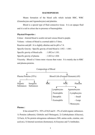
- 1. HAEMOPOIESIS Means formation of the blood cells which include RBC, WBC (Granulocytes and Agranulocytes) and platelets. Blood is a special type of fluid connective tissue. It is an opaque fluid and it is red in colour due to presence of haemoglobin. Physical Properties : Colour : Arterial blood is scarlet red and venous blood is purple. Volume : volume of blood is a normal adult is 5 litres. Reaction and pH : It is slightly alkaline and its pH is 7.4. Specific Gravity : Specific gravity of total blood is 1.052 – 1.061. Specific gravity of blood cells : 1.092 to 1.101. Specific gravity of plasma : 1.022 to 1.026 Viscosity : Blood is 5 times more viscous than water. It is mainly due to RBC and plasma proteins. Composition of Blood Plasma Proteins (55%) Blood Cells (Formed Elements) (45) Organic Inorganic RBC WBC Platelets Substances Substances Lymphocytes Agranulocytes - Neutrophils - Lymphocytes - Basophils - Small - Eosinophils - Large - Monocytes Plasma : It has around 91% - 92% of H2O and 8 – 9% of solid organic substances. 1) Proteins (albumin), Globulin and Fibrinogen), 2) Carbohydrates (Glucose), 3) Facts, 4) No protein nitrogenous substances (NH3 amino acids, creatine, uric acid etc), 5) Internal secretions (hormones), 6) Enzymes and 7) Antibodies. 1
- 2. Inorganic Substances : 1) Calcium 2) Sodium 3) Potassium 4) Magnesium 5) Chloride 6) Iodide 7) Iron 8) Phosphates 9) Copper Functions of Blood : 1) Nutrient function : It helps in absorption of nutritive substances like glucose, A.A., lipids and vitamins from GIT and carried to different parts of body. 2) Respiratory function : Transport of respiratory gases is done by blood O2 and CO2 are transported through blood. 3) Excretory function : Waste products due to various metastasis are removed and carried to the excretory organs through this medium. 4) Transport of Hormones : Hormones and few enzymes are carried by blood to different parts of body. 5) Regulation of H2O balance : H2O content of blood is freely interchangeable with interstitial fluid which helps in regulation of water content of body. 6) Acid base balance : Plasma proteins and Hb acts as buffers and helps in regulation of A-base balance. 7) Regulation of body temperature : Due to high specific heat of blood, helps in maintaining thermoregulatory mechanism. 8) Storage function : serves as a ready source of substance like water, glucose, Na and K which are taken from blood during starvation and fluid loss etc. 9) Defense functions : Due to various cells present in it. Blood cells are basically divided into 3 categories ; - Red blood cells 2
- 3. - White blood cells - Platelets Red Blood Cells : They are circular, biconcave cell without a nucleus be considered as a kind of a living bag containing Hb, RBC. Mainly carry oxygen from lungs to the tissues. White Blood Cells : They are basically divided into ; i) Granulocytes o Neutrophils o Eosinophils o Basophils ii) Agranulocytes o Monocyte o Lymphocyte i. Small ii. Large They are basically divided based on the presence or absence of granules in the cells. They all are nucleated cells with granules or of granular. They are mainly involved in the body’s defence mechanism against various bacteria and even some foreign material. Platelets : They are small colourless, non-nucleated and moderately refractive bodies. These formed elements of blood are considered to be the fragments of cytoplasm. They are spherical / oval shaped and become oval shaped when inactivated. Plates play key role in the reaction of hemostasis. All the blood cells including lymphocytes, originate from a single class of primitive cells, the pluripotent stem cell. But however lymphocytes differ from other blood cells, which arise normally arise after fetal life only within the 3
- 4. bone marrow and referred to as marrow cells. Lymphocytes although originating from the marrow precursor cells, mature and proliferate outside of the marrow in the thymus and peripheral lymphoid tissue. Erythrocytes, neutrophils, eisinophils, monocytes and platelets have finite life span and therefore fraction of these cells must be replaced daily. Approximate concentration and Daily Production in Peripheral Blood Cells : Mean concentration / Microitre Daily Production Rate / Kg Body Wt. Erythrocytes – Males - Females 5.4 x 10⁶ 4.8 x 10⁶ 3.8 x 10⁶ WBC - Neutrophils - Monocytes - Basophils - Eosinophils - Lymphocytes 4,500 300 40 150 2500 1.6 x 10⁹ 1.7 x 10⁸ - Variable - Platelets 2.5 x 10⁶ 2.8 x 10⁹ In addition to this bone marrow must respond intermittently to increased demands for specific cells like Eg.Granulocyte to tight bacterial infection or for erythrocytes after acute blood loss. HAEMOPOIETIC ORGANS: During embryogenesis (Development of fetus),the site of haeamopoiesis changes for several times. The first blood cells which is formed i.e. the nucleated erythrocyte appear in the blood islands in Yolk sac and “these yolk sac” heamopoiesis is produced during early development i.e. 3rd week – 34d month gestation period (IUL). The embryonic erythrocytes (yolk sac derived) do not loose their nuclei during maturation and they contain heamoglobin molecules of α and β family which are different from those founding later fetal / adult erythrocytes. As the fetus develops, the organic formation takes place. This stage that is during the mid-gestation period the main organ for heamopoiesis is fetal liver. Even the spleen also participates in the formation. During this period β- 4
- 5. chains of heam are present which are different from the adult β-globin chains of heamoglobin. During, about 20th embryonic week, heamopoiesis begins in bone marrow. It is fully active in RBM by 7-8th month of fetal life. As fetus matures heamopoiesis increases in the RBM and decreases in the liver and spleen. At birth practically whole of bony skeleton contains active marrow. After birth all blood cells except lymphocytes are generally made in RBM. Bone marrow is supplied by nutrient arteries which widely branch into capillaries which in turn enter sinuses and sinusoids. Between the sinuses and sinusoids the stem cells and precursors cells which by process of multiplication and differentiation form the erythrocytes, granulocytes and platelets. Only once they are matured they enter the circulation through the apertures in sinus walls. Sites : In young children, active heamopoietic marrow is found throughout both the axial skeleton (cranium ribs, vertebra and pelvis) and the bone of extremities. But in adults heamopoietic marrow is confirmed to axial skeleton and proximal end of femur and humors. In an adult heamopoietic marrow, it normally consist of islands of cellular active marrow separated and supported by fat. In old age, proportion of active marrow decreases and thereby fat increases. In some pathological conditions like heamolytic anaemias, thalassaemias, the marrow cellular activity increases at the expense of fat and sometimes cellular changes also takes place. In certain rare pathological conditions like myeloidmetaplasia, the heamopoietic activity runs back to extramedullary sites like spleen and liver. 5
- 6. GROWTH FACTORS : These are cytokines which control growth, differentiation and function of blood cells. Several different growth factors exist each capable of supporting a particular spectrum of hemopoietic cell types. Physiochemically they are acid glycoproteins which define these ability to alter growth and function of progenitor cells and differentiated cells. These heamopoietic growth factor are present in tissue fluids and blood in concentration of 10-10 to 10-11 . they effect the target cells through binding to specific cell surface receptors. Growth factors has 2 types of actions : 1) Induction of cell growth and differentiation in immature cells. 2) Modulation of effector functions in mature cells. Some growth factors act synergistically and multiple growth factor act simultaneously upon the precursor cells. Macrophage play a central role in controlling heamopoiesis through secretion of cytokine, IL-1. IL-1 stimulation growth of T-lymphocytes with elevates IL3 , and stem fibroblasts, endothelial cells to secrete GM-CSF and G-CSF. IL3 and GMCSF inturn stem, early cell division of progenitors of most, if not all the myeloid cell lineages. IL1- also acts upon mature blood cells, stimulation of release of neutrophils from RBM. IL3 acts upon myeloid cell progenitors along with GM-CSF. It acts particularly on earlier progenitor cell subset expanding and preparing for subsequent exposure to GM-CSF. GM-CSF 1st record to its ability to stimulate growth of gran, macrocytes and eosinophils. They also stimulate early division of erythrocyte progenitor and megakaryocytic progenitors. It also acts on mature granulocytes and macrophages including enhancement of granulocyte responses to chemotactic stimuli and ability of macrophages to kill intracellularly. 6
- 7. For full development such factors (IL3 and GMCSF) are not enough, but also simultaneous / sequential presence of 2nd growth factor acting specifically on more differentiated precursors. For neutrophilic gram it is Ci-CSF Erythrocytes Erythropoietin In addition to growth factor a variety of humoral agents have been identified that inhibit myeloid cell growth. They include TGF (transforming growth factor), granulocytic protein lactoferrin, acidic isoferritius (certain intracellular iron storage proteins) and Esthetics prostaglandins. Growth Stages Growth Factors 1) Factors that act on multilineage progenitors. 2) Granulocyte and macrophage growth factor 3) Differentiation of neutrophils, monocytes and eosinophils 4) Megakaryocyte growth factor 5) β – Lymhocytes 6) Erythroid cells GM-CSF, G-CSF, CSF-1, IL-3, 4,6,11,12 GM-CSF (monocytes and granulocytes) G-CSF, CSF-1, IL-5 CFU-MK, IL-3, IL-6 CFU-Pre B, IL-7 BFU-B, CFU-E, IGF-1, IL-3 Heamopoietic Stem Cells : They can be said as “stem cells represent a cell type which, in adult organic can maintain its non numbers in spite of physiological / artificial removal of cells from the population. Means “maintaining its own number”. They possess found props. i) An ability by cell division to give rise to new stem cell (self renewal). ii) An ability to different mature specialized blood cells. - Frequency of in bone marrow established : 1-2 /1000 nucleic stem cells and small number also circulate in blood. 7
- 8. - Specific supporting tissues like bone marrow (man) permit growth and differentiation of stem cells and they are referred as “heamopoetic microenvironment”. - When multitude of cells arises from a single cell, or when a single stem cell gives rise to a spleen colony that multitude of cells is called a CLONE. - In normal heamopoesis, mature blood cells originate from multiple stem cells, so normal heamophoisis is polyclonal. - In hematological malignancies, the progency of single abnormal stem cell may overgrow and suppress normal stem cells and here heamopoiesis is monoclonal. - As the stem cells differentiations, it losses its ability to give rise to all blood cells and becomes committed to production of one / more cell lines. Different mechanism have been postulated to regenerate commitment. a) Probability of differentiation increases with each division of a stem cells. b) Extra cellular inductive factor perhaps the heamopoetic growth factor / cell surface molecules in the heamopoietic microenvironment determine the changes within a stem cell that leads to commitment c) Others says, due to some series of unknown factors influence the probabilities for self renewal / commitment. It is said that, functionally 3 different stem cells. a) Pluriopotent stem cell b) Myeloid stem cell (which gives rise to erythrocytes, granulocytes of all types, monocytes and platelets0. c) Lymphocytes stem cells. 8
- 9. - As commitment progress, the capability of the stem cells for self renewal diminishes and when it is markedly limited / lost, the cell is no longer called a stem cell but called as PROGENITOR CELL. - This progenitor cell are recognized by their ability to give rise to clonal aggregates colonies) of differentiated cells on cultures. - Progenitor cells are more numerous than stem cells and like stem cells present in both bone marrow and circulating blood. Blast Cells : - The immediate offsprings of progenitor cells are the blast cells. For the erythroid series, there are blast cells like the pronormoblasts, early, intermediate and late normoblasts. - In case of granulocytic series, they are the myeloblasts and similarly there are lymphoblasts, monoblasts and megakaryoblasts. - Blast cells are immature cells. Stages of Development : Except for 1st 3 months of fetal life, all erythroid development are extravascular. When erythroids cells is almost mature, it enter the sinusoids from the extravascular space. The most primitive cell is called the pluripotent stem cell. This cell divides and differentiates, whereas the pluripotent stem cell can give rise to erythrocytes, granulocyte, monocyte, lymphocyte, platelet, that is why it is called pluripotent stem cell (Pluripotent – Capable of producing many / different cells). But the daughter cells are not capable of producing all the types of cells they can produce either ; - Myeloid series - Erythroid series More they advance in lineage, more they become restricted in the plurality of the patency. 9
- 10. The pluripotent stem cells divides and differentiated to give rise to committed stem cell. Here there are two types, one type of committed stem cells gives rise to myeloid series (Gram, no, RBC, platelets) but not lymphocytes. Whereas other type of committed stem cell gives rise to lymphocyte (T and B). Cells committed to produce myeloid series now divide and differentiate to produce the daughter cells called “Progenitor cells’. Several type of progenitor cells develop. One type of PC gives rise to cells of erythroid series, another granulocyte. Monotype and other megakaryocyte, and last eosinophil. Progenitor cells are usually called “Colony Units” (CFU) and designated as CFU-E, CFU, CFU-EO, CFU-Mega. RBC : There are 2 P.C. a) BFU-E b) CFU-E From CFU-0 : Pronomoblast cell (1st cell in the which is morphologically recognizable cell in erythro series) develops. Nucleus is strongly basophilic which very scanty cytoplasm and no Hb found. From pronormoblast early neuroblast – also called change basophilic neuroblast intermediate / chromatophilic normoblast late normoblast ortho chromatic normoblast. Hb appears in intermediate normoblast and so but poly chromatophilic and nucleus becomes smaller. In late neuroblast plenty of cytoplasm which is eosinophilic with fair amount of Hb, nucleus very small and deep pyknotic. Late Normoblast : Reticulocyte (matures for ½ in RBM and then enters peripheral blood, where it not a still a fully matured RBC Then it matures from mature RBC. Nucleus becomes smaller as cell matures from pronormo to late normoblast and here the pyknotic nucleus is extend to form reticulocyte. 10
- 11. As cell mature s- more and abundant cytoplasm. Hb-appears in intermediate normoblast and develops. Cell is capable of division to intermediate normoblastic. After this stage only maturation no division. PRODUCTION OF MYELOID CELL LINES : ERYTHROPOIESIS : Erythroid Progenitor Cells (EPC) : The EPC are of 2 types ; Burst forming Units – Erythrocyte (BFU-E) Colony forming units – Erythrocyte (CFU-E) - BFU-E are large, multilobulated colonies whereas the CFU-E are much smaller colonies. - Actually the BFU-E gave rise to CFU-E through continuum of intermediate forms and it is a most differentiated erythroid progenitors. - Growth of BFU-E requires presence of one / more growth factors which are collectively refer to as Burst Promoting Activity (BPA) which is general present in IL-3 and GM-CSF. - In contrast CFU-E is independent of BPA but require hormone erythropoietin for proliferation and maturation to heamoglobin containing erythrocytes. - Neither BFU-E nor CFU-E have morphological features permitting their distinction from each other or their recognition as members of erythroid series. MATURATION OF NORMOBLASTS : - The earliest morphologically recognizable member of erythroid series is the Pronormoblast / erythroblast. - This appears as cell of moderate – large size with a round nucleus occupying most of the cell and containing fine punctuate chromatin and blue nucleoli and with a rim of cytoplasm which is deeply basophilic. The deep blue colour of the immature cells is due to the presence of 11
- 12. large amounts of RNA and which is associated with active protein synthesis. - Subsequently as the cell matures the nuclei become smaller and denser, losing their nucleoli and then cell is then called normoblast and subsequently the size of normoblast diminishes progressively, the nucleus shrinks into a purple black pyknotic mass that is finally extended. Any remaining nuclear material is very effectively removed by a process of pitting during passage through the splenic sinus walls and eventually the nuclei become pyknotic and structureless before being extruded from the cell after which they are phagocytosed. - The cytoplasm also matures, the dense blue colour gradually changes through intermediate shade to orange pink as increasing amount of hemoglobin are synthesized. - The successive cytoplasmic change from blue to orange pink, various adjectives are given as – Basophilic, Polychromatic, Orthochromatic. - 3-5 cell divisions takesplace during pronormoblast- normoblast maturation. - Others are pronormoblast gives rise to form 8-32 RBC cell division stops at the late normoblast stage. - The normal time required for an erythroid cell to mature from a pronormoblast to a newly released circulating RBC is generally 5-7 days. - Heamoglobin synthesis has 3 requisites. a) Adequate amounts of mRNA for polypeptide chains of globin. b) Cell must synthesize normal amounts of tetrpyrole porphyrin. c) Iron must be available for incorporation into protoporphyrin to form HEME. Plasma transferring brings the iron to the developing erythroid cells. - When lack of body iron prevents transferring from bringing sufficient iron to developing RBC’s for heamoglobin synthesis erythropoiesis is impaired leading anaemiabesis. 12
- 13. MATURATION OF RETICULOCYTE : When first formed by extrusion of nucleus of the late normoblast, a non nucleated RBC which is larger than the mature RBC and does not yet have its full complement of heamoglobin but still possess some cytoplasmic RNA, mitochondria and surface transferring receptors i.e. the synthetic machinery required to continue to make heamoglobin. This newly differentiated immature RBC is called as reticulocytes, which is named because the staining procedures show the cells residual RNA, giving rise to beads and strands of dark blue material that form reticulin network within cytoplasm. Reticulocyte normally matures for 1-2 days before entering the circulation, during which time heamoglobin synthesis continues and the cells size decreases. Regulation and Assessment of Erythropoiesis : Although the growth factors with BPA may help in the earliest stage of erythropoiesis, the ERYTHROPOIETIN functions as a major regulate of erythropoiesis. ERYTHROPOIETIN : This is produced by the cells of the kidney that sense tissue hypoxia. This tissue hypoxia may result from any of the 3 cases. i) Decreased blood hemoglobin content (Anaemia) ii) Failure to oxygenate Hb adequately in the lungs (lung disease, certain congenital heart diseases, high attitude). iii) Impaired release of O2 from Hb at a normal tissue O2 tension (as occurs when blood C.O. level is elevated). - Normal erythropoiesis requires a basal level of stimulation of erythroid progenitor cells by erythropoietin. 13
- 14. Factors Influencing Erythropoiesis : 1) Erythropoietin 2) BPA – For proliferation of BFU-E 3) Vitamins – B12, folic acids important other like Vitamin B6 and C 4) Iron – Required for Hb synthesis 5) Miscellaneous Life Span and Destruction : Total life span is 120 days. As age advances, the enzymes which protect the erythrocyte from damaging effects of O2 begin to loose their efficiency. Therefore oxidative damages begin to appear and RBC, becomes rather rigid, spheroidal and fragile. During circulation, RBC pass through spleen whose diameters are very narrow and when such fragile RBC pass through these, they are ruptured. But young and healthy RBC with sufficient degree of biconcavity can survive. Normally spleen is the important slaughter house for RBCs, but when erythemia are diseased, fragility increases, they break down when passing through other structures also particularly liver. Functions of RBC : 1) RBC mainly help in carrying of the oxygen from the lungs to the peripheries. 2) It also helps in carrying CO2 from the peripheries to the lungs. The haemoglobin in the RBC combines which CO2 and form carbheamoglobin. About 30% of CO2 is transported in this form. 3) Hb in RBC also function as a good buffer by this action, it regulates the H+ ion concentration and thereby takes part in the maintenance of the acid- base balance. 14
- 15. 4) RBC carry the blood group antigens like A.agglutinogenous, B.agglutinogen and Rh factor which helps in determination of blood group. - In end stage kidney disease / after bilateral nephrectomy, the basal secretion of erythropoietin is eliminated, patients become severely anaemic unless they are infused with exogenous recombinant erythropoietin. In chronic steady state anemia’s, the plasma Hb concentration rises in inverse proportion to degree of reduction of blood haemoglobin concentration. - When erythropoietin secretion is increased, CFU-E and their more mature progency increase in number. - Total marrow cellularity and relative proportion of erythroid precursors to granulocytic forms are augmented. Reticulocytes are released without first maturing in the marrow. - The reticulocyte count will be elevated, reflecting both an elevated rate of RBC production and persistence for a longer period n the circulating blood of reticulocytes released early from the marrow. Erythropoiesis Count : - Total erythropotsis – estimated from the number of nucleated RBC in bone marrow. - Effective erythropoiesis – erythropoiesis producing RBC’s that circulate which can be estimated from the absolute reticulocyte count correlated for the early release of marrow reticulocytes. - In effective erythropoiesis – erythropoiesis which gives rise to defective cells that do not circulate but are phagocytosed by macrophages within the marrow. This can be evaluated by comparing the number of nucleated RBCs seen in marrow with the corrected, absolute reticulocyte counts. 15
- 16. - In healthy persons only negligible fraction of erythropoiesis is ineffective, but in certain disorders of erythorpoiesis, like pernicious anaemia, in effective erythropoiesis may predominate. - When erythropoiesis is stem by erythropoesis and large amounts of iron for Hb synthesis are readily available from the breakdown of RBC’s, s occurs in most heamolytic disorders, effective erythropoiesis may increase 6-8 fold. - Whereas erythropoiesis stem by erythropoiesis, but only limited amounts of iron are readily available from normal body stores like in acute blood loss, effective erythropoesis increases only about 2-3 fold. LEUKOPOIESIS : - It is defined as development and maturation of leukocytes. - The leukocytes are of 2 main varieties based on their presence of granules. i) Those with granules – Granulocytes – 2 types a) One with multi lobed nuclei - Polymorphonuclear leukocytes 9Eosonophils, Basophils, Neutrophils). b) Other has a single sound / lobulated nucleic monocytes. ii) The non granular leukocytes – lymphocytes. Formation / Leukopoiesis : The bone marrow and blood contains progenitor cells that give rise to colonies of granulocytes macrophages or both. The progenitor cells cannot be distinguished morphologically from other undifferentiated marrow and blood cells. The earliest morphologically identifiable member of granulocyte series is he myeloblast. It has a large sound nucues containing fine stippled chromatin and from 1-5 nucleoli, cytoplasm bulue and of any granules. 16
- 17. As the cell matures is nucleus undergoes condensation and finally regeneration. It cytoplasm acquires 3 types of granules – large azurophilic primary granules (appropriate), Two types of granules called specifies tertiary granules. NEUTROPHIL : They are generally found in peripheral blood, bone marrow and even in tissues. So it is said that neutrophils are present in 3 pools. a) Bone marrow pool 90% of neutralization b) Pool of peripheral blood 3% - Actual circulation and Marginal pool. c) Tissue pool 7% Formation : The red bone marrow pool - - Mitotic Pool and Post-mitotic pool - In mitotic pool – neutrophil are dividing as well as maturing (3-5 days) and it consists of myeloblasts, promylocytes and myelocytes. In post mitotic pool – cells no longer divided but only mature. - The committed stem cells in the myeloid series are the main cells which divide and differentiate to form different colony forming units which later give rise to different blood cells. - The progenitor cells for neutrophils being the CFU-GM which gives rise to both granulocytes or neutrophil and monocyte. - The CFU GM undergoes gradual change to form myeloblast which is the precursor of neutrophil. - Myeloblast divides and matures to promyelocyte and this and divides and matures to form myelocyte. - Till here the division takes place in the mitotic pol. From myeloblast myelocyte it takes -3 – 4 days. - The myelocyte enters the post mitotic pool where only matures but not divides and is converted to the neutrophil. This takes about 6 days in post mitotic pool. - In short journey of neutrophil from birth to grave. 17
- 18. - Myeloblast Myelocyte Neutrophil entry into intra vascular pool (6-7 hours) Emigration into tissue pool (1-4 days) Death. - Total life span from the stage of myeloblast which is a recognizable state is 12-14 days. - The most primitive cell as the CFU-GM : it is a common ancestor for both neutrophil and monocyte. But it is not recognizable. - CFU-GM divides and differentiates to myeloblast which is the first cell (precursor) of neutrophil which can be identified by the microscope. Nucleus is large, contains many nuclei and cytoplasm takes the bluish stain and contains no granules. - Myeloblast matures further and become promyelocyte where there is only one nucleolus in nuclei and the cytoplasm contain primary granules (azurophilicgram). - Promyelocyte develops to myelocyte which has radish nucleus with some indentation and abundant cytoplasm and cytoplasm contains pink staining granules (here primary granules no longer visible) no nucleolus. - Next stage of development is metamyelocyte where the nucleus shows indentation and cytoplasm contains large number of secondary granules. - Metamyelocytes matures to form band cell where the indentation in nucleus further deeps and acquires almost a horse shoe shape. - Finally the nucleus is segmented and becomes lobed. Uptill this stage it was in bone marrow now it is released into circulation by forming young neutrophil. Factors Influencing : i) Colony stimulating factor : Required for growth of CFU-GM. Produced by mononuclear cells ii) Prostaglandin-E : Inhibits CFU-GM (inhibit check mechanism (feed back much) by which excess CSF activity is prevented. 18
- 19. iii) Acidic lactoferrin : Produced by monocytes. Inhibits the normal CFU GM columns in both marrow but not the leukemic ones. iv) Lacrtoferrin : Produced by granules of neutrophils in whites CFU GM. - The life span is shortened example, in acute infections the immature forms (i.e. even metamyelocytes) are released into blood to meet demands of excessive needs of neutrophils. - The normal bone marrow reserve for neutrophil is 3 days and for children it is more less. Most neutrophil which are destured to die enter GIT where they die and are excreted out. EOSINOPHIL : - These are also present in different pools like neutrophils i.e. i) Bone marrow pool – 5.5 days ii) Intravascular pool : 8-12 hours Circulatory Marginal iii) Tissue pool – Death - Eosin are born and matured in RBM where their total stay is about 5.5 days. Then they enter the circulation i.e. the intra vascular pool and stay for about 8-12 hours. Sizes in the marginal and the actual circulatory pool is they are almost equal. From the circulatory pool they migrate to the tissues where they die. Formation : - Origin and development of eosinophil is similar to that of the neutrophil. - The most primitive cell for eosin is CFU-EO which cannot be identified by microscopic method forms myeloblast. - The myeloblast (more specific eosinophilic myeloblast) matures to form eosinophilic myelocyte. 19
- 20. - This further matures to form the meta myelocyte and then finally the mature eosinophil. - This is released into the peripheral blood. Factors effecting the Origin /Development / Release : i) CSF Required for growth of CFU-E. ii) Eosinopoitin : Increases the eosinophil count in the peripheral blood. Especially when there is eosinopenia. iii) Parasitic invasions and allergic conditions causes rise of eosiniophil count. iv) Cortisol : Causes eosinopenia (fall of eosinophil count). Eosinophil (Matureform) : - The mature eosinophil contains bilobed nucleus and distinctive large granules which stains orange red. - Material called Major Basic Protein (MBP) and make sup 50% of large granule has a key role to play in the eosinophils ability to damage the larva tissue stage. - 50% of large granule has a key role to play in the eosinophil’s ability to damage the larva issue stage of heliminthic parasite and is extremely potent tissue toxin. - A potent bactericidal and tissue toxin called (a) Eosinophilic cationic protein (ECP), (b) a neurotoxic protein and (c) Eosinophilic peroxidase are also present in large granules. - Along with these it also contain Aryl Sulphatase enzyme which hydrolyses the S-O ester linkages and which can inactive the sulfur containing leukotrienes that tissue mast cells liberate in immediate hypersensitivity. - For these reasons the blood eosinophils, increased count rise in helminthic infestations and in allergic states. Even when blood eosinophil counts are normal eosinophils are usually increase din 20
- 21. number at tissue sites of allergic reactions, whereby degrading mast cells thereby decreasing the clinical mean of allergic responses. - Administration of adrenal lucocorticoids causes blood eosinophil levels to fall within about 2 hours, presumably because of marignation and sequestration of the circulating cells. Continued administration leads to eosinopenia and impaired release of eosinophils from bone marrow. Functions : - They have antibacterial function due to MPO granules. - It has anti parasitic roles – act against the larva of the parasites. This is due to presence of MBP. - It also helps in allergic reaction. BASOPHIL : - They constitute around 1% of total WBC’s - The basophiles of blood develop in the ed bone marrow - The basophils have a very closed relation to the mast cells, but it is exactly not known. - Basophils are produced by RBM and present within the blood but whereas the must cells are present outside the circulation / blood within the tissue, that are produced in the RBM. - The functions of both being some due to same constitution and same morphology. The Development : The development stages of basophil are basically same as that of he other granulocytes. - The first cell which can be secondary is the blast cell. - This differentiate and divides to form promyelocyte. This again divides and differentiates from myelocyte. - This myelocyte differentiates to form the metamyelocyte. - The granules being to appear in the myelocyte stage. 21
- 22. - The meta myelocytes mature to form into a band cell. - This band cell matures fully to form a basophil. - This basophil is released into the blood where they stay for couple of hours and then migrate into the tissues. - After this what happens is unknown. Mature Basophil : Diameter varies from 10-12 µ. Nucleus is irregular and often ‘S’ shaped. Cytoplasmic granules are coarse and basophil and often so present or the nuclear zone that the nucleus appears partly covered by the granules. The granules of basophil (its counterpart in the tissue mast cell) contain 1) Histamine, 2) Heparin, 3) Acid peptides, 4) Acid hydrolases, 5) Neutral proteolytic enzyme. Functions : 1. It reacts in the allergic conditions. 2. It has an antibacterial effect. 3. It also helps in phagocytosing of the foreign cells, i.e. it acts as a defence mechanism in various conditions. MONOCYTES : - They constitute around 2% - 8% of total WBC and the target of the WBC’s (12-20 µ). - like other WBCs it is also distributed into different pols of the body. i) Marrow pool ii) Intravascular pool – 8-71 hours iii) Tissue pool – RES - There is a controversy regarding the marginal pool whether it is present /not. Some say its size is over 3 times of the circulatory pool and some say it is totally absent. Development 22
- 23. - The most primary cell being the CFU-GM as said earlier and cannot be identified by the staining procedure. - This differentiates to form promonocyte which can be identified and which has nucleus rounded/ slightly indented and cyto deeply basophilic and contains very few granules. - This promonocyte differentiates to form the monocyte. Uptil here the development occur sin RBM. - Monocyte is released into blood circulation where it stays for some time (exactly not known). - It enters the tissue and becomes tissue macrophage and member of RES. Here it undergoes further differentiation in tissues to give rise to cells of the mononuclear phagocyte system. Tissue macrophage. - Life span of it is unknown. Mature Monocyte / macrophage : - Nucleus not lobed, commonly ovoid and very often indented, the indentation is so deep than it acquires a horse shoe shaped appearance. - Although it is categorized as anon granulocyte the cytoplasm yet contains very fine granules. Functions : i) Formation of tissue macrophage : main function is to remove various undesirable substances by phagocytosis in various tissues like liver, spleen, lungs and so forth. ii) Role in acute and chronic infection: In any site of infection, neutrophils emigrate and phagocytose them, therefore in first 24 hours bacterial invasion neutrophils preponderate, whereas after that monocytes emigrate and attack them but no granules are present in them, but digest by production of H2O2. Neutrophils – 1st line of defence Monocyte s- 2nd line of defence 23
- 24. In chronic infection, macrophage engulf the bacteria but does not kill them / digest which is seen typically in incubation period of typhoid fever. iii) Role in lymphocyte mediated immunity : In this macrophages produces monokines which causes death of invading antigen and well as innocent host cells. iv) Monocytes produce – CSP, prostaglandin and acid lactoferrin to control production of neutrophils. LYMPHOCYTES : - This constitutes around 25-30% of the WBC, the lymphocytes play an important role in immunity. Functionally these lymphocytes are classified into 2 types – T.lymphocytes and B.lymphocytes. T- lymphocytes respective for cellular immunity. B lymphocytes – respect for humoral immunity. Development : - The development starts in the bone marrow directly from the stem cells. - The pluripotent stem cell gives origin to the CFU and lymphoid STEM CELLS (LSC). - In the RBC the most primitive cells is the pluripotent stem cell, which gives rise to committed tem cells. One type of CSC give rise to cells of myeloid series (erythrocytes, granulocytes, monocytes and platelets) and other type gives rise to lymphocytes (either B/T lymphocytes). Pluripotent stem cells Committed stem cells Myeloid series and Lymphoid series. Formation of T.lymphocytes : - From the RBM the committed stem cells of lymphocyte migrate to cortex of the thymus where they greatly proliferate in number by mitotic division. - From cortex of thymus these newly formed cells go to the medulla of thymus where no further divisions take place. 24
- 25. - In medulla of cortex they undergo further processing and after such processing they become immunologically competent T.cells and now they are released into circulation. FORMATION OF β-LYMPHOCYTES : - The committed stem cells (committed to produce β-lymphocytes) migrate to bursa of fabricus (in humans the homologous organ for this being lymphoid tissues of gut. Eg. Tonsils, Payers patches of intestine and appendix and they are collectively known as gut associated lymphoid tissue (GALT) and nowadays they say that homologous to it is the RBM) where they are further processed and after rule processing the mature β-lymphocytes are released into the circulation. - Thymus and Bursa Fabricus (GALT and RBM) are termed the central lymphoid tissue. - From the central lymphoid tissue, the immunologically competent T- cells and B cells come out and enter the peripheral blood, stay for couple of hours in the blood and later enter the peripheral lymphoid tissue. - In primary lymph tissue they stay for couple of hour sand come and again come back to the peripheral blood and from where it again goes back to peripheral lymphoid tissue thereby making, back and forth shutting between blood and peripheral lymphoid tissue. - The life span of lymphocytes is variable, some have unusually long life span (yes together). - While these lymphnodes are staining lymphnode, if the person is suddenly challenged by antigen, the lymphocytes become lymphoblast and multiply vigorously with the lymphnodes. - Thymus regresses in adult life and in old age it regresses remarkably. However T-lymph continue to multiply within lymph nodes (Peripheral 25
- 26. lymphoid tissue) and T.lymphocytes can increase eve in old age when necessity arises. - In case of RBC, development occurs only in RBM in the post natal life. Btu for lymphocytes, these reside in the lymph nodes and help in development. Lymphocytes belong to different clones when an invasion occurs by specific antigen, members of specific clone undergo blast transformation and proliferation. Ti is in this way, the lymphocyte supply to the peripheral blood is maintained. - Therefore the deepening of bone marrow in adult life, the absolute count of lymphocytes count in peripheral blood should not fall, although that of WBC’s belonging to myeloid series should fall. - It also explains that although thymus regresses and becomes atrophied in adult life, the supply of lymphocytes to the peripheral blood is not hampered. - But if thymus development is poor in the et al stage, the T.lymphocytes do not develop and child dies early. Functions : - They helping the specific immunity of the body, removes the various antigen like bacteria / virus / tumor cells. - Tissues when transplanted to our body from foreign persons are usually rejected and this phenomenon is called as tissue rejection. This mechanism specifically plays a vital role in tissue rejection of the cancer tissue. - NK and K cells kill cancer cells. PLATELET / THROMOCYTE : There are three types of formed elements of which thrombocytes are the one which assists in hemostasis (prevention of blood loss) by various processes including the helping in the coagulation. Normal platelet count is between 150,000 – 400,000/cmm. When the count goes below 1,00,000 – thrombocytopenia is diagnosed and when it goes 26
- 27. below 40,000 hemorrhagic manifestations occurs. Thus 40,000 platelet count is called the critical count. Development : - Platelets are derived from the giant cells in RBM i.e. megakaryocytes, whose diameter is usually around 100 µm. - A single megakaryocyte can give rise to about 1000 platelets. The platelets are produced in the RBM. The most primitive cell in line of platelet production as usual is the pluripotent stem cells. - This stem cell divides and differentiates to CFU megakaryocyte, which gives rise to promegakaryoblast (small sized cell). - The promegakarblast matures to form a megakaryobalst which inturns matures to from mega keraocyte. - They are readily recognizable by heir large size, multi lobulated nucleus, abundant cytoplasm. - Normal marrow contains about 1 megakal/500 nucleated red blood cells. - Analysis of DNA content of megakaryoblasts suggest that they contain even number of multipliers of the normal human DNA content (Polyploidy) Zn, Gn, 8n etc. This results from nuclear duplication of cell division, a process called entoreduplication. - The pseudopods of megakaryocyte are thrown inside of the sinusoids of bone marrow and then the tip of the pseudopoid is detached; this detached part of pseudopoid which does not contain any nucleus become a platelet. - A single megakaryocyte can give rise to about 1000 platelets and after this, the remnant of megakaryocytic is attached by the macrophages of the bone marrow, engulfed and digested. - Normally bone marrow contains only about one days reserve of platelets. Therefore human begins are susceptible to develop thrombocytopenia more quickly than granulocytopenia/ erythrocytopenia. 27
- 28. Morphology : - Platelets are very small borders consisting of cytoplasm encased within a membrane. They are non nucleated structures. - When inactive state i.e. circulating in blood they have disc like structure but when there is an injury/ bleeding they become activated and have a spherical structure. - Platelets contain mitochondria and golgi apparatus and apart from these some special structures like granules, tubules, filamentous structures made of actinomycin and various chemicals like adrenalin, serotonin, ADP and thrombospondin. The platelet does not contain DNA / RNA. Factors Influencing Thrombopoisis : i) Thrombopoietin : Substances which stimulates thrombopoiesis. ii) TGFβ : Released from α granules of platelets and cause deepening of platelet count by suppressing (-ve feed back mechanism). Functions of Platelets : - When here is any injury to blood vessels, these platelets adhere (to injured site), to aggregate (large number adhere to one another) and release many chemicals and pulp in hemostatic plug formation. - Platelets and release adrenaline and serotonin, thus helping in the vasoconstriction (which is necessary for hemostasis). - They also release some chemicals which help the blood to coagulate and they are called or coagulants. - They contain actinomyosin which helps in contraction and retraction of the platelet plug. - They also help in fixation of fibrin and platelets so that ultimately clot formation occur sonly where there is platelet plug. - They also release some substances which oppose the hemostasis and blood coagulation. Life Span : 28
- 29. - Normal life span is about 7-12 days. - Most of platelets are destroyed in spleen. About 1/3rd of platelet reside in spleen. Therefore spleenectomy causes a rise in blood platelet count. Applied Physiology: - Thrombocytopenia – condition where platelet count is low which leads to purpura but not coagulation defect. - After surgical operation and child birth number of platelets increase this produces risk of intravascular clotting and to prevent this early ambulation is advocated. CLINICAL CONSIDERATIONS : Anaemias : means deficiency of RBCs which can be caused ; 1) Too rapid blood loss 2) Too slow production of RBCs Types : a) Blood loss anaemia To rapid loss of blood – Acute and Chronic. b) A plastic anemaia : Lack of bone marrow functioning, which may be due to - Gamma ray radiation - Excessive x-ray exposure - Certain industrial chemicals - Drugs c) Megaloblastic anaemia : Loss of any of these i.e. vitamin B12, folic acid, zinc and intrinsic factor. d) Hemolytic anaemias : Due to early destruction / excessive destruction of RBCs anaemia occurs. - Hereditary spherocytosis Cells are small and spherical, so they can’t compress themselves when they pass through spleen, so easily rupture. 29
- 30. e) Sickle cell anaemia : Due to abnormal shape of Hb, cells becomes elongated and thereby fragile and rupture easily. Thalassaemias – Very little hb is present (which is due to deficiency) cells are abnormal in shape and fragile. Erythroblasts fetalis – Rh (+)ve antibodies of fetus are attached by Rh(-)ve mother and leads to destruction of RBC’s and severe anaemia. Effect of Viscosity : Normally the viscosity of blood is 3 times that of water, due to anaemia, it falls around 1.5 times that of H2O. So this decreases the resistance to blood flow in peripheral vessels, so that greater quantities of blood return to heart. Moreover hypoxia due to diminished transport of O2 by blood causes the tissue vessels to dilate, allowing further increase in return of blood to heart. So one of the major effects of anaemia is greatly increased work load on heart. POLYCYTHEMIA : When tissues become hypoxic due to little O2 in atmosphere (high altitudes, etc) the blood forming organs produce large quantities of RBC’s. This is physiological polycythemia. Here increases upto 6-8 mil/cm3 Polycythemia Vera : RBC as high as 7-8 mil/m3 and hematocrit increases as high as 60-70%. - It is a tumours condition of organs that produce blood cells. - Also causes excess production of WBCs and platelets. - Here total blood volume increases by 2 times which results in entire vascular system engorged and many capillaries become plugged by viscous blood, because the viscosity increases by 10 times to that of H2O. DENTAL MANAGEMENT : In patients with G-6-PD deficiency where there is hemolysis due to alteration of cell membrane and thereby fragile and leading to rupture of cells. 30
- 31. Blockage of Hexose monophosphate shunt path way in individuals with G-6- PD deficiency allows accumulation of oxidans in RBCs. These substances which generally produce wet hb and denatured Hb forms Heinz bodies which attach to cell membranes thereby altering the cell membranes which leads to hemolysis of cells. Such patients have increased incidence of drug sensitivity with sulfonamides, aspirin and chloremphenicol, penicillin, streptomycin and isoniazid etc. Drugs containing phenacetin is avoided in these patients. Sickle Cell anaemias : - Arrange short appointments - Avoid long and complicated procedures - Maintain good dental repair - Institute aggressive preventive dental care a) Oral hygiene instruction b) Diet control c) Tooth brushing and flossing d) Fluoride gel application - Avoid oral infection; treatment aggressively when present - Use L.A. (avoid G.A.) / epinephrine for routine dental care and for surgical procedures use 1:1,00,000 epinephrine in LA. - Avoid Barbiturates and strong narcotics; sedation with Diazepam can be used. - Use prophylactic antibiotics for surgical procedures. - Avoid liberal use of salicylates. Pain control with acetaminophen and codeine. - Use N2O – O2 with great care. WBC : 1) Agranulocytosis : Where bone marrow stops producing WBC’s leaving body unprotected agonist bacteria and other agents which invade body. 31
- 32. General human body lives in symbosis with many bacteria and all mucous membranes of body are constantly exposed to large number of bacterial, i.e. mouth, GIT, eyes, urethra and vagina. Therefore any decrease in number of WBC imm. Allows invasion. So within 2 days after RBM stops producing WBCs, ulcers appear in mouth and colon and develops respiratory infection. Bacteria from these enter surrounding tissues and invade the body and blood. Treatment death often ensues 3-6 days after acute agranulocytosis begins. Due to irradiation of γ-rays/exposure to drugs and chemical (benzene / anthracene) cause aplasia of RBM. So after irradiation injury where all stem cells and other cells are destroyed in RBM, sufficient time should be given for the regeneration to occur before doing any dental treatment. DENTAL MANAGEMENT OF LEUKEMIC PATIENT : New dental patients under medical treatment for leukemias, lymphoma and multiple myeloma must be identified by their health history and current status established by consultation with the physician. Patient who is in a state of remission can receive mot indicated dental treatment. Patient with acute signs and symptoms of disease should receive only cons. Emergency dental care until infection has been received and patient has returned to normal state. Patient with advanced disease and when prognosis is limited as in many cases of acute leukemia and multiple myeloma, should receive only emergency care, complex restorative procedure and extensive dental restorations are usually not indicated. If any procedure is to be done a platelet count / bleeding time should be obtained and if abnormal platelet replacement is indicated. For recently diagnosed leukemic patient dentist should be involved early on during the treatment planning stages of cancer therapy. For example, in 32
- 33. addition to providing oral prophylaxis and hygiene instrument he may recommend RCT /extension of teeth susceptible to acute exacerbations. CONCLUSION : As good as the saying “if it weren’t for the rocks in its bed the stream would have no song’. So likewise if no heamopoiesis, there is no formation of blood, as blood is the medium of life. 33