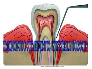
Endo perio interrelation /certified fixed orthodontic courses by Indian dental academy
•
24 gostaram•3,769 visualizações

The Indian Dental Academy is the Leader in continuing dental education , training dentists in all aspects of dentistry and offering a wide range of dental certified courses in different formats.

Recomendados
Recomendados
Mais conteúdo relacionado
Mais procurados
Mais procurados (20)
Semelhante a Endo perio interrelation /certified fixed orthodontic courses by Indian dental academy
Semelhante a Endo perio interrelation /certified fixed orthodontic courses by Indian dental academy (20)
Mais de Indian dental academy
Mais de Indian dental academy (20)
Último
Último (20)
Endo perio interrelation /certified fixed orthodontic courses by Indian dental academy
- 2. INDIAN DENTAL ACADEMY Leader in continuing dental education www.indiandentalacademy.com www.indiandentalacademy.com
- 3. CONTENTS INTRODUCTION DEFINITION CLASSIFICATION ETIOLOGY CONTROVERSIES REGARDING THE COMBINED LESION PATHWAYS OF SPREAD COMPARISION OF CLINICAL PRESENTATION B/W APICAL & MARGINAL PERIODONTITIS DIFFERENTIAL DIAGNOSIS EFFECT OF PULP & ITS TREATMENT ON PERIODONTIUM EFFECT OF PERIO. DISEASE & TREATMENT ON PULP LESIONS DIAGNOSIS TREATMENT REFERENCES CONCLUSION www.indiandentalacademy.com
- 4. DEFINITION An isolated, usually narrow, deep probing depth of pulpal or periodontal origin. Lesion with sub marginal or intrabony periradicular bone loss of pulpal and/or periodontal origin that communicates with the oral cavity via probing defect. A localized periodontal probing depth of pulpal or periodontal origin. www.indiandentalacademy.com STOCK
- 5. COHEN • • • • • • • Primary endodontic lesion Primary endodontic lesion with secondary periodontal involvement Primary periodontal lesion Primary periodontal lesion with secondary endodontic involvement True combined lesion Concomitant pulpal & periodontal lesion www.indiandentalacademy.com
- 6. WEINE Type I - Tooth in which symptoms clinically and radiographically simulate periodontal disease but are due to pulpal inflammation Type II - Tooth that has both pulpal and periodontal disease concomitantly Type III - Tooth has no pulpal problem but require endodontic therapy plus root amputation to gain periodontal healing Type IV - Tooth that clinically and radiographically simulate pulpal or periapical disease but infact have periodontal disease www.indiandentalacademy.com
- 7. LESIONS REQUIRING ENDODONTIC TREATMENT ONLY GROUP I necrotic pulp and apical granulomatous tissue replacing periodontium with or without sinus tract Chronic periapical abscess with sinus tract Longitudinal and horizontal root fractures Pathologic and iatrogenic root perforations Teeth with incomplete apical root development Endodontic implants / replants / transplants Teeth that require hemisection Root submergence GROSSMAN www.indiandentalacademy.com
- 8. LESIONS REQUIRING PERIODONTAL TREATMENT ONLY GROUP II Occlusal trauma causing reversible pulpitis Occlusal trauma plus gingival inflammation resulting in pocket formation and reversible pulpitis Suprabony or infrabony pocket formation treated with overzealous root planning and curettage leading to pulpal sensitivity Extensive infrabony pocket formation extending beyond the root apex and sometimes coupled with lateral or apical resorption yet with pulp that responds with in normal limits to clinical testing www.indiandentalacademy.com
- 9. LESIONS REQUIRING COMBINED ENDO – PERIO TREATMENT GROUP III Any lesion in Group I That results in irreversible reactions in the attachment apparatus and requires periodontal treatment Any lesion in Group II that results in irreversible reactions to the pulp tissue and also requires endodontic treatment www.indiandentalacademy.com
- 10. ATYPICAL ANATOMIC FACTORS Malaligned tooth Multirooted teeth / additional root Additional canal Cervical enamel projection Large lateral / accessory canal TRAUMA With gingival inflammation Tooth fracture Pulp / perio involvement + sinus tract Cellular changes - resorption www.indiandentalacademy.com
- 11. MISCELLANEOUS Iatrogenic systemic SINUS TRACT INFRABONY POCKET •From canal •From gingival crevice •Narrow •wide www.indiandentalacademy.com
- 12. Causes : ( Stock ) Root fractures – crown / root ( vital / non vital ) Root canal infection Root resorption Anatomical anomalies ( palatogingival groove,enamel pearls , root division , fused teeth , invagination ) Root perforation Orthodontic treatment Localized periodontal disease Transplantation & replantation www.indiandentalacademy.com Poorly designed restorations
- 13. Multiple endo perio lesion •Isolated lesion upon gen. periodontitis •Chronic periodontitis •Aggressive periodontitis www.indiandentalacademy.com
- 14. CONTROVERSIAL ASPECT CONCERNING THE COMBINED LESION PULPAL PERIODONTAL Chacker Massler Czarnecki & Schilder PERIODONTAL PULPAL ? Venous blood flow outward Drawback Lateral / accesory canal - flow bothways Seltzer & bender Stahl www.indiandentalacademy.com
- 15. Physiologic : • Apical foramen • Lateral canals • Dentinal tubules • Periodontal ligament • Alveolar bone • Neural pathways • Vasculolymphatic pathway • Palatogingival grooves • Cementum defect Iatrogenic : • • Vertical root fractures Perforations www.indiandentalacademy.com
- 16. COMPARISION MARGINAL PERIODONTITIS APICAL PERIODONTITIS Cervical Apex Plaque Pulpal inflammation Horizontal / Vertical bone loss - Seldom bone loss – localized generalized & deep Open Contained www.indiandentalacademy.com
- 17. Attachment loss asso. with Anatomic defect on root Nature of pathogenic flora Necrotic & infected pulp Host defense mechanism defect. Aggresiveness asso with Lateral & apical foramen Nature of flora Apical host defense Periodontal probing & radiographic examination Radiographic examination www.indiandentalacademy.com
- 18. DIFFERENTIAL DIAGNOSIS PULPAL PERIODONTAL CLINICAL Cause pulp infection periodontal Vitality non vital vital Restorative deep or extensive not related Plaque /calculus not related primary cause Inflammation acute chronic Pockets single and narrow multiple and wide pH value acidic alkaline Trauma primary or secondary contributing factor Microbial few coronally www.indiandentalacademy.com complex
- 19. RADIOGRAPHIC Pattern Bone loss Periapical Vertical bone loss localized wider apically radiolucent no generalized wider coronally not related yes HISTOPATHOLOGY Junctional epithelium Granulation tissues Gingival no apical migration apical (minimal) normal present coronal (larger) recession TREATMENT Therapy RCT www.indiandentalacademy.com Periodontal therapy
- 20. Problems in diagnosis : Vertical root fracture: varied radiographic picture Different angulations Surgical exposure lateral condensation excessive Post placement Cause Extensive restorations Older patients Gingival sulcus & pocket area Single rooted teeth multirooted teeth Developmental grooves In doubt ? – Biopsy / Histological analysis Systemic diseases mimic lesion on radiograph : Scleroderma Metastatic carcinoma Osteosarcoma www.indiandentalacademy.com
- 21. EFFECT OF PULP AND ITS TREATMENT ON PERIODONTIUM Periodontal inflammation & bone loss Sub marginal bone loss Horizontal bone loss Vertical intrabony pockets Furcation involvement Periodontal wound healing Traumatized necrotic pulp RC infection – compromised healing Gingival tissue thickness Alveolar bone level Surgical trauma to flap Effective flap repositioning Root canal treatment Doubtful pulpal status Iatrogenic problems www.indiandentalacademy.com
- 22. EFFECT OF PERIODONTAL DISEASE & ITS TREATMENT ON PULP Periodontal disease & pulp •Limited •Channels closed + dystrophic calcification- chronic •Sufficient viurlence – pulpal disease •Poor prognosis •Extraction / Root resection Periodontal treatment & pulp •Scaling & root planing – excessive cementum removal •Compromised pulp www.indiandentalacademy.com
- 23. PRIMARY ENDODONTIC LESION Caries / trauma / restorative procedure Pulp Inflammation Apical / lateral / Furcation / Attachment apparatus Pain , swelling , tenderness , marginal gingiva swelling Suppurative process – Sinus tract Pdl / Patent channels Multirooted Teeth Gr. III thru & Thru Furcation defect Diagnosis : Necrotic / Vitality test Treatment : RCT www.indiandentalacademy.com Ging. Sulcus ( GP / Probe to apex)
- 24. PRIMARY ENDODONTIC WITH SECONDARY PERIODONTAL Unchecked endo lesion Periapical alveolar bone destruction Interradicular area Drainage Hard / soft tissue Plaque / Calculus Apical attachment migration ( perio disease) Diagnosis : Necrosis / Calculus accumulation Treatment : Both www.indiandentalacademy.com
- 25. PRIMARY PERIODONTAL LESION Sulcus Plaque / Calculus Inflammation Apex Alv. Bone / Pdl Clinical attachment loss acute Abscess Lateral root / Furcation / TFO ( isolated lesion ) Diagnosis : Tooth mobility positive pulp test Broad based pocket / Plaque & calculus Generalized Treatment : Periodontal therapy www.indiandentalacademy.com osseous defects
- 26. PRIMARY PERIODONTAL & SECONDARY ENDODONTIC Periodontium Pulp Dentinal tubules Lateral canals Diagnosis : Deep pocket H/O extensive periodontal disease Past treatment Treatment : Both TRUE COMBINED LESIONS CONCOMITANT LESIONS www.indiandentalacademy.com Oral cavity
- 27. DIAGNOSIS OF ENDO PERIO LESIONS History of dentinal / pulpal pain History of periodontal symptoms (bleeding, recur. Infection , mobility) - nature / duration - risk factors Signs and symptoms of pulpal / periapical disease (vitality) Periodontal charting (probing profile) - Recession - Mobility - Furcation involvement - Attachment loss www.indiandentalacademy.com
- 28. Clinical signs of pocket formation : Bluish red marginal gingiva / vertical zone extending from marginal to attached gingiva. “Rolled” edge separating gingival margin form tooth surface. Enlarged edematous gingiva. Bleeding, suppuration, loose extruded teeth. www.indiandentalacademy.com
- 29. Symptoms of pocket formation Usually painless Localized or radiating pain or sensation of pressure after eating which gradually diminishes. Foul taste in localized areas. Sensitivity hot and cold Tooth ache in absence of caries are present www.indiandentalacademy.com
- 30. BIOLOGIC DEPTH PROBING DEPTH FORCE : 0.75N POCKET DEPTH LEVEL OF ATTACHMENT GINGIVAL RECESSION 6 POINT CHARTING DISTOPALATAL MID PALATAL www.indiandentalacademy.com MESIOPALATAL
- 32. LONG NARROW POCKETS: ENDODONTIC ORIGIN LATERAL ENDODONTIC ABSCESS WIDE AND DEEP POCKET “BLOW OUT” LESION www.indiandentalacademy.com
- 33. RADIOGRAPHIC PATTERN OF BONE LOSS •Apical extent of bone loss •Definite Pdl space absent •Shape of bone defect ( angularity / marginal bone ) Bone defect contributed by pulp infection : - Periodontal intrabony defect – 2/3 root length - Horizontal bone loss - 2/3 root length - periodontal bone loss involving root end Acute pain generally absent in endo perio – open nature 30 – 60 % spirochaetes 0 – 10 % spirochaetes - perio origin - endo origin www.indiandentalacademy.com
- 34. Causes: o Endo o Perio o Fracture o Resorption o Anatomy Endo perio lesion usually isolated, narrow localized pocket Check endodontic status Root treated Not root treated Evaluate adequacy Vitality tests Preparation: Obturation: oUnder prepared oOver prepared oPerforation oZipping oledges oUnder filled oOverfilled oPoor adaptation Is root canal re-treatment feasible? www.indiandentalacademy.com MANAGEMENT
- 35. Feasible re-treatment? No Yes Try OHI + debridement OHI Resolution? Resolution? No Yes No Yes oDo first stage endo oClean and shape canals oDress with calcium hydroxide Extract Resolution? Yes www.indiandentalacademy.com Extract No
- 36. Vitality tests Negative Positive Root canal treatment Perio treatment Resolution? Resolution? Yes No No Yes Check OHI and perio Check vitality again: If in doubt- do RCT Still no resolution: look for other causes Extract, resect , hemisect www.indiandentalacademy.com
- 37. TREATMENT ALTERNATIVES ROOT RESECTION REGENERATIVE TECHNIQUES ROOT RESECTION : “ Sectioning & removal of one or two roots of a multirooted teeth with accompanying odontoplasty.” ROOT AMPUTATION : “Removal of one or more roots of a multi rooted tooth while the others are retained.” HEMISECTION : “Removal or separation of root with its accompanying crown portion of mandibular molars.” www.indiandentalacademy.com
- 38. RADISECTION : “Newer terminology for removal of roots of maxillary molars .” BISECTION / BICUSPIDIZATION : “Separation of mesial and distal roots of mandibular molar along with its crown portion, where both segments are then retained individually.” www.indiandentalacademy.com
- 39. ROOT RESECTION Furcation involvement. ( Maxillary / Mandibular - 3 point / Nabers probe ) Classification of degree of Furcation involvement Class I - Horizontal loss of periodontal support< one third of tooth width Class II - Horizontal loss of periodontal support> one third but not encompassing the total width of the tooth Class III - Horizontal through and through destruction of the periodontal tissue in the furcal area www.indiandentalacademy.com
- 40. INDICATIONS FOR RESECTIONS Periodontal indications Severe vertical bone loss involving only one root of a multi rooted tooth Through and through furcation destruction Unfavorable proximity of roots of adjacent teeth Severe root exposure due to dehiscence www.indiandentalacademy.com
- 41. Restorative and endodontic indications: Prosthetic failure of abutments within a splint Endodontic failure: perforations, over extension , obstructed canals, separated instrument , root resorption Vertical fracture of one root Restorative reasons: sub gingival caries, erosion of large part of crown and root, traumatic injury Combination of these www.indiandentalacademy.com
- 42. Contraidications Root fusion making separation impossible Angulation or position of tooth in the arch Root morphology Improperly shaped occlusal contact www.indiandentalacademy.com
- 43. Poor prognosis Retained roots SURGICAL CONSIDERATIONS Buccal + Palatal flaps Releiving incision Intracrevicular incision Full thickness flap Undersurface of crown - bevelled . www.indiandentalacademy.com
- 44. Envelop Type Flaps Little Or No Attached Gingiva Flap Edges - Sutured Full Flap - Periodontal Disease - Scaling, Curettage Or Osseous Contouring Procedures www.indiandentalacademy.com
- 45. REGENERATIVE TECHNIQUES GTR – Differential tissue development Barrier Resorbable Collagen Synthetic Non resorbable Enamel matrix derived protein Barrier – principle - stiff www.indiandentalacademy.com
- 46. ANTIBIOTICS FOR ENDO PERIO LESION Tetracycline 250 mg (qid) Doxycycline 100 mg ( bd / od ) Metronidazole 250 mg ( tid for 7 days) Chlorhexidine www.indiandentalacademy.com
- 47. REFERENCES The use of guided tissue regeneration principles in endodontic surgery for induced chronic periodontic-endodontic lesions: a clinical, radiographic, and histologic evaluation J Periodontol. 2005 Mar;76(3):450-60. Pathologic interactions in pulpal and periodontal tissues. J Clin Periodontol. 2002 Aug;29(8):663-71. The influence of endodontic treatment upon periodontal wound healing. J Clin Periodontol. 1997 Jul;24(7):449-56. www.indiandentalacademy.com
- 49. www.indiandentalacademy.com Leader in continuing dental education www.indiandentalacademy.com