Bleaching/ cosmetic dentistry courses by indian dental academy
•
25 gostaram•2,114 visualizações

Indian Dental Academy: will be one of the most relevant and exciting training center with best faculty and flexible training programs for dental professionals who wish to advance in their dental practice,Offers certified courses in Dental implants,Orthodontics,Endodontics,Cosmetic Dentistry, Prosthetic Dentistry, Periodontics and General Dentistry.

Recomendados
Mais conteúdo relacionado
Mais procurados
Mais procurados (20)
Destaque
Destaque (20)
Semelhante a Bleaching/ cosmetic dentistry courses by indian dental academy
Semelhante a Bleaching/ cosmetic dentistry courses by indian dental academy (20)
Mais de Indian dental academy
Mais de Indian dental academy (20)
Último
Último (20)
Bleaching/ cosmetic dentistry courses by indian dental academy
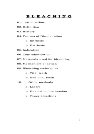
- 1. B L E A C H I N G 01. Introduction. 02. Definition 03. History 04. Factors of Discoloration a. Intrinsic b. Extrinsic 05. Indication 06. Contraindication 07. Materials used for bleaching 08. Mechanism of action 09. Bleaching techniques a. Vital teeth b. Non vital teeth 10. Other methods a. Lasers b. Enamel microabrasion c. Power bleaching 1
- 2. INTRODUCTION: Bleaching is an age old treatment, which has been performed over a century. The current trend towards cosmetic dentistry has generated more interest in bleaching as patient’s are asking for whiter and more beautiful teeth. Our society tends to dislike yellowing of teeth that comes with age or by various stains. White teeth are not only considered attractive but are also indicative of nutritional health, self esteem, hygiene pride and economic status. Discoloured teeth on the other hand have a far reaching effect on an individual both socially and psychologically. The different treatment modalities available are: Bleaching. Veenering. Jacket crowns. Micro abrasion etc. Out of all these bleaching is the most commonly used technique, least expensive with maximum conservation of tooth structure. DEFINITION: Whitening of a tooth through the application of chemical agents to oxidize / reduce the organic pigmentation in the tooth is termed as bleaching. 2
- 3. HISTORY: For vital tooth: 1877: 1st publication for bleaching by Chappel who used oxalic acid. 1884: Harlen used hydrogen peroxide for the 1st time, which he called as hydrogen dioxide. 1911: Rossentel suggested ultraviolet waves. 1918: Abbot introduced the combination of H2O2 with heat and light. 1989: Heywood and Heymann published an article on night guard vital bleaching using 10% carbomide peroxide. For non-vital tooth: 1848: Non-vital bleaching with chloride of lime. 1864: Trumann used chlorine from a solution of calcium hydrochloride and acitic acid commercial name was ‘Labarraque solution’. 1895: Garretson published 1st report of bleaching non- vital tooth. Early 1900’s: Superoxol (30% H2O2) was introduced. 1958: Pearson packed superoxol in the pulp chamber. 1967: “Nutting and Poe” demonstrated walking bleach using superoxol and sodium perborate to achieve prolonged effect in pulp chamber. Discoloration: Normal colour of primary teeth – bluish white. Normal colour of permanent teeth – Yellowish white. 3
- 4. Colour of enamel is determined by the translucency of enamel, which shows the colour of underlying dentin. Any alteration in colour may be Endogenous / Exogenous Physiologic / Pathologic Refractory index of teeth is 1.5. Causes for discolouration: Extrinsic Intrinsic 1. Coffee and tea stains 2. Cigarette 3. Diet 4. Tobacco 5. Nasmyths membrane (seen in children caused due to reduced enamel epithelium) 1. Tetracycline stains a) 1st degree b) 2nd degree c) 3rd degree d) 4th degree 2. Fluorosis stains 3. Trauma to tooth 4. Systmic condition a) Erythoblastosis foetalis b) Jaundice c) Amelogenesis imperfecta d) Enamel hypoplasia due to deficiency of vitamin A,C,D. 5. Iatrogenic discoloration a) Amalgam b) Intracanal medicaments (Kerr root sealer, Grossman sealer, Procosol sealer). Tetracycling staining: Teeth are more susceptible to tetracycline discolouration during their formation i.e. during the second trimester in vitro to roughly 8 years after birth. 4
- 5. Tetracycline molecules chelate with calcium and gets incorporated into the hydroxyapatite crystals. It involves predominantly the dentin. Severity of stains depends on the time and duration of drug administration. 4 Degrees: 1st Degree - Light yellow, brown stains - Uniformly distributed - No banding or localized concentration - Responds to bleaching in 2 or 3 session 2nd Degree - Dark gray stains - Extensive than 1st degree - Responds to bleaching in 4-6 session 3rd Degree - Dark gray stains with banding. - Responds to bleaching best bands will be evident. 4th Degree - Does not respond to bleaching. Fluorosis: High concentration of fluoride in more than 4ppm cause moderate to severe discoloration. Prevalence – Premolars, 2nd molars and mandibular and maximum incisors. Types Mild fluorosis - Brown pigmentation on a smooth enamel surface - Responds well to bleaching Moderate - Opaque fluorosis appear gray with white flecks on enamel surface. Severe - With pitting and dark pigmentation with surface defects. - Does not respond to bleaching 5
- 6. Traumatic Injuries: Causes rupture of blood vessel in the pulp. Causing diffusion of blood into dentinal tubules. Dark pink immediate after trauma and changes to pinkish brown after some days. Causes: Haemoglobin degrades into hemin, hematin, hematoiden and haemosidrin. Hydrogen sulphide produced by bacteria combines with hemoglobin to dark colour to tooth. Systemic Condition: 1) Erythroblastosis foetalis: (Rh factor incompatibility between mother and foetus) characterized by – breakdown of erythrocytes. 2) Jaundice: Bluish green or brown stains in dentin caused by bilirubin or biliverdin. 3) Amelogenesis imperfecta: is a genetic condition which interface with the normal enamel matrix formation. 4) Enamel hypoplasia: caused by deficiency of vitamins i.e. A, C, D and calcium and phosphorus. Iatrogenic discolouration: a] Trauma during pulp extirpation – hemorrhage b] Failure to removal of all pulpal remnants. c] Amalgam restoration cause – dark gray. d] Gold – dark brown when combined with products of decay. e] Break down of restoration i.e. acrylic, silicate and composite resins can cause the tooth to look grayer and discolored. f] Silver containing root canal sealers i.e. “Kerr root”, “grossman sealer”. g] Volatile oils → yellowish brown stain. h] Pins cause → bluish grain stains. 6
- 7. Indications: 1) Discolouration of anterior teeth – after R.C.T. 2) Tetracycline stains (mild) 3) Fluorosis 4) Haemorrhagic discolouration. 5) Discolouration due to ageing 6) Medication discolouration Contraindications: 1) Sensitive teeth 2) Opaque or white spots 3) Pregnancy 4) Lactation 5) Deep microcracks staining 6) Extensive silicate, acrylic or composite restoration. Materials used for bleaching: 1) H2O2 (30-35%) eg. Superoxol 30%; Starbrite 35%. (1-10%) Eg. Brite-smile; Natural white. 2) Carbamide peroxide 10% with carbopal, E.g. Ultralite; Opalescance. 10% without carbopal E.g. Gly-oxide; white and brite 10% Carbamide peroxide, Eg. Nu-Smile 3) Sodium perborate Available as granules, which are powdered later for use. S.P. + 30/H2O2 Walking bleach (Paste) S.P. + H2O (New) (Liquid). Bleaching mechanism: The mechanism is oxidation / reduction process called as “Redox process”. In this process the oxidizing agent has a free radical with unpaired electrons, which it gives up, becoming 7
- 8. reduced. The reducing agent (i.e. the substance being bleached) accepts the electrons and becomes oxidized. Reducing agent Oxidising agent Tooth Bleaching material After the process Tooth is oxidized Bleaching material is reduced (Organic pigmentation of tooth oxidized) Mechanism of each materials: Hydrogen peroxide in various concentration is the primary material currently used by the profession in the bleaching process. Current inoffice techniques for vital teeth and walking bleach technique use 30-35% H2O2. H2O2 is produced and regulated in the body and after involves in wound healing in low concentrations, also seen in eyes, spleen and liver etc i.e. it is more biocompatible. In high concentration it is bacteriostatic. In very high concentration it is mutagenic possibly by disrupting the DNA strand. H2O2 H2O+[O] or H2O2 H + OOH or H2O2 OH+OH; HOO + OH H2O +[O] and OOH [O]+H Action of H2O2 in tooth bleaching is considered to be oxidation, although the process is not well understood. It is felt that the oxidizers remove some unattached organic matter from the tooth without dissolving the enamel matrix but also may change the discoloured portion to a colourless state. (Continued long-term treatment will result in dissolution of the enamel matrix). 8
- 9. Tetracycline stains are more resistant to oxidation because the molecule is tightly bound to the mineral in the enamel prism matrix, during formation and hence is less accessible to immediate action. Therefore treatment takes a prolonged time. H2O2 diffuses through the organic matrix of enamel and dentin. Because the radicals have unpaired electrons, they are unstable and will attack other organic molecules for stability. Carbamide peroxide: The majority product in the market for night guard bleaching use 10% C.P. C.P 3% H2O2 + 7% urea H2O2 H2O + 2[O] and Urea NH3 + CO2. H2O2 is the active ingredient whereas urea raises the pH of the solution. Sodium perborate: For non-vital tooth bleaching sodium perborate is used with H2O2 called ‘walking bleach’ technique. a) Sodium perborate is a white powder which decomposes into sodium metaborate and H2O2. S.P. S. Metaborate + H2O2. b) When S.P. is mixed with H2O2 it decomposes into sodium metaborate, H2O and O2. SP + H2O2 S.m.Borate + H2O + O2. c) S.P + H2O S.M.Borate + H2O2. McInnes solution: 9
- 10. Old: Contain 30% H2O2 – Bleaches the enamel 36% HCl – Etches the enamel 0.2% Ether removes surface debris 5:5:1 H2O2 bleaches the enamel by a process of oxidation. HCl increases the penetration of the solution and helps in faster action. But HCl has deleterious effect such as; a) Loss of contour b) Irritation to gingiva c) Sensitivity of teeth So Chen, Xu and Shing (1993) replaced HCl by 20% NaOH. NaOH is highly alkaline in nature and therefore dissolves calcium at a slower rate. The results suggest that 1:1 mixture of H2O2 with 20%. NaOH is an effective as old McInnes solution and the calcium dissolved is much less with the new McInnes solution. A study by Dr.Versha Nangrani showed that the use of old McInnes solution resulted in loss of contour of teeth. The time taken by New McInnes solution was double than that of old McInnes solution but it did not show loss of contour of the teeth. Dr.Shadwala studied the amount of calcium dissolution with opalescence night guard vital bleaching and old McInnes solution and found that old McInnes 10
- 11. solution caused less calcium dissolution as compared to Night guard vital bleaching. The reason for this was night guard and vital bleaching uses bleaching action, which lasts for 6 hours for 2 weeks where as McInnes solution has to be used only for 5 minutes to 20 minutes. Treatment planning: 1) Record keeping and photographs 2) Careful diagnosis – Which included: Vitality tests; any restorations i.e. composite, acrylic etc. 3) Oral prophylaxis and polishing: To get rid of surface stains and calcium. 4) Preparation of teeth to be bleached i.e. a. Rubber dam b. Protection of soft tissue by Vaseline or Orabase. c. Floss to ligate interdentally to present seepage of bleaching agents into gums. d. Gauze saturated with cold water is placed under the rubber dam. e. Protective glass Bleaching of vital teeth 1) In office bleaching a. Thermocatalytic - Light; Heat b. McInnes solution – Old; New 2) Nigh guard vital bleaching 3) Over the counter 4) White strips 11
- 12. In office bleaching a) Thermocatalytic using light / heat After the rubber dam placement Teeth is covered with 35% H2O2 with Gauze The peroxide solution is activated by light / heat. Keep the light about 30 cms (13 inches) from the teeth and direct the beam to the surface to be bleached temperature ranges from 115°-140°F. When heat is used temperature is 46°-60°C. Gauze is kept wet by dispensing fresh bleaching solution. Bleaching agent should be kept in contact of light / heat for 30 minutes. After removing the Gauze, wash with copious amount of warm water and then polish. Repeat the procedure till desired shade is obtained. (Neutralizing the solution with 5.25% NaOCl) Tungsten halogen curing light. McInnes solution: Old New 30% H2O2 – Bleaching 30% H2O2 36% HCl - Etching 20% NaOH 0.2% Ether – Removal of surface debris 0.2% Ether – Removal of surface debris - Rubber dam is applied - Apply the paste on teeth for 5mts and repeated after 1 minute in level with cotton applicators. - Old McInnes solution is neutralized with baking soda. 12
- 13. - Copious irrigation of warm water. - Polish . Disadvantages of old McInnes solution overview. 1) Loss of contour 2) Sensitivity 3) Irritation to gingiva Ca dissolution is more Night guard bleaching (10% carbamide peroxide): Make primary impression and pour a cast. Make reservoirs on the cast on the labial aspect. Prepare the polyresin tray with the help of vacuum form machine. The tray is trimmed and extended 1mm short of the gingival margin on the labial aspect to avoid irritation of gingiva by the bleaching material. Tray is then tried on the patient’s mouth to check for fit. Instructions are given to the patient to load the tray with the gel only on the labial aspect and wear the tray at night during sleep for 6-7 hours for 2 weeks. If patient any sensitivity he can discontinue wearing the tray. Patient should be instructed not to bite the tray. After tray removal of the tray the patients brushes off the material. Weekly check up should be done. Disadvantages of night guard bleaching. - More lab procedures - Patient complains of gag reflex while using tray 13
- 14. White strips: Which is a thin flexible polyetheline strips which contains 5.3% hydrogen peroxide in gel form. The strips are used for 30 minutes twice daily for 14 days. Non vital bleaching: 1) In office bleaching. 2) Walking bleach (out of office bleaching). Establish a lingual opening of sufficient size to provide access to the pulp chamber and orifice of root canal. The root canal filling should be removed to a depth of 2-3 mm apical to cervical line. Zinc polycarboxylate or cavit cement should be used to refill, 1-2 mm coronally to the CEJ. Bleaching should never be attempted on any tooth that does not have a complete seal in the root canal. 1) In office bleaching: Rubber dam is placed. The pulp chamber is filled loosely with cotton fibres and the labial surface is covered with a few strands of cotton fibre to form a matrix for retaining the bleaching solution. 35% H2O2 is used to saturate the cotton inside the pulp chamber and on the labial surface. Excess should be wiped immediately. A thin tapered instrument can be heated and inserted into the pulp chamber for 5 minutes in a sequence of 1mt on 15 seconds off. It has been established by Caldwell that a non-vital tooth can with stand a temperature of 73°C without causing patient discomfort. 14
- 15. An alternative to activate H2O2 is the use of light and heat from a bleaching light. The tooth is subjected to 5 minutes exposure and one replenishes the bleaching agent at frequent intervals. The heating instrument and cotton can be removed. Process can be repeated for 4-6 times or 20-30 minutes each time placing a new cotton fibre. This technique can be used alone or in combination with walking bleach. 2) Walking bleach (by Nutting & Poe in 1963): This procedure consists of filtering the prepared chamber as described previously with a paste of 35% H2O2 and sodium perborate. When sealed into the pulp chamber, it oxidizes and discolours the stain slowly continuing its activity for a larger period. A small pledget of cotton wool in placed over the paste and the cavity is sealed with polycarboxylate or cavity cement. Patient should return in 4-7 days. Modified walking bleach: (Leonard and Stettem Brim et.al. 1997) Fabrication of the study model. Light cured composite are placed on the tooth or teeth to be treated. This acts as a reservoir to be created in a vacuum processed mouth guard whose thickness is 0.2- 03 inch. Mouth guard is trimmed at the cervical margins on the labial and lingual portions and tried on patients mouth. The G.P. in the root canal is sealed off from the pulp chamber at the cervical line and sealed with GIC. 15
- 16. Patient is taught how to inject 10% carbomide peroxide into the canal orifice and into the mouth guard with a syringe. Patient can use the mouth guard while sleeping. At the end of the daily treatment, patient rinses the mouth and then places a cotton pellet to prevent the food from getting into the opening. Treatment proceeds for 3-4 days. Anderson and L.A.F Punenta (1999) Spassier suggested the use of sodium perborate and H2O as a walking bleach technique instead of H2O2 to prevent cervical resorption. Sodium perborate breaks down to sodium metaborate and H2O2. This showed satisfactory results. Enamel micro abrasion: In 1916 Dr.Walter Kane of Colorado springs used 18% HCl with a warm instrument to successfully remove stains associated with endemic fluorosis. In 1984 McCloskey found that brown fluorosis stains can permanently be removed by rubbing the enamel with 18% HCl acid soaked cotton pellet wrapped around amalgam condenser. Two year later Croll and Cavananaugh developed a similar techniques that involves pressure application of 18% HCl with pumice to achieve colour modification. This was called enamel micro abrasion. Dr.Croll believed that the acid abrasive action of the compound gives the enamel surface a superfine polishing as a microscopic layer of enamel is removed. The freshly polished surface then develops or shiny glass like texture. 16
- 17. Jacobson – Hunt (1988) reported 30 second application of the acid abrasive compound using a mandrel and gear reduction handpiece on extracted human teeth which resulted in a enamel loss of less than 200 µm. In 1889 Kendell reported 5 second application of HCl acid pumice mixture removes 46 µm of enamel which should be considerably tolerable. Important concern of HCl pumice abrasion is the low viscosity and high concentration of 18% HCl. To eliminate this problem and ensure safety the viscosity of the acidic solution is increased by mixing 18% HCl acid with quartz particles so that the solution takes on a water soluble gel like form. This came to be known as ‘modified 18% HCl acid quartz – pumice abrasion technique’. Procedure: Gingiva is protected by a layer of petroleum jelly. Tooth is isolated will rubber dam. Teeth is dried and the paste consisting 18% HCl acid quartz – pumice particles are applied with cotton tip application to the stained areas of enamel. Paste is allowed to remain for 5 seconds and then for 10 seconds the enamel microabrasion is effectuated with a cotton swab pressure. After 10 seconds a marked degree of success is obtained and stain was removed. After 15 seconds, the enamel of the teeth turned to a normal shade. 17
- 18. At the end of treatment teeth is washed and dried with a neutral sodium gel. In this process the quartz particles convert the acid into a gel form and functions as an additional abrasive agent. Patient is reviewed after 6 months. Advantages: Relatively economical; No lab procedures Lasers: The action is to stimulate the catalyst in the chemical. There is no thermal effect and less dehydration of enamel. Argon laser of 488 nm wave-length for 30 seconds to evaluate the activity of bleaching gel. As the laser energy is applied, the gel is left in place for 3-4 minutes and then removed. This procedure is repeated for 4-6 times. Another product uses Ion laser technology. Argon laser is used as described before. Then CO2 laser is employed with another peroxide solution to provide penetration of the bleaching agent into the tooth to provide bleaching below the surface. Argon laser is in the form of blue light and is absorbed by dark colour. It is an ideal instrument to be used in tooth whitening when used with 50% H2O2. The affinity to dark colour ensures that the yellow brown colour can be easily removed. CO2 laser no colour requirement. It is unrelated to the colour of tooth and energy is emitted in the form of 18
- 19. heat. It is invisible and penetrates only 0.1 mm into water and H2O2 where it is absorbed. This energy can enhance the effect of whitening after the initial argon laser process. Power bleaching: This is done using xenon plasma arc curing light or a laser. This is a light cured resin gel that is applied on the gingiva and areas of the teeth that you do not wish to bleach. (Rubber dam is not required) Advantages – It is faster Disadvantages – unlike rubber dam, lips and soft tissues are not protected. Diode laser light: A true laser light produced from a solid-state source. It is ultra fast, taking 3-5 seconds to activate the bleaching of agent. This type of laser produces no heat. Complications of internal bleaching: 1) Cervical resorption - H2O2 percolates from the access cavity to the root surface through the acid treated patent dentinal tubules. - This stimulates an inflammatory response tending to dentin resorption. 2) Spillage of bleaching agents - Oxidising agents is easy to handle as a paste than a solution. 19
- 20. - Use rubber dam. - Any spillage is diluted immediately with copious volume of H2O. 3) Failure to bleach - Commonest is discolouration by mild ions in silver amalgam. - H2O2 improperly stored or expired. - In complete removal of composite or GIC which prevents the bleaching agents to penetrate into dentinal tubules. 4) Brittleness of crown - Bleaching causes coronal tooth structure to become brittle, this may be caused due to removing of all discoloured dentin rather than using the bleaching agent the discolour dentin. FERENCES: 1. “Bleaching Teeth” – Ronald Fieman, Roland Goldstein, David Garber, Quintessence Publishing Company, Inc., 1987. 2. History, safety and effectiveness of current bleaching technique and application of the night guard vital bleaching technique, Haywood Van B. Quintessence International, 1992; 23; 471-488. 3. Estimation of dissolution of calcium by old McInnes solution and new McInnes solution, R.Nageswar Rao and Nangrani V., Indian Endodontic Journal, 1998; 50- 53. 20
- 21. 4. “Bleaching teeth” new materials and new role, Roland Goldstein, JADA, 1987; 43-52. 5. Historical development of whiteners “clinical safety and efficacy”, Haywood Van B., Dental Update, 1997, April. 6. Laser assisted bleaching: An Update, JADA, 1998; 129; 1484-1487. 7. Non-vital tooth bleaching: A two-year case report, A.T.Hara, L.A.F. Pimenta, Quintessence International, 1999; 30; 748-754. 21