Neuraxial anesthesia
•Transferir como PPTX, PDF•
76 gostaram•13,630 visualizações

for anesthesia students

Recomendados
Mais conteúdo relacionado
Mais procurados
Mais procurados (20)
Semelhante a Neuraxial anesthesia
Semelhante a Neuraxial anesthesia (20)
Último
Último (20)
Neuraxial anesthesia
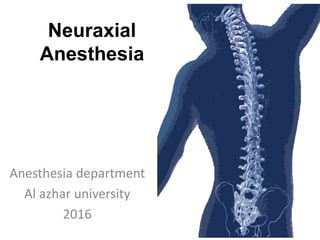
- 1. Neuraxial Anesthesia Anesthesia department Al azhar university 2016
- 2. Neuraxial Anesthesia • Neuraxial anesthesia is a type of regional anesthesia that involves injection of anesthetic medication in the fatty tissue that surround the nerve roots as they exist the spine (also known as an epidural and caudal) or into the cerebrospinal fluid which surrounds the spinal cord (also known as a spinal).
- 3. HISTORY • 1885 - J. Leonard Corning – first spinal anesthetic was administered accidentally The needle was made of gold • 1898 - August Bier - first planned spinal anesthesia for surgery • In 1921, Spanish military surgeon Fidel Pagés (1886– 1923) developed the modern technique of lumbar epidural anesthesia • Robert Andrew Hingson (1913–1996), working at the United States Marine Hospital in New York, developed the technique of continuous caudal anesthesia.
- 4. Advantages of Regional Anesthesia over GA Safe, reliable technique in patients at risk of apnoea, bradycardia, desaturation, cardiac or respiratory complications after GA Good alternative for day care surgeries Minimal risk of postoperative respiratory depression Limited stress response to surgery Cost effective
- 5. Spinal anesthesia Spinal anesthesia involves the use of small amounts of local anesthetic injected into the subarachnoid space to produce a reversible loss of sensation and motor function. The anesthesia provider places the needle below L2 in the adult patient to avoid trauma to the spinal cord. Spinal anesthesia provides excellent operating conditions for – Hernia (Inguinal or epigastric). – Haemorrhoidectomy , fistula , fissure. – cystectoscopy – Transurethral resection of the prostate and transurethral resection of the bladder tumors. – Abdominal and vaginal hysterectomies – Caesarean sections. – Lower limb surgery(orthopedic,plastic,vascular)
- 6. Vertebral Anatomy • Each vertebra consists of a pedicle, transverse process, superior and inferior articular processes, and a spinous process. • Each vertebra is connected to the next by intervertebral disks. • There are 2 superior and inferior articular processes (synovial joints) on each vertebra that allows for articulation. • Pedicles contain a notch superiorly and inferiorly to allow the spinal nerve root to exit the vertebral column.
- 7. VERTEBRA 33 Vertebrae ◦ 7 Cervical ◦ 12 Thoracic ◦ 5 Lumbar ◦ 5 Sacral ◦ 4 Coccygeal
- 8. Vertebral Anatomy- Top View Transverse Process Vertebral Body Spinal Canal Spinous Process Lamina
- 9. Ligaments that support the vertebral column Ventral side: Anterior and posterior longitudinal ligaments Dorsal side: Important since these are the structures your needle will pass through!
- 10. Spinal Cord • Spinal Cord – Adult • Begins: Foramen Magnum • Ends: L1 – Newborn • Begins: Foramen Magnum • Ends: L3 – Terminal End: Conus Medullaris – Filum Terminale: Anchors in sacral region – Cauda Equina: Nerve group of lower dural sac
- 11. Termination of Spinal Cord In adults usually ends at L1. Infants L3 There are anatomical variations. For most adults it is generally safe to place a spinal needle below L2 unless there is a known anatomic variation.
- 12. Sagittal Section Through Lumber Vertebrae Supraspinous Ligament (Outer most layer) Intraspinous Ligament (Middle layer) Ligamentum Flavum (Inner most layer)
- 13. CSF • Clear fluid that fills the subarachnoid space • Total volume in adults is 100-150 ml • Volume found in the subarachnoid space is 25-35 ml • Continually produced at a rate of 450 ml per 24 hour period replacing itself 3-4 times • Reabsorbed into the blood stream by arachnoid villi and granulations • Specific gravity is between 1.003-1.009 (this will play a crucial role in the baracity of local anesthetic that one chooses)
- 14. Membranes that surround the spinal cord • Pia mater- highly vascular, covers the spinal cord and brain • Arachnoid mater- non vascular and attached to the dura mater. Principal barrier to the migration of medications in and out of the CSF • Dura mater (“tough mother”)- extension of the cranial dura mater, extends from the foramen magnum to S2 (ending at the filum terminale)
- 15. Adapted with permission from “Unintended subdural injection: a complication of epidural anesthesia- a case report”, AANA Journal, vol. 74, no. 3, 2006.
- 16. CONTRAINDICATIONS Absolute Patient Refusal Infection At The Site Of Injection Coagulopathy And Other Bleeding Disorders Severe Hypovolemia Increased Intracranial Pressure Severe Aortic Stenosis Severe Mitral Stenosis
- 17. CONTRAINDICATIONS Relative Sepsis Uncoperative Patient Preexisting Neurological Deficits Severe Spinal Deformity Controversial Prior Surgery At The Site Of Injection Complicated Surgery Prolonged Operation Major Blood Loss
- 18. Spinal Technique • Preparation & Monitoring – EKG – NBP – Pulse Oximeter • Patient Positioning – Lateral decubitous – Sitting – Prone (hypobaric technique)
- 19. Landmark • landmark: iliac crest spinous processL4-5
- 20. PATIENT POSITIONING • SITTING POSITION
- 21. PATIENT POSITIONING • LATERAL DECUBITUS
- 22. Positions for Spinal Tap Procedures
- 24. • Midline Approach – Skin – Subcutaneous tissue – Supraspinous ligament – Interspinous ligament – Ligamentum flavum – Epidural space – Dura mater – Arachnoid mater • Paramedian or Lateral Approach – Same as midline excluding supraspinous & interspinous ligaments Anatomic Approach
- 26. Complications of Spinal Anaesthesia • Hypotension • Bradycardia • Total Spinal Anesthesia • Neurological Complecations – Cauda Equina Syndrome • Post Dural Puncture Headache • Infection • Backache
- 28. Introduction: • Epidural anesthesia is a central neuraxial block technique with many applications. • The epidural space was first described by Corning in 1901, and Fidel Pages first used epidural anaesthesia in humans in 1921. • In 1945 Tuohy introduced the needle which is still most commonly used for epidural anesthesia. • it can be used as an anesthetic, as an analgesic adjuvant to general anesthesia, and for postoperative analgesia in procedures involving the lower limbs, perineum, pelvis, abdomen and thorax.
- 29. Indications: • Epidural anaesthesia can be used as sole anaesthetic for procedures involving the lower limbs, pelvis, perineum and lower abdomen. • It is possible to perform upper abdominal and thoracic procedures under epidural anaesthesia alone, but the height of block required, with its attendant side effects, make it difficult to avoid significant patient discomfort and risk. • The advantage of epidural over spinal anaesthesia is the ability to maintain continuous anaesthesia after placement of an epidural catheter, thus making it suitable for procedures of long duration.
- 31. Technique of Epidural Anesthesia Loss of resistance technique to identify the epidural space. – 0.5% Bupivacaine (mainly) or lidocaine (2.0%) is usually used to produce epidural anaesthesia. – Local anaesthetic solutions are deposited in the peridural space between the dura mater and the periosteum lining the vertebral canal. – The peridural space contains adipose tissue, lymphatics and blood vessels. – The injected local anaesthetic solution produces analgesia by blocking conduction at the intradural spinal nerve roots. 32
- 32. 33
- 33. 34
- 34. Indication and Contraindication: • The same of spinal anaesthesia. • Additional indication is the post operative Pain management using the epidural catheter technique. • Complications: the same of spinal anaesthesia, except the postdural puncture headache. 35
- 35. Detail Technique of Epidural Anaesthesia • Using local anaesthetic raise a subcutaneous wheal at the midpoint between two adjacent vertebrae. • Inflitrate deeper in the midline and paraspinously to anaesthetise the posterior structures. • Insert epidural needle to the skin at this point, and advance through the supraspinous ligament, with the needle pointing in a slightly cephalad (upward) direction. • Then advance the needle into the interspinous ligament, which is encountered at a depth of 2-3 cm. • Until distinct sensation of increased resistance is felt as the needle passes into the ligamentum flavum.
- 37. With 5-10ml of air in the syringe, attach it to the hub of the needle once it has entered the interspinous ligament. The plunger is gently pressed, and if there is resistance ("bounce"), the needle is very carefully advanced, with the dorsum of both hands resting against the back to provide stability. After 2-3mm, the plunger is again gently pressed, and this procedure is repeated as the needle is carefully advanced through the tissues. The distinctive decrease in resistance when the needle enters the ligamentum flavum is felt, and the process is continued in 2mm increments. There is usually a distinctive "click" when the needle enters the epidural space, and provided great care is taken, and the needle only advanced in 2mm increments, the needle should stop before it reaches the dura. At this point air can be injected into the epidural space very easily. The syringe is removed and the catheter threaded
- 38. • Remove the syringe and thread the catheter gently via the needle into the epidural space. • The catheter has markings showing the distance from its tip, and should be advanced to 15-18cm at the hub of the needle, to ensure that a sufficient length of catheter has entered the epidural space. • Remove the needle carefully, ensuring that the catheter is not drawn back with it. • The markings on the needle will show the depth of the needle from the skin to the epidural space, and this distance will help determine the depth to which the catheter should be inserted at the skin. • For example, if the needle entered the epidural space at a depth of 5cm, the catheter should be withdrawn so that the 10cm mark is at the skin, thus leaving approximately 5cm of the catheter inside the epidural space, which is an appropriate length.
- 39. Choice of drugs: • The choice of drugs administered epidurally depends on the indication for the epidural: • Surgical anaesthesia:Requires dense sensory block and usually moderate to dense motor block. To achieve this, concentrated local anaesthetic preparations are required. The most commonly used local anaesthetics in this setting are 2% lignocaine 10-20ml (with or without adrenaline 1:200 000) or 0.5% bupivacaine 10-20ml. The latter has a longer duration of action, but a slower onset time, compared with lignocaine. • For analgesia during labour: 0.1-0.25% bupivacaine 5-10ml is more popular, as it produces less motor block. • Postoperative analgesia: weaker concentrations of bupivacaine, e.g. 0.1-0.166% with or without added low dose opioids.
- 40. Differences between Spinal and Epidural Anesthesia Spinal anaesthesia Extradural Anaesthesia Level: below L1/L2, where the spinal cord ends Level: at any level of the vertebral column. Injection: subarachnoid space i.e, puncture of the dura mater Injection: epidural space (between Ligamentum flavum and dura mater) i.e ,without puncture of the dura mater Identification of the subarachnoid space: When CSF appears Identification of the Peridural space: Using the Loss of Resistance technique. Doses: 2.5- 3.5 ml Bupivacaine 0.5% heavy Doses: 15- 20 ml Bupivacaine 0.5% Onset of action: Rapid (2-5 min) Onset of action: Slow (15-20 min) Density of block: More dense Density of block: Less dense Hypotension: Rapid Hypotension: Slow Headache: Is a probably complication Headache: Is not a probable complication. 41
- 41. Complications and Side Effects • Hypotension • Inadvertent high epidural block • Local anaesthetic toxicity • Total spinal block
- 43. Introduction: • Caudal anesthesia has been used for many years and is the easiest and safest approach to the epidural space. When correctly performed there is little danger of either the spinal cord or dura being damaged. • It is used to provide peri and post operative analgesia in adults and children. • It may be the sole anesthetic for some procedures, or it may be combined with general anesthesia.
- 44. Indications: 1. Anesthesia and analgesia below the umbilicus 2. Obstetric analgesia :For the 2nd stage or instrumental deliveries. Care should be taken as the foetal head lies close to the site of injection and there is real risk of injecting local anesthetic into the foetus. 3. Chronic pain problems relating to lower limbs and lower abdominal pains.
- 45. Contraindications: 1. Infection near the site of the needle insertion. 2. Coagulopathy or anti coagulation. 3. Congenital abnormalities of the lower spine or meninges, because of the unclear or impalpable anatomy.
- 46. Anatomy: • The caudal epidural space is the lowest portion of the epidural system and is entered through the sacral hiatus. • The sacrum is a triangular bone that consists of the five fused sacral vertebrae (S1- S5). • It articulates with the fifth lumber vertebra and the coccyx.
- 47. • The sacral hiatus is a defect in the lower part of the posterior wall of the sacrum formed by the failure of the laminae of S5 and/or S4 to meet and fuse in the midline. • The sacral canal is a continuation of the lumbar spinal canal which terminates at the sacral hiatus.
- 48. Choice of drugs & dosage: • Drugs that are commonly used include : Lignocaine 1% and Bupivacaine 0.25%.
- 49. Technique: The patient is prepared as for general anaesthesia: (1) He/she should be fasted (2) All appropriate equipment for resuscitation must be available. (3) An intravenous cannula should always be inserted in an upper limb, in case of accidental intravenous injection, or profound sympathetic blockade from a high epidural block. (4) The procedure must be carried out with a strict aseptic technique. The skin should be thoroughly prepared and sterile gloves worn.
- 50. (5) There are three main approaches: • The prone, the semi-prone, and the lateral. The choice depends on the preference of the anesthetist and the degree of sedation of the patient. The caudal space is made more prominent by asking the patient to internally rotate their ankles. The semi-prone position is preferred for the anesthetised or heavily sedated patient as the airway is easier to control in this position, while still allowing reasonably easy access to the sacral hiatus. The lateral position is often used in children, as the landmarks are easier to find than in adults. Care should be taken to avoid over flexing the hips (as for lumber epidurals) as this can make the landmarks more difficult to palpate
- 51. 6) The landmarks are palpated. The sacral hiatus and the posterior superior iliac spines form an equilateral triangle pointing inferiorly. • The sacral hiatus can be located by first palpating the coccyx, and then sliding the palpating finger in a cephalad direction until a depression in the skin is felt.
- 53. 7) Once the sacral hiatus is identified the area above is carefully cleaned with antiseptic solution, and a 22 gauge cannula or needle is directed at about 90 degree to skin and inserted till a "click" is felt as the sacro- coccygeal ligament is pierced.
- 54. Care should be taken not to insert the needle too far as the dura lies at or below the S2 level in the child. The needle should be aspirated looking for either CSF or blood. The injection should never be more than 10 ml/30 seconds • Further tests to confirm the correct position include: Introduction of a small amount of air will not produce subcutaneous emphysema, and will be heard as a "woosh" sound if a stethoscope is place further up the lumbar spine. There should be no local pain during injection.
- 55. (9) A small amount of local anaesthetic should be injected as a test dose (2-4mls). It should not produce either a lump in the subcutaneous tissues, or a feeling of resistance to the injection, nor any systemic effects such as arrhythmias or hypotension. If the test dose does not produce any side effects then the rest of the drug is injected, the needle removed and the patient positioned for surgery. In the post-operative period, motor function must be checked and the patient should not be allowed to try and walk until complete return of motor function is assured. The patient should not be discharged from hospital until he/she has passed urine, as urinary retention is a recognized complication.
- 56. Complications: Intravascular or intraosseous injection:This may lead to grandmal seizures and/or cardio-respiratory arrest. Dural puncture: Extreme care must be taken to avoid this as a total spinal block will occur if the dose for a caudal block is injected into the subarachnoid space. If this occurs then the patient will become rapidly apnoeic and profoundly hypotensive. Perforation of the rectum: Contamination of the needle is extremely dangerous if it is then inserted into the epidural space. Sepsis:This should be a very rare occurrence if strict aseptic procedures are followed. Urinary retention. Haematoma
Notas do Editor
- To show the landmarks that are helpful during the technique