Asbestos is a naturally occurring mineral that was commonly used in building materials for its heat resistance and fireproof properties. However, inhalation of asbestos fibers can cause serious illnesses like asbestosis, lung cancer, and mesothelioma. Asbestosis specifically refers to scarring of the lungs caused by asbestos exposure. Symptoms include shortness of breath. Diagnosis is made through chest imaging and history of asbestos exposure. There is no cure for asbestosis, so treatment focuses on relieving symptoms and managing complications.
1. Manilyn J. IlanEntrep II-ASubject: NatscieTeacher: KathrinaPobreFebruary 26, 2011Time 1:00pm
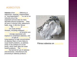
2. aSBESTOS Asbestos (from Greekἄσβεστος or asbestinon, meaning "unquenchable" or "inextinguishable")[1][2] is a set of six naturally occurring silicate minerals exploited commercially for their desirable physical properties.[2] They all have in common their asbestiform habit, long, (1:20) thin fibrous crystals. The inhalation of asbestos fibers can cause serious illnesses, including malignant lung cancer, mesothelioma (a formerly rare cancer strongly associated with exposure to amphibole asbestos), and asbestosis (a type of pneumoconiosis). Long exposure to high concentrations of asbestos fibers is more likely to cause health problems, as asbestos exists in the ambient air at low levels, which itself does not cause health problems.[3] The European Union has banned all use of asbestos[4] and extraction, manufacture and processing of asbestos products.[5] Fibrous asbestos on muscovite
3. Asbestos became increasingly popular among manufacturers and builders in the late 19th century because of its sound absorption, average tensile strength, and its resistance to heat, electrical and chemical damage. When asbestos is used for its resistance to fire or heat, the fibers are often mixed with cement or woven into fabric or mats. Asbestos was used in some products for its heat resistance, and in the past was used on electric oven and hotplate wiring for its electrical insulation at elevated temperature, and in buildings for its flame-retardant and insulating properties, tensile strength, flexibility, and resistance to chemicals. Asbestos
4. Types and associated fibers Six minerals are defined by the United States Environmental Protection Agency as "asbestos" including that belonging to the serpentine class chrysotile and that belonging to the amphibole class amosite, crocidolite, tremolite, anthophyllite and actinolite. There is an important distinction to be made between serpentine and amphibole asbestos due to differences in their chemical composition and their degree of potency as a health hazard when inhaled. However asbestos and all commercial forms of asbestos (including chrysotile asbestos) are known to be human carcinogens based on sufficient evidence of carcinogenicity in humans.[ Chrysotile asbestos
5. SerpentineWhite is obtained from serpentinite rocks which are common throughout the world. Its idealized chemical formula is Mg3(Si2O5)(OH)4. Chrysotile fibers are curly as opposed to fibers from amosite, crocidolite, tremolite, actinolite, and anthophyllite which are needlelike.[8] Chrysotile, along with other types of asbestos, has been banned in dozens of countries and is only allowed in the United States and Europe in very limited circumstances. Chrysotile has been used more than any other type and accounts for about 95% of the asbestos found in buildings in America.[9] Applications where chrysotile might be used include the use of joint compound. It is more flexible than amphibole types of asbestos; it can be spun and woven into fabric. The most common use is within corrugated asbestos cement roof sheets typically used for outbuildings, warehouses and garages. It is also found as flat sheets used for ceilings and sometimes for walls and floors. Numerous other items have been made containing chrysotile including brake linings, cloth behind fuses (for fire protection), pipe insulation, floor tiles, and rope seals for boilers.[citation needed] Asbestos fibers
6. Asbestosis is a chronic inflammatory and fibrotic medical condition affecting the parenchymaltissue of the lungs caused by the inhalation and retention of asbestos fibers. It usually occurs after high intensity and/or long-term exposure to asbestos (particularly in those individuals working on the production or end-use of products containing asbestos) and is therefore regarded as an occupational lung disease. People with extensive occupational exposure to the mining, manufacturing, handling or removal of asbestos are at risk of developing asbestosis.[1] Sufferers may experience severe dyspnea (shortness of breath) and are at an increased risk for certain malignancies, including lung cancer but especially mesothelioma.[2] Asbestosis specifically refers to interstitial (parenchymal) fibrosis from asbestos, and not pleural fibrosis or plaquing.
7. Signs and symptoms The signs and symptoms of asbestosis do not manifest until after an appreciable latency (time since first exposure), often several decades under current conditions in the US.[3] The primary symptom of asbestosis is generally the slow onset of dyspnea, especially on exertion.[4] Clinically advanced cases of asbestosis may lead to respiratory failure. On auscultation of the lungs, the physician may hear inspiratoryrales. The characteristic pulmonary function finding in asbestosis is a restrictive ventilatory defect. [5] This manifests as a reduction in lung volumes, particularly the Vital Capacity (VC) and Total Lung Capacity (TLC). The TLC may be reduced through alveolar wall thickening; however this is not always the case.[6] Large airway function, as reflected by FEV1/FVC, is generally well preserved.[3] In the more severe cases, the drastic reduction in lung function due to the stiffening of the lungs and reduced TLC may induce right-sided heart failure (corpulmonale).[7][8] In addition to a restrictive defect, asbestosis may produce reduction in Diffusion Capacity and arterial hypoxemia.
8. Diagnosis According to the American Thoracic Society (ATS)[3], the general diagnostic criteria for asbestosis are: Evidence of structural pathology consistent with asbestosis, as documented by imaging or histology Evidence of causation by asbestos as documented by the occupational and environmental history, markers of exposure (usually pleural plaques), recovery of asbestos bodies, or other means Exclusion of alternative plausible causes for the findings The abnormal chest x-ray and its interpretation remain the most important factors in establishing the presence of pulmonary fibrosis.[3] The findings usually appear as small, irregular parenchymal opacities, primarily in the lung bases. Using the ILO Classification system, "s", "t", and/or "u" opacities predominate. CT or high-resolution CT (HRCT) are more sensitive than plain radiography at detecting pulmonary fibrosis (as well as any underlying pleural changes). More than 50% of people affected with asbestosis develop plaques in the parietal pleura, the space between the chest wall and lungs. Once apparent, the radiographic findings in asbestosis may slowly progress or remain static, even in the absence of further asbestos exposure.[10] Rapid progression suggests an alternative diagnosis. Asbestosis resembles many other diffuse interstitial lung diseases, including other pneumoconioses. The differential diagnosis includes Idiopathic Pulmonary Fibrosis (IPF), Hypersensitivity pneumonitis, sarcoidosis, and others. The presence of pleural plaquing may provide supportive evidence of causation by asbestos. Although lung biopsy is usually not necessary, the presence of asbestos bodies in association with pulmonary fibrosis establishes the diagnosis.[11] Conversely, interstitial pulmonary fibrosis in the absence of asbestos bodies is most likely not asbestosis.[3] Asbestos bodies in the absence of fibrosis indicate exposure, no
9. Diagnosis According to the American Thoracic Society (ATS)[3], the general diagnostic criteria for asbestosis are: Evidence of structural pathology consistent with asbestosis, as documented by imaging or histology Evidence of causation by asbestos as documented by the occupational and environmental history, markers of exposure (usually pleural plaques), recovery of asbestos bodies, or other means Exclusion of alternative plausible causes for the findings The abnormal chest x-ray and its interpretation remain the most important factors in establishing the presence of pulmonary fibrosis.[3] The findings usually appear as small, irregular parenchymal opacities, primarily in the lung bases. Using the ILO Classification system, "s", "t", and/or "u" opacities predominate. CT or high-resolution CT (HRCT) are more sensitive than plain radiography at detecting pulmonary fibrosis (as well as any underlying pleural changes). More than 50% of people affected with asbestosis develop plaques in the parietal pleura, the space between the chest wall and lungs. Once apparent, the radiographic findings in asbestosis may slowly progress or remain static, even in the absence of further asbestos exposure.[10] Rapid progression suggests an alternative diagnosis. Asbestosis resembles many other diffuse interstitial lung diseases, including other pneumoconioses. The differential diagnosis includes Idiopathic Pulmonary Fibrosis (IPF), Hypersensitivity pneumonitis, sarcoidosis, and others. The presence of pleural plaquing may provide supportive evidence of causation by asbestos. Although lung biopsy is usually not necessary, the presence of asbestos bodies in association with pulmonary fibrosis establishes the diagnosis.[11] Conversely, interstitial pulmonary fibrosis in the absence of asbestos bodies is most likely not asbestosis.[3] Asbestos bodies in the absence of fibrosis indicate exposure, noT.
10. Close-up asbestosis right lower zone ILO 2/2 S/S Lateral Chest X-ray in asbestosis shows plaquing of the diaphragm.
11. Treatment There is no curative treatment for asbestosis.[12]Oxygen therapy at home is often necessary to relieve the shortness of breath and correct underlying hypoxia. Supportive treatment of symptoms includes respiratory physiotherapy to remove secretions from the lungs by postural drainage, chest percussion, and vibration. Nebulized medications may be prescribed in order to loosen secretions or treat underlying Chronic Obstructive Pulmonary Disease. Immunization against pneumococcal pneumonia and annual influenza vaccination is administered due to increased sensitivity to the diseases. Patients are at increased risk for certain malignancies. If the patient smokes, cessation reduces further damage. Periodic PFTs, chest x-rays, and clinical evaluations, including cancer screening/evaluations, are given to detect additional hazards.
![aSBESTOS Asbestos (from Greekἄσβεστος or asbestinon, meaning "unquenchable" or "inextinguishable")[1][2] is a set of six naturally occurring silicate minerals exploited commercially for their desirable physical properties.[2] They all have in common their asbestiform habit, long, (1:20) thin fibrous crystals. The inhalation of asbestos fibers can cause serious illnesses, including malignant lung cancer, mesothelioma (a formerly rare cancer strongly associated with exposure to amphibole asbestos), and asbestosis (a type of pneumoconiosis). Long exposure to high concentrations of asbestos fibers is more likely to cause health problems, as asbestos exists in the ambient air at low levels, which itself does not cause health problems.[3] The European Union has banned all use of asbestos[4] and extraction, manufacture and processing of asbestos products.[5] Fibrous asbestos on muscovite](data:image/gif;base64,R0lGODlhAQABAIAAAAAAAP///yH5BAEAAAAALAAAAAABAAEAAAIBRAA7)