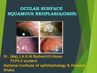
3. OCULAR SURFACE denotes involvement
of the conjunctiva or cornea
SQUAMOUS excludes other epithelial
cells such as basal cells and melanocytes
NEOPLASIA includes both dysplastic and
carcinomatous lesions.
4. Definition
The term Ocular Surface Squamous
Neoplasia [OSSN] presently refers to the
entire spectrum of dysplastic, pre-invasive
and malignant squamous lesions of the
conjunctiva and cornea
5. Lee and Hirst classified OSSN as:-
I. BENIGN DYSPLASIA
• Pseudoepitheliomatous hyperplasia
• Benign hereditary intraepithelial dyskeratosis
II. PREINVASIVE OSSN
• Conjunctival/corneal carcinoma in situ
III. INVASIVE OSSN
• Squamous carcinoma
• Mucoepidermoid carcinoma – aggressive
6. Epidemiology
Third most common ocular tumour after melanoma
and lymphoma
Caucasians
older age group(6-7 decade)
Males >females
Patiens with HIV and Xeroderma pigmentosum
present earlier
All young patients with OSSN should be screened for
HIV.
7. Risk factors
Ultraviolet light
Immunosuppression/ HIV
Human papillomavirus (HPV)- Type 16 & 18
Mutation or deletions of tumor suppressor
gene p53
8. Clinical Features
Patients may be asymptomatic or present with
chronic redness and irritation of the eye.
Visual acuity is not affected unless there is
extensive corneal involvement
In most cases, patient has the history of
several months.
9. Location
OSSN normally occurs in
Interpalpebral region arising from the
limbal stem cells, involving the bulbar
conjunctiva, the cornea or both of these
structures
10. Clinically :
The lesions are described as being
slightly elevated, variably shaped,
relatively sharply demarcated from the
surrounding normal tissues.
Accompanied by feeder blood vessels
Color vary from pearly gray to reddish
gray depending on the vascularity of the
tumor .
12. In clinical practice, gelatinous type
is the commonest. These lesion can be
Circumscribed, which are most common
Nodular variety, which has a propensity
for rapid growth
Diffuse variety, the least common,
which can masquerade as chronic
conjunctivitis
13. Diagnostic Tests
Diagnosis is most often made clinically.
Fluorescein or Rose Bengal are often used to
highlight & delineating the extent the lesion.
Rose bengal stain of corneal epithelial dysplasia
14. a. Anterior Segment Optical Coherence
Tomography (ASOCT)
b. Impression cytology
c. Confocal microscopy
d. High frequency ultrasound
e. Histopathology
Diagnostic Tests
15. a. Anterior Segment Optical Coherence Tomography
(ASOCT) :
Distinctive features of OSSN
hyper reflectivity
thickened epithelium
abrupt transition from normal to abnormal tissue
17. c.Confocal microscopy
- helpful in guiding treatment since it is able
to reveal cellular details.
- difficulte of use and limited field of view
18. d. Histopathology
DYSPLASIA:
Mild - less than a third thickness occupied by
atypical cells
Moderate - three quarters thickness occupied
by atypical cells
Severe - nearly full thickness occupied by
atypical cells
19. d. Histopathology
CARCINOMA IN SITU: as above with loss of the
normal surface layer
INVASIVE SQUAMOUS CELL CARCINOMA: as above
with basal epithelial layer has been breached and
invasion of the substantia propria has occurred.
24. Pterygium can be differentiated by
younger age
more triangular in shape
flatter rather than gelatinous
more linear blood vessels
Cause more symptoms
25. Papilloma may occur
younger patients
anywhere on the conjunctiva
may be sessile or pedunculated
has a punctate vascular pattern
Pedunculated exophytic
conjunctival papilloma
26. Malignant melanoma has a
regular smooth surface,
lacks gelatinous or leukoplakic surface
may be ulcerated
33. MITOMYCIN C
Most commonly used
A non cell cycle specific ALKYLATING AGENT
that acts by alkylating the cross-linked DNA
and inhibits DNA, RNA, and protein synthesis
0.04% four times a day for 1 week with two to
three cycles in alternate weeks
Success rates ranging from 87 to 100% have
been reported.
34.
35. 5-FU
Pyrimidine analogue that acts by
integrating with the DNA during S phase.
It also interferes with RNA synthesis.
It is used as 1% topical solution four times
a day for 1 week , followed by 30 days or
1–2 weeks off
Side effects are similar to MMC
36. Interferons
For OSSN: Topical IFN- α2b.
1 million IU/ml as 4 times a day until
resolution, and a month thereafter
More expensive than MMC and 5 FU
Requires prolonged treatment but has a
better safety profile
39. [2] Suspected OSSN 3 – 6 clock hours –
A diagnostic biopsy is required
Pre-invasive lesions
topical chemotherapy
Invasive lesions
surgery + cryotherapy is done after
chemoreduction with 4 to 6 cycles of topical
chemotherapy.
40. [3] OSSN > 6 clock hours –
A diagnostic biopsy is
required.
Pre-invasive lesions
Topical chemotherapy
Invasive
Surgery + cryotherapy is done after
chemoreduction with 4 to 6 cycles of topical
chemotherapy.
If there is no response to chemotherapy
Palliative radiotherapy or extensive surgery like
enucleation / exenteration may be required.
41. Metastasis
Regional and systemic metastases are also
uncommon.
Common sites of metastasis include pre-
auricular , submandibular and cervical lymph
nodes, parotid gland, lungs, and bone.
42. Recurrence
Ranges from 15-52%, average 30%
Higher in case of inadequate excision margins
More aggressive behaviour
43. Conclusion
Good clinical exam is sufficient for diagnosis.
Excision with cryotheraphy is successful but
can be associated with recurrence rates
Chemotherapeutic agents are usefull
alternative specially in recurrent, corneal &
annular lesion.
Notas do Editor
Gelatinous lesion with surface vessels ; leukoplakic lesion ; Papillomatous lesion ; extensive with corneal involvement
first is exfoliative cytology by using spatula scrapings or a cytobrush to collect the sample, and
second is impression cytology by using the collecting devices to collect the sample by contact with the
surface of the lesions. cellulose acetate paper (CAP)
Mild dysplasia; the basal cells are disordered with increased nuclear sizes and coarse nuclear chromatin.
B. Severe dysplasia; the epithelial cells are varied in shapes and sizes with large pleomorphic nuclei.
C. Carcinoma in situ: the entire thickness of the epithelium is composed of dysplastic cells bearing pleomorphic nuclei.
D. Invasive squamous cell carcinoma; the invasive nest in the stroma is composed of bizarre cells similar to those in the epithelium. The nuclei are plemorphic with thick nuclear membranes and prominent nucleoli (Hematoxylin and Eosin stain.
Once the lesion is removed en bloc, the specimen is marked in the proper orientation with sutures and then transferred to a piece of pencilmarked
cardboard. The specimen is sent to pathology in formalin.
MMC can be stored at room temperature (22°C) for up to 1 week; if refrigerated (4°C), it can maintain
90% of its activity for longer.
the one week on, one week off regimen prevents damage to more slowly dividing epithelial cells and limbal stem cells, allowing them to repair their DNA. Allowing time for complete epithelial healing before application of MMC is important in avoiding the more serious complications such as corneal epitheliopathy, scleral ulceration, uveitis, cataract, and glaucoma
This one week on and one week off drug regimen has the added advantage of good efficacy and better tolerance.
More aggressive because of the tissue disruption associated with the primary excision theoretically enhancing the ability of the tumor cells to enter the eye