Cerebral palsy
•Transferir como PPTX, PDF•
181 gostaram•18,455 visualizações

short yet comprehensive coverage of the topic of cerebral palsy and management

Recomendados
Mais conteúdo relacionado
Mais procurados
Mais procurados (20)
Semelhante a Cerebral palsy
Semelhante a Cerebral palsy (20)
Mais de Harjot Gurudatta
Último
Último (20)
Cerebral palsy
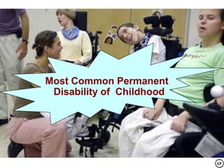
- 1. Most Common Permanent Disability of Childhood
- 2. CEREBRAL PALSY By: Harjot Singh Gurudatta Moderator: Dr. Rohit Sharma
- 3. William John Little (18101894) • In 1860s, known as "Cerebral Paralysis” or “Little’s Disease” • After an English surgeon wrote the 1st medical descriptions
- 4. CEREBRAL PALSY (CP) • Cerebral“- Latin Cerebrum; – Affected part of brain • “Palsy " -Gr. para- beyond, lysis – loosening – Lack of muscle control
- 5. CEREBRAL PALSY • A motor function disorder – caused by permanent, non-progressive brain lesion – present at birth or shortly thereafter. (Mosby, 2006) • Non-curable, life-long condition • According to a recent definition CP “describes a group of permanent disorders of the development of movement and posture,causing activity limitation, that are attributed to non-progressive disturbances • that occurred in the developing fetal or infant brain. The motor disorders of cerebral palsy are often accompanied by disturbances of sensation, perception,cognition, communication, and behaviour; by epilepsy; and by secondary musculoskeletal problems”
- 6. CEREBRAL PALSY A Heterogenous Group of Movement Disorders
- 7. In CP • Muscles are unaffected • Brain is unable to send the appropriate signals necessary to instruct muscles when to contract and relax
- 9. An insult or injury to the brain – Fixed, static lesion(s) – In single or multiple areas of the motor centers of the brain – Early in CNS developmentSevere deprivationon of oxygen or blood flow to the brain – Hypoxic-ischemic encephalopathy or intrapartal asphyxia
- 10. Etiology Prenatal(MC) • I, iron def.,poor –nut. • Inf, UTI, high fever • Chorioamniotis • HTN, DM • Teratogens • Poor ANC • Rh ? • Twins • Fetal vasculopathy • Maternal drugs/smoking(>30) Perinatal • Birth asphyxia • Breach/vacuum/forc • Premature / LBW(>60/1000) • IUGR • Hyperbilirubenemia • Intraventricular hemorrrhage • Sepsis, pneumonia, meningitis • Develop. Malformation, • abruptio Postnatal •CNS infections •Head injuries •Seizures •Hypoxic damage •Hyperpyrexi a damage •Stroke
- 12. Classification of CP According to: 1. Neurologic deficits 2. Type of movement involved 3. Area of affected limbs
- 13. 1. Accdg. to Neurologic Deficits • Based on the - extent of the damage - area of brain damage • Each type involves the way a person moves
- 14. 3 MAIN TYPES 1. PYRAMIDAL - originates from the motor areas of the cerebral cortex 2. EXTAPYRAMIDAL - basal ganglia and cerebellum 3. MIXED
- 16. Spastic CP • Increased muscle tone, tense and contracted muscles – Have stiff and jerky or awkward movements. – limbs are usually underdeveloped – increased deep tendon reflexes • most common form • 70-80% of all affected
- 17. Types of Spastic CP A/C LIMB INVOLVED • Paraplegia • Diplegia LL>UL , mc, normal iq,walks eventually,though delayed • Hemiplegia UL>LL, seizures+, sensory changes in ul, limb length discrepancy • Quadriplegia only slight H N control loss,low iq, highest hip subluxation,cognitive deficiency, • Monoplegia –one limb (extremely rare) • Whole body with seizure, drooling hn control loss
- 18. Diplegia/ Paraplegia •both legs w/ slight involvement Elsewhere May also have Contractures of hips and knees and talipes equinovarus (clubfoot). •both legs
- 19. Hemiplegia limbs on only one side
- 20. • Hemiplegia on right side – Hip and knee contractures – Talipes equinus (“tip-toeing” - sole permanently flexed) – Asteriognosis may be present. (inability to identify objects by touch)
- 21. Quadriplegia
- 22. • Spastic Quadriplegia Characteristic “scissors” positions of lower limbs due to adductor spasms.
- 23. Athetoid/ Dyskinetic CP • Fluctuating tone – involves abnormal involuntary movements – that disappear during sleep and increase with stress. – Interferes with speaking, feeding, reaching, grabbing, and any other skills • 20% of the CP cases, Wormlike movements • Slow, uncontrolled motion, writhing or twisting in character in the face, extremities, and torso. • Dystonia - when held as a prolonged posture – Grimacing, drooling and dysarthria. – Adductor spasm
- 24. Movements may become choreoid (rapid, irregular, jerky) and dystonic (disordered muscle tone, sustained muscle contractions) especially when stressed and during the adolescent years.
- 25. Ataxic CP • Poor balance and lack of coordination – Wide-based gait – Depth perception usually affected. – Tendency to fall and stumble – Inability to walk straight line. – Least common 5-10% of cases
- 26. MIXED CP • A common combination is spastic and athetoid • Spastic muscle tone and involuntary movements. • 25% of CP cases, fairly common
- 27. DEGREE OF SEVERITY 1. Mild CP- 20% of cases 2. Moderate CP50% - require self help for assisting their impaired ambulation capacity. 3. Severe CP30%; -totally incapacited and bedridden and they always need care from others.
- 28. The Peabody Development Motor Scales • In-depth assessment • 6 Subtests include: – – – – – – Reflexes Stationary Locomotion Object Manipulation Grasping, Visual-Motor Integration. – The subtests yield a gross motor quotient – a fine motor quotient – a total motor quotient. • Ages covered: from birth through five years of age
- 29. Denver Test II • Developmental Screening Test • Cover 4 general functions: – personal social (eg. smiling), – fine motor adaptive (eg. grasping & drawing) – language (eg. combining words) – gross motor (eg. walking) Ages covered: from birth to 6 years
- 31. History + Exam – Include all that may predispose an infant to brain damage or CP • Risk factors • Psychosocial factors • Family adaptation • Often admitted to hospitals for corrective surgeries and other complications. – – – – – – Respiratory status Motor function Presence of fever Feeding and weight loss Any changes in physical state Medical regimen P osturing / Poor muscle control and strength O ropharyngeal problems(drooling?swallowing?feeding?) S trabismus/ Squint T one (hyper-, hypotonia)Muscle strength testing E volutional maldevelopment R eflexes (e.g. increased deep tendon) *Abnormalities 4/6 strongly point to CP
- 33. Early Signs Infancy (0-3 Months) • Stiff or floppy posture • Excessive lethargy or irritability/ High pitched cry • Poor head control • Weak suck/ tongue thrust/ tonic bite/ feeding difficulties
- 34. neonatal reflexes Moro’s reflex Startle response; Startle reflex; Embrace reflex when the infant feels as if it is falling,1st abduct ,2nd adduct,ie embrace Asymmetric tonic neck reflex "fencing position“ -- head to one side, arm & leg on that side extended, opposite limbs flexed Placing reflex When the dorsal (back) side of the hand or foot is placed on the edge of a surface, such as a table, the infant will lift the extremity and place it on the flat surface. Landau reflex When the infant is held in a horizontal prone position, the infant will lift head and extend the neck and trunk. When the neck is passively flexed, the entire body will flex 3m-2y
- 35. Parachute Reflex • When held around the waist in a horizontal prone position and then lowering the infant slowly, head first to the surface. By age 6 to 8 months the infant should respond by extending the arms and hands to break the “fall”. If this response is asymmetrical it indicates an unilateral motor abnormality.
- 36. CHILD with CP
- 37. Late infancy • Inability to perform motor skills as indicated: – Control hand grasp by 3 months – Rolling over by 5 months – Independent sitting by 7 months • Abnormal Developmental Patterns: – – – – Hand preference by 12 months Excessive arching of back Log rolling Abnormal or prolonged parachute response
- 38. Abnormal Developmental Patterns after 1 year of age: • “W sitting” – knees flexed, legs extremely rotated • “Bottom shuffling” Scoots along the floor • Walking on tip toe or hopping • Bleck ”not sitting independently @4 yrs, not walking @ 8 yrs bad”
- 39. ASSO. CONDITIONS • Hearing and visual problems • • • • • Bladder and bowel control problems, digestive problems Sensory integration (gastroesophageal reflux) problems Failure-to-thrive, Feeding • Skeletal deformities, dental problems problems • Mental retardation and Behavioral/emotional learning disabilities in difficulties, some Communication • Seizures/ epilepsy disorders
- 40. DIAGNOSIS • • • • Physical evaluation, Interview MRI, CT Scan EEG Laboratory and radiologic work up Assessment tools – i.e. Peabody Development Motor Skills, Denver Test II
- 42. - No treatment to cure cerebral palsy. - Brain damage cannot be corrected. • Crucial for children with CP: –Early Identification; –Multidisciplinary Care; and –Support
- 43. I. Nonphysical Therapy “The earlier we start, the more improvement can be made” -Health worker
- 44. A. General management - Proper nutrition and personal care B. Pharmacologic Intrathecal, Baclofen - control muscle spasms and seizures, Glycopyrrolate -control drooling Pamidronate -may help with osteoporosis.
- 45. Baclofen • Oral delivery was common but sedation is more, so Intrathecal Baclofen Using a pump Delivered directly to the spinal fluid • GABA agonist – inhibits release of excitatory neurotransmitter at level of spinal cord • It is commonly used in a starting dose of 1.25 - 2.5 mg BD orally To avoid brain effects
- 46. BOTOLUNIM TOXIN The toxin is produced by the anaerobic sporeforming bacterium Clostridium botulinum, of which eight immunologically distinct serotypes – A, B, C1, C2, D, E, F, and G – have been identified. Type A and B are used clinically in variousconditions like focal tissue spasms, blepharospam, dystonia, achalia, urology, cerebral palsy, cosmetic surgeries, toe clawing. Relief of athetosis and dystonia - is difficult occasionally levo-dopa for severe athetosis and carbamezepine for dystonia may be helpful. Thalamotomy for athetoid CP, stereotactic dentatomy and chronic cerebellar stimulation via implanted electrode . Multiple injection sites to avoid toxicity. acetylcholine-blocking chemical denervant Requires adjunctive therapy like splints, orthoses, casting, manual stretch, posturing The children in CT receive bilateral Botox injection @12 u/kg with topical anaesthesia and analgesic cover/ sedation
- 47. C. Surgery -To loosen joints, -Relieve muscle tightness, - Straightening of different twists or unusual curvatures of leg muscles - Improve the ability to sit, stand, and walk.
- 48. Selective posterior rhizotomy • Best for : spastic diplegia, 4-8 yrs, no previous surgery, no contractures, no extra pyramidal signs • Not done in hemi/ quadriplegia
- 49. How it Works • The Sensory nerve fibers are exposed, then stimulated and the responses of the leg muscles are observed. Those that have an abnormal or excessive response are severed. Those with a normal response are left intact. • Intensive rehabilitation is required after the surgery, usually up to six weeks, followed by physical therapy on an ongoing basis
- 50. Walker cp D. Physical Aids Orthosis, braces and splints - Keep limbs in correct alignment - Prevent deformities. - Ankle foot orthosis, knee ankle foot orthoses Positioning devices -Enable better posture Walkers, special scooters, wheelchairs - make it easier to move about. Active exercises - Passive ROM exercises - Passive stretching - Bracing Spiral thigh brace
- 51. E. Special Education Rehabilitation - To meet the child's special needs - Improve learning. - Vocational training can help prepare young adults for jobs H. Other Treatment - Therapeutic electrical stimulation, Acupuncture, Hyperbaric therapy Massage Therapy might help
- 52. Cerebral palsy Orthopedic Procedures Usually multiple deformities at different joints Knowledge of complex effects each deformity has on other lower extremity joints Diiferentiate between primary deformity : needs treatment compensatory deformity : can improve without intervention
- 53. Cerebral palsy Prerequisites for effective surgery • Type : spastic • Extent : hemiplegics / diplegics : good results quadriplegics : minimal improvement • Age : 3- 12 years • IQ : good • Good upper limb function : for walking • Underlying muscle power : not weak • Walker / non-walker :walker surgery hardly changes state but improves gait, to get him better from current state
- 54. Cerebral palsy Timing For Orthop Surgery • For structural changes : Early e.g. Hip subluxation , usually <5 years • To improve function ( gait ) : defer until walking ( independently / with aids ) until gait pattern develops and could be assessed walking : 18 – 21 months in hemiplegia 3 – 4 years in spastic diplegia • Optimum time of lower extremity surgery 5 – 7 years: can analyze and observe gait pattern
- 55. Avoid birthday surgeries ?ETA Equinus ? Hams Crouch ? Psoas Flexion ? Rectus Femoris Stiff Knee Ok
- 56. Cerebral palsy Hip Deformities • Dynamic deformities • Tight adductors – scissoring • Tight flexors – with pelvic inclination compensatory knee / ankle/ trunk deformities • Hip subluxation / dislocation : not common in walking patients with diplegia • Wind-swept deformity : in quadriplegics • Internal rotation : in spastic hemi & diplegics
- 57. Adductor Deformity Superficial layer - Pectineus Adductor longus gracilis • Needs to be corrected if gait is disturbed<20 abduction and scissoring. • Do open Adductor tenotomy of Adductor longus.Gracilis and some add. Brevis can be released • Its important not To include much of adductor Brevis and spare post. Obt. Nerve • Long leg casts given 3 week
- 58. Thomas test for ruling out knee ffd Holts test for hamstring tightness Adductor and medial hamstring?? Explained later Ely test for RF Spasm • Ilio-psoas ( the main and most powerful ) • • • • Sartorius Tensor fascia lata Rectus femoris Adductors FLEXION DEFORMITY Ascertain the site of primary anomaly Hip flexion deformity > 20o needs surgery <30DEG Iliopsoas Recession and Z plasty In ambul. >3o Rectus femoris, Sartorius, Glutei ant. Fibre
- 59. Cerebral palsy Hip Problems in Spastic Quadriplegia Hip at risk (valgus, anteverted, acet. Dysplasia, increased NS angle,flexion contracture>20, abduction <30) Femoral Anteversion more than true Valgus
- 60. • When a hip at risk is identified,a program of aggressive physical therapy and abduction splinting typically is started, later soft tissue release of contracted tendons is indicated. • In Subluxated hip, younger children, soft tissue releases alone may be sufficient, but most patients with hip subluxation require osteotomy in addition to soft tissue release. Operative correction of femoral valgus and anteversion and acetabular dysplasia is necessary at this stage to prevent further subluxation and dislocation. osteotomies described by Salter, Pemberton, Dega, Ganz, and Steel and salvage-type osteotomies such as the Chiari and shelf may be done
- 61. Cerebral palsy Spastic Diplegia – The Knee • Crouched gait : - tight hamstrings : needs hamstring release - could be secondary to weak triceps surae • Type of surgery : - - lengthening : best ( medial first , ? then lateral) Ascertain primary deformityThe Hamstring Test Holt’s method • • Hip flexed 90 degrees Popliteal angle degrees less than full extension More hip flextion increases flexion
- 62. Fractional medial hamstring lengthening Lengthening hamstrings : reduces hip extension power increases hip flexion Add hip flexor ( Psoas ) lengthening if concomitant hip flexion is present Add distal rectus femoris release (ELY test) if concomitant cospasticity(stiff knee gait) of quadricep and hamstrings +nt exacerbates knee hyperextension OR transfer RF to Sartorius, smt, iliotibial band
- 63. Cerebral palsy Spastic Diplegia – The Foot & Ankle Toe Walking • Dynamic : Treat by : Bracing Spasticity reduction Surgery ( careful ! ) • Fixed : Treat by : Serial casting Surgery SILVERSKOLD
- 64. LENGTHENING OF THE ACHILLES TENDON For equinus Indications : - fixed deformity - tight TA Problems : over lengthening esp in z Weak push off Types : z -plasty sliding : - percutaneous - open
- 65. Cerebral palsy Spastic Diplegia – The Foot & Ankle Equino - varus • Not very common but more disabling and Most Imp. Is Tibialis post contracture or Tibi. Ant weakness • Treatment : - split transfer of tib. post. tendon lateral half, posterior to interosseous memb., to peroneus brevis laterally - elongation of tib. Post. and split transfer of tib. ant. Laterally Dwyer closing wedge osteotomy of calcaneus for varus heel. Full transfer can cause weakness
- 66. • • • • • Equino - valgus More frequently seen in di/ quadri Tight TA and Talonavicular subluxation No perfect muscle balancing procedure The gastrocnemius-soleus acts as the primary deforming force and TA lenghthening is must Treatment : ETA and subtalar arthrodesis Osteotomies
- 67. Intoeing • Usually caused by femoral ante-version • Internal tibial torsion adds to intoing • If severe : Derotation osteotomy Delay to late childhood if possible • Derotation osteotomy of femur might cause tightening of medial hamstrings ( might need lengthening ) Femur anteversion
- 68. "Time and gravity are enemies of very aging body, especially mine." - Adult with CP “A disabled child has the right to enjoy a full and decent life, in conditions which ensure dignity, promote self-reliance and facilitate the child’s active participation in the community.” -UN Convention on the Rights of the Child. 1989. THANK YOU FOR PATIENTLY LISTENING!!!