Thyroid gland
•Transferir como PPTX, PDF•
37 gostaram•16,405 visualizações

thyroid gland anatomy

Recomendados
Mais conteúdo relacionado
Mais procurados
Mais procurados (20)
Semelhante a Thyroid gland
Semelhante a Thyroid gland (20)
Mais de Hamzeh AlBattikhi
Mais de Hamzeh AlBattikhi (20)
Último
Último (20)
Thyroid gland
- 2. Size & Shape • One of the largest endocrine glands. • Its weight is about 25 grams. • Its butter-fly shapped organ.
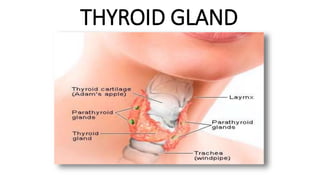
- 3. Structure Its formed of the following parts: • 2 Lateral lobes each lobe is pear-shaped. Each lobe is pyramidal in shape, with its apex directed upward and its base directed downward. • A narrow median isthmus connecting the 2 lateral lobes. • A small pyramidal lobe, (28%-55% of population), It is of conical shape and extends from the upper part of the isthmus, up across the thyroid cartilage to the hyoid bone.
- 4. Location of the gland • Its Situated on the anterior side of the neck, lying against and around the larynx and trachea, reaching posteriorly the oesophagus and carotid sheath. It starts cranially at the oblique line on the thyroid cartilage (just below the laryngeal prominence, or 'Adam's Apple'), and extends inferiorly to approximately the fifth or sixth tracheal ring.
- 5. Capsules of the thyroid gland It has 2 Capsules: • True Fibrous capsule enclosing the gland. • False fascial capsule derived from the pretrachial fascia.(Thickend laterally forming the lat. Ligament of berry which fixes the gland to the cricoid cartilage)
- 6. Relations of the gland 1. Relations of the isthmus. 2. Relations of the lateral lobes.
- 7. 1. Relations of the isthmus. The isthmus has 2 surfaces (Ant. & Post.) & 2 borders (Upper & lower) Anterior Posterior Upper Lower 1. Skin & superficial fascia. 2. Ant. Jugular veins. 3. Deep fascia. 4. Sternohyoid & sternothyroid muscles. 1. Trachea (2nd, 3rd & 4th rings) 1. Related to the sup. Anastomotic a. (between rt & lft sup. Thyroid aa) 2. The pyramidal lobe may project upwards from the isthmus. 1. Give rise to the Rt & Lt Inf. Thyroid veins. 2. The thyroid ima a. enters the lower border. 3. An Inf. Anasomotic a. runs along the lower border of the isthmus.
- 8. Anterior 1. Skin & superficial fascia. 2. Ant. Jugular veins. 3. Deep fascia. 4. Sternohyoid & sternothyroid muscles.
- 9. Lower 1. Give rise to the Rt & Lt Inf. Thyroid veins. 2. The thyroid ima a. enters the lower border. 3. An Inf. Anasomotic a. runs along the lower border of the isthmus. Upper 1. Related to the sup. Anastomotic a. (between rt & lft sup. Thyroid aa) 2. The pyramidal lobe may project upwards from the isthmus.
- 10. 2. Relations of the lateral lobes. each lobe has 3 surfaces: Posterior Medial Anterolateral 1. 2 Parathyroid glands ( sup. & inf. ) embedded in the post. Surface. 1. 2 arteries: A- Common Carotid a. ( inside the carotid sheath) B- Inf. Thyroid a. before it enters the gland. 1. Its upper part related to: A- Layrnx (thyroid & cricoid cartilages & cricothyroid m.) B- Pharynx (Inf. Constrictor m.) C- External laryngeal n. 2. Its lower part is related to: A- trachea B- oesophagus C- Recurrent laryngeal n. in between 1. Skin, superficial fascia & deep fascia. 2. Its upper part is crossed by sup. Belly of omohyoid. 3. Its middle part is covered by sternohyoid & sternothyroid m. 4. Its lower part is overlapped by the ant. Border of sternomastoid.
- 11. Posterior 1. 2 Parathyroid glands ( sup. & inf. ) embedded in the post. Surface. 1. 2 arteries: A- Common Carotid a. ( inside the carotid sheath) B- Inf. Thyroid a. before it enters the gland.
- 12. Medial 1. Its upper part related to: A- Layrnx (thyroid & cricoid cartilages & cricothyroid m.) B- Pharynx (Inf. Constrictor m.) C- External laryngeal n. 2. Its lower part is related to: A- trachea B- oesophagus C- Recurrent laryngeal n. in between
- 13. Anterolateral 1. Skin, superficial fascia & deep fascia. 2. Its upper part is crossed by sup. Belly of omohyoid. 3. Its middle part is covered by sternohyoid & sternothyroid m. 4. Its lower part is overlapped by the ant. Border of sternomastoid.
- 14. Arterial supply to the gland • The thyroid is supplied with arterial blood from the: 1. superior thyroid artery, a branch of the external carotid artery. 2. inferior thyroid artery, a branch of the thyrocervical trunk. 3. sometimes by the thyroid ima artery, branching directly from the subclavian artery.
- 15. Venous Drianage • venous blood is drained via: 1. superior thyroid veins, emerges from the apex of each lateral lobe, draining in the internal jugular vein. 2. Midlle thyroid viens, emerges from the lower part of each lat. Lobe. 3. inferior thyroid veins, emerges from the isthmus & lower part of the lat. Lobe. draining via the plexus thyreoidea impar in the left brachiocephalic vein.
- 16. Lymphatic drainage • The lymphatic of the gland drain into: 1. Prelaryngeal L.Ns, infront of cricothyroid memb. 2. Pretracheal L.Ns, infront of trachea. 3. Paratracheal L.Ns, alongside the trachea. 4. Upper & lower deep cervical L.Ns, along the I.J.V . 5. Brachiocephalic L.Ns.
- 17. Thank You Done by: Dr. Hamzeh Al-Battikhi